Tumor-targeting bacteria are becoming increasingly more attractive as a theranostic platform. They can be genetically engineered to localize tumors and trigger a therapeutic effect while carrying detectable labels for additional diagnostic imaging 1, 2, 3. Current imaging strategies for the development of bacteria-mediated cancer therapy in small animals rely largely on optical methods like bioluminescence and fluorescence for visualization 4, 5. These optical approaches are incapable of providing additional anatomical or physiological information, nor can they offer the resolution necessary to non-invasively analyze the details of bacterial cancer targeting and efficacy 6, 7. Radiological methods like magnetic resonance imaging (MRI) or positron emission imaging (PET) have also been explored, but these techniques suffer from limitations in sensitivity and resolution, respectively. Furthermore, the high cost associated with these methods and the use of radioisotopes (PET) makes them unsuitable for routine applications. Altogether, these challenges limit the possibilities for detailed studies of novel theranostic agents, their mechanisms of action, and the responses of tumors and their hosts to treatment.
Optoacoustic (OA) imaging, based on optical excitation and ultrasound detection, represents a promising solution to the above limitations, as it is a powerful molecular imaging modality owing to the unique combination of high spatio-temporal resolution, deep penetration and spectrally-enriched contrast 7. This method therefore offers many possibilities for labeling strategies. Moreover, because the detection of ultrasound is not limited by photon scattering, it enables scalable high-resolution imaging deep in the tissue (20–200 µm) 7. In particular, multispectral optoacoustic tomography (MSOT) was proven to be effective for preclinical imaging of tumors in small animals and has been implemented in a handheld clinical system for tumor assessment 7, 8, 9. MSOT is regularly used to detect contrast from endogenous absorbers such as hemoglobin, lipids and water, or from exogenous absorbers such as dyes or nanoparticles 8. Labeling bacteria with transgene fluorescent or chromoproteins for detection in OA has however thus far proven unsuccessful due to weak acoustic signals and often poor photostability, limiting sensitivity and observation time 10; photo-switching chromoproteins provide a promising alternative but their development and use is still in its infancy 11. In contrast, a particularly strong OA signal can be generated by expressing the gene encoding tyrosinase, which is the rate-limiting enzyme in the production of melanin, a strong contrast agent for OA imaging 12, 13.
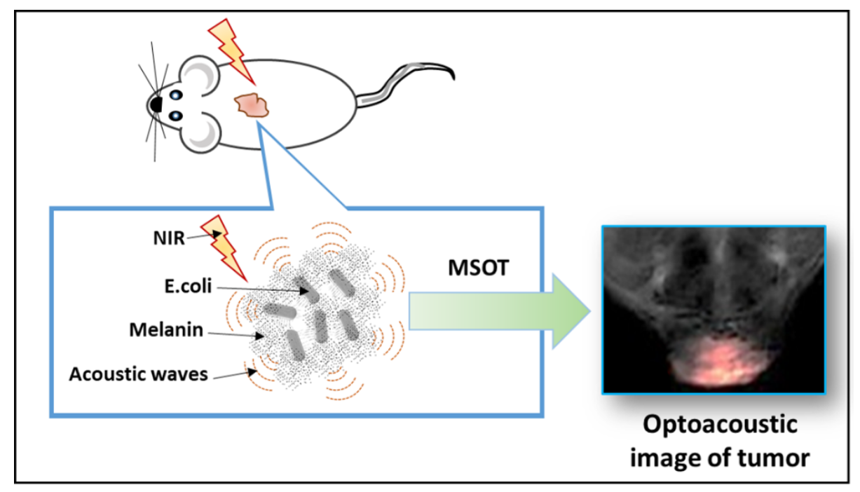
In the presence of the substrate l-tyrosine, cells produce melanin which in turn allows their detection with great sensitivity by MSOT 12, 14, 15. Here, we use MSOT to monitor bacterial targeting, infiltration and proliferation specifically in the tumor microenvironment based on E. coli that express the transgene for tyrosinase. After injection into mouse tumors, the enzyme is secreted out of the bacterial cells and converts l-tyrosine in the surrounding environment into melanin. We were able to track accumulation of the bacteria in the tumor, which was also confirmed using histology via labelling of melanin and bacteria. This study suggests that tyrosinase-expressing E. coli may be an effective optoacoustic imaging agent with potential utility in bacterial therapy. To our knowledge, this is the first report of optoacoustic imaging of melanin contrast in tumor-targeting bacteria in-vivo.