Our work has identified active foci of S. mansoni transmission in six collection sites from the municipalities of Franciscópolis, Jequitinhonha, Joaíma, Malacacheta, and Ponto dos Volantes, confirming that these are endemic areas in the state of Minas Gerais. Molecular approaches enabled the detection of infected snails with higher accuracy than parasitological methods, reinforcing the need for additional tools to precisely map and monitor endemic areas, and, in the future, achieve schistosomiasis control and elimination.
The transmission of S. mansoni has been reported so far in 19 Brazilian states. The state of Minas Gerais includes around 70% of the endemic areas, being the subject of many studies over the years (3, 40–44). The Mucuri and Jequitinhonha Valleys are both very poor regions within the state of Minas Gerais. The Brazilian Institute for Geography and Statistics (IBGE) (45) calculated, in 2010, the human development index adjusted for the context of each municipality (IDHM), and estimated that Franciscópolis, Jequitinhonha, Joaíma, Malacacheta, and Ponto dos Volantes had indices ranging from 0.587 to 0.618, which means a low to medium level of socio-economic development, according to the United Nations Development Program (UNDP) (46). Poverty contributes to increased contact of individuals with contaminated water, as populations from underprivileged areas usually seek natural watercourses for work and leisure activities, raising schistosomiasis transmission rates (41, 43). In addition to knowledge regarding socio-economic aspects and habits, biological and ecological features are relevant for understanding the transmission process in each region (47). The temperature from the water body is a determining factor for the development of both the snail and the parasite, and prospective studies have predicted the future impact of climate change on the dynamics of transmission (48). Specific vegetation and parasitism in snails can affect not only their populational density but also cercarial abundance (49). Rain volume has a close relationship with the density of Biomphalaria snails and the positivity rate found among them for S. mansoni infection. According to Calasans et al (47), the abundance of Biomphalaria snails is negatively related to the rainfall, while Biomphalaria infection rises during wet periods of the year. Historically, the Mucuri and Jequitinhonha Valleys have low rainfall levels in all seasons (50). Between July and August 2019, when our surveys were undertaken, the accumulated rainfall ranged from 10–30 mm3 according to the Meteorology National Institute (INMET) (51), being the lowest pluviometric measurements in that year. This indicates that the density of infected Biomphalaria snails found in our survey might have been underestimated due to the characteristics of rainfall while it was undertaken.
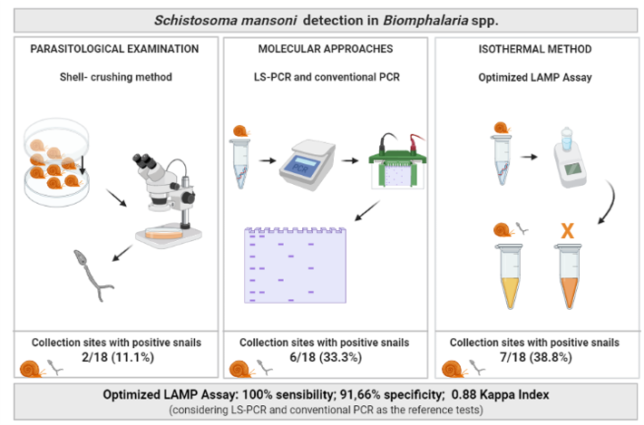
In this study, a total of 1,001 snails were collected from 18 sites in 5 municipalities of the Mucuri and Jequitinhonha Valleys in the state of Minas Gerais, Brazil. Parasitological examination of the collected snails by the shell-crushing method has enabled detection of trematode larvae in 16.6% (3/18) of the collection sites, with most of them being due to infection with S. mansoni (2/3).
PCF-RFLP enabled the identification of B. glabrata in 72.2% (13/18) of the surveyed sites, indicating that these are potential areas for the transmission of schistosomiasis, as this snail species is the main intermediate host of the parasite in Brazil due to its high compatibility with S. mansoni and wide distribution throughout the country (3, 47, 52, 53). Biomphalaria kuhniana (Clessin, 1883) were found in 27.8% (5/18) of the surveyed sites. Although this species does not have importance in the transmission of schistosomiasis, the high morphological similarity showed between B. kuhniana and the intermediate host B. straminea, highlights the importance of the correct identification of these snails in order to properly map potential foci for schistosomiasis (54–56).
Part of the snails collected in this study was deposited in the Medical Malacology Collection (Fiocruz-CMM). This collection was founded in 1993, and since then has accumulated a large number of representative specimens, especially with regard to Biomphalaria snail hosts in Brazil, having currently more than 16,800 snails from all over the world (57, 58). According to the database of Fiocruz-CMM available on the CRIA website (59), the presence of Biomphalaria snails has been previously reported in 14 of the 23 municipalities included in the Mucuri Valley. Biomphalaria glabrata, B. straminea, and Biomphalaria schrammi (Crosse, 1864) have been collected in 9, 11, and 2 municipalities, respectively. The last survey in Fransciscópolis happened in 2013, and in Malacacheta in 2015. In our study, we have identified for the first time the presence of B. kuhniana in Malacacheta and B. glabrata in Franciscópolis. The Jequitinhonha Valley comprises 55 municipalities, and Biomphalaria snails have been reported in 33 of them. Biomphalaria straminea, B. glabrata, B. kuhniana, and B. tenagophila have been previously collected in 21, 18, and one municipality, respectively. The most recent surveys were conducted in Jequitinhonha in 2006, in Joaíma in 2014, and in Ponto dos Volantes in 2012. The Jequitinhonha Valley results from our study matches the data obtained from previous ones. We have found that in almost all the areas where we conducted our surveys, the presence of B. glabrata has been maintained over the years. Data from Fiocruz-CMM combined with our findings reinforce the importance of constant monitoring of these areas.
The presence of trematode infection in snails was investigated using a multiplex PCR protocol that enables the differentiation of four important families commonly found parasitizing Biomphalaria snails (37, 60–62). Schistosomatidae species were detected in 44.4% (8/18) of the study sites, while Echinostomatidae and Strigeidae were each found in 5.5% (1/18). Snails from the collection sites MV 41 and JV 04 that were found shedding S. mansoni cercariae in the parasitological examination had their infection confirmed by multiplex PCR due to the amplification of 140 bp target corresponding to the Schistosomatidae family. In five further sites from the Mucuri Valley, and one from the Jequitinhonha Valley, the presence of Schistosomatidae infection in snails was also detected. Even though the amplification of Schistosomatidae DNA does not necessarily mean the presence of S. mansoni itself, this result raises concern that these areas might be potential foci for schistosomiasis. As expected, no amplification was observed in the snails collected at the JV 03 site, since the set of primers used does not cover the Spirorchiidae family isolated from this location during the parasitological examination of snails. As the primers used by the multiplex PCR only amplify four trematode families, it is not possible to confirm that snails from the remaining collection sites are not infected by other trematode families.
LS-PCR and conventional PCR were both able to detect the presence of S. mansoni in 33.3% (6/18) of the surveyed sites. The optimized LAMP assay developed in this work revealed the presence of in snails from 38.8% (7/18) of the collection sites, when amplification was visualized using polyacrylamide gels, having an “almost perfect” Kappa agreement with LS-PCR and conventional PCR, with 100% sensitivity, and 91.66% specificity. When the chosen method to check the result was visual inspection of reaction tubes after the addition of an intercalating dye, amplification was detected in six collection sites, presenting the same result obtained when LS-PCR and conventional PCR were used. A very weak amplicon corresponding to the Schistosomatidae family was detected in snails from the collection site MV 03 but no amplification was detected with LS-PCR and conventional PCR using this sample as a template. The apparent LAMP product from this sample, when visualized using a polyacrylamide gel, raises the hypothesis that the LAMP assay was more sensitive than LS-PCR and conventional PCR in detecting S. mansoni infection in snails. However, the limit of detection of each method indicates that this is not the case, since LS-PCR can detect up to 1 pg of S. mansoni DNA (18), conventional PCR up to 0.01 pg (Additional file 3) and optimized LAMP assay presented a limit of detection of 0.1 ng. Therefore, LAMP is less sensitive than the other evaluated methods, suggesting that snails from the site MV 03 are not infected with S. mansoni. Although several trematode samples have been used to test the specificity of the optimized LAMP assay, samples from other species that belong to the Schistosomatidae family have not been used. The analysis of the results from the LAMP assay combined with multiplex PCR suggests cross-reactivity between members of the same family. In the Brazilian context, other schistosomes do not have much relevance to human health, as only S. mansoni causes schistosomiasis in this country. Avian schistosomes have been reported causing cercarial dermatitis in countries from the Northern Hemisphere, but the occurrence of this condition has not been reported so far in Brazil (63–65). As an alternative to overcome this false-positive result, we suggest that the visual inspection of reaction tubes by the naked eye should be prioritized, instead of running the LAMP products in gels. This visualization strategy not only reduces the possibility of false results, but also makes the assay more applicable directly in the field in low infrastructure conditions.
The LAMP assay was first described in 2010 (66), and since then this technique has been used to detect many pathogens, including S. mansoni, but mostly in human samples (38, 67–71). The applicability of LAMP for screening snails to characterize transmission areas is very promising (49), and has been tested by several authors (26–30). Molecular techniques can detect the presence of S. mansoni even when snails are not shedding cercariae, which would provide valuable information for surveillance services, as in many endemic areas collected snails rarely shed cercariae even though schistosomiasis transmission remains present. This failure to find cercarial shedding can be misleading, often giving the false impression of low or even absent transmission (49). Although molecular methods such as conventional PCR, DNA sequencing, and qPCR can properly fulfill this gap, these techniques are inappropriate for laboratories with limited resources, as they require expensive machinery and technical expertise, raising the associated costs of each reaction. Among all advantages of the using isothermal assays, the possibility of performing the test directly in the field in laboratories with limited infrastructure is undeniable. Our group optimized the LAMP assay using the primers described by Fernández-Soto et al (38). When following the exact conditions described by Gandasegui et al (29), non-specific amplification was detected in trematode samples belonging to the families Diplostomidae and Spirorchiidae (Additional file 5). Even though the optimization resulted in a reduction in the analytical limit of detection (from 1 fg to 0.1 ng), our findings confirmed that the amount of S. mansoni DNA that can be detected by the assay is sufficient to detect, by visual inspection alone, the presence of the parasite 7 days after exposure of the snails to 8 miracidia, and in pooled samples.
By the use of the optimized LAMP assay, we detected three times more the infection by S. mansoni in snails when compared to parasitological examination using shell-crushing method, revealing six active transmission areas for schistosomiasis in both Mucuri and Jequitinhonha Valleys. Molecular methods also allowed the mapping of potential transmission foci through the identification of B. glabrata in much of the surveyed area, as demonstrated in the maps generated by this study.