General rules for chemical synthesis
1H NMR and 13C NMR spectra were recorded using TMS as the internal standard on a Bruker BioSpin GmbH spectrometer at 400 and 101 MHz, respectively. The high-resolution mass spectra were obtained using a Shimadzu LCMS-IT-TOF mass spectrometer. Reagents used in the synthesis were obtained commercially and used without further purification, unless otherwise specified. The reactions were monitored by thin layer chromatography (TLC) on glass-packed precoated silica gel plates and visualized in an iodine chamber or with a UV lamp. Flash column chromatography was performed using silica gel (200−300 mesh) purchased from Qingdao Haiyang Chemical Co. Ltd. The purity of the samples was determined by high-performance liquid chromatography (HPLC), conducted on a Shimadzu LC-20AT series system with a TC-C18 column (4.6 mm × 250 mm, 5 μm), and the samples were eluted with a 40:60 acetonitrile/H2O mixture, at a flow rate of 0.5 mL/min.
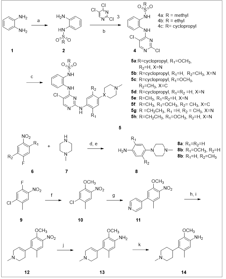
The synthesis of the target compounds.
Synthesis of intermediate 2.
To a solution of 1,2-phenylenediamine (2.16g, 20 mmol) in 15ml of anhydrous acetonitrile, methyl sulfonic anhydride (3.83g, 22 mmol), DMAP (0.122g) and triethylamine (2.1ml) were added. The reaction was refluxed at 80℃ for 8 hours. The solvent was evaporated under reduced pressure and the residue was extracted with dichloromethane (30ml *4). The solvent was removed and the crude product was purified by silica gel column chromatography (petroleum ether: ethyl acetate, 10:3, then, 2:1) afford to intermediate 2.
Synthesis of intermediate 4.
Intermediate 2a (2.5g, 13.4 mmol) was dissolved in 17ml of anhydrous DMF, sodium hydride (60%, 1.2g) was added at 0℃. After stirred for 15 minutes, 2,4,5-trichloropyrimidine (3.2g) in 13 ml of anhydrous DMF was added in dropwise. The ice bath was removed and the reaction was continued for 2-3 hours at the room temperate. Water 15ml was added to quench the reaction and then extracted with 30ml of ethyl acetate for 3 times. The combined organic phase was dried with anhydrous sodium sulfate, concentrated under reduced pressure to provide the crude product which was purified by silica gel column chromatography (petroleum ether: ethyl acetate, 4:1).
1H NMR for N-(2-((2,5-dichloropyrimidin-4-yl)amino)phenyl)methanesulfonamide: 1H NMR (400 MHz, Chloroform-d) δ 8.38 (s, 1H), 8.23 (s, 1H), 8.04 (d, J = 9.1 Hz, 1H), 7.47 – 7.34 (m, 2H), 3.06 (s, 3H).
N-(2-((2,5-dichloropyrimidin-4-yl)amino)phenyl)ethanesulfonamide: 1H NMR (400 MHz, Chloroform-d) δ 8.19 (s, 1H), 7.37 (d, J = 7.8 Hz, 2H), 7.21 (ddt, J = 10.5, 7.4, 2.4 Hz, 2H), 3.12 (q, J = 7.4 Hz, 2H), 1.41 (t, J = 7.4 Hz, 3H).
1H NMR for N-(2-((2,5-dichloropyrimidin-4-yl)amino)phenyl)cyclopropanesulfonamide: 1H NMR (400 MHz, Chloroform-d) δ 8.00 (s, 1H), 7.48 – 7.29 (m, 3H), 7.22 – 7.18 (m, 1H), 1.33 (s, 1H), 1.14 – 1.08 (m, 2H), 1.03 – 0.94 (m, 2H).
Synthesis of amine intermediates 8b-c.
A solution of 4-fluoro-2-methoxy-1-nitrobenzene (3.42g, 20 mmol) in 27ml of anhydrous acetonitrile was added N-methylpiperazine (3.0g, 30 mmol), anhydrous potassium carbonate (4,41g, 32 mmol). The reaction was refluxed at 80 ℃ for 8-10 hours. The solvent was removed under the reduced pressure. Water 30 ml was added to the residue and then extracted with dichloromethane (50ml *2). The organic phase was dried over Na2SO4, and concentrate to give yellow oily for the next step without purification. The above yellow oily intermediate 5.9g was dissolved in 60ml of dichloromethane/methanol (1/1), stannous chloride (17g) and 14ml of concentrated hydrochloric acid were added. The reaction was heated at 50 ℃ for 4-8 hours. After the reaction is complete monitored by TCL, ammonia hydroxide and Na2CO3 were added to the reaction under an ice bath, filtered with diatomite, washed with dichloromethane/methanol (1/1). The organic layer was separated and the water layer was extracted with dichloromethane. The combined organic phase was dried over Na2SO4. The solvent was removed to afford the crude product which was purified by silica gel column chromatography (CH2Cl2/CH3OH (1/1).
8b. 1H NMR (400 MHz, Chloroform-d) δ 6.62 (d, J = 8.3 Hz, 1H), 6.51 (d, J = 2.3 Hz, 1H), 6.40 (dd, J = 8.3, 2.4 Hz, 1H), 3.82 (s, 3H), 3.40 (s, 2H), 3.16 – 3.01 (m, 4H), 2.69 – 2.53 (m, 4H), 2.34 (s, 3H).
8c. 1H NMR (400 MHz, Chloroform-d) δ 6.87 (d, J = 8.3 Hz, 1H), 6.52 (d, J = 2.8 Hz, 1H), 6.47 (dd, J = 8.3, 2.8 Hz, 1H), 3.43 (s, 2H), 2.84 (t, J = 4.8 Hz, 4H), 2.54 (s, 4H), 2.33 (s, 3H), 2.22 (s, 3H).
Synthesis of amine intermediate 14.
A solution of 1-chloro-2-fluoro-5-methyl-4-nitrobenzene (7.0g, 37mmol) in methanol (68ml) was added anhydrous potassium carbonate (5.2g, 37.7mmol). After refluxed at 66 ℃ for 4-6 hours, the reaction was filtered and removed the solvent to give the product for the use of next step directly. 1H NMR (400 MHz, Chloroform-d) δ 7.78 (s, 1H), 7.09 (s, 1H), 3.94 (s, 3H), 2.36 (s, 3H).
The above intermediate (16g, 79.6 mmol) was dissolved in 1,4-dioxane (100ml), and then 4-pyridylboric acid (4.56g, 37 mmol), anhydrous potassium carbonate (10.2g, 74 mmol), bis triphenylphosphine palladium dichloride (0.78g, 1.11 mmol), 25ml of water were added. The reaction was refluxed at 101 ℃ under nitrogen for 8 hours. After the solvents were removed under the reduced pressure, dichloromethane was added. The extracts was washed with brine, dried over sodium sulfate, concentrated and purified with silica gel column chromatography (Petrolem: ethyl acetate, 4:1) to give the intermediate 11. 1H NMR (400 MHz, Chloroform-d) δ 8.72 (d, J = 5.8 Hz, 2H), 7.78 (s, 1H), 7.29 (d, J = 5.9 Hz, 2H), 6.96 (s, 1H), 3.96 (s, 3H), 2.25 (s, 3H).
To a solution of intermediate 11 in 60ml of acetonitrile, iodomethane (6.31g, 44.4mmol) was added and the mixture was heated at 50 ℃ for about 4 hours. The solvent was removed to afford the crude product witch was directly used for the next step without purification. The above crude product was dissolved in 200ml of methanol; sodium borohydride (8.4g, 222mmol) was added in batches under an ice bath. After the reaction finished (monitored by TLC), aqueous sodium hydroxide solution was added to adjust pH to 10-12, and the mixture was extracted with dichloromethane. The organic phase was concentrated and the crude product was purified by silica gel column chromatography (dichloromethane: methanol, 20:1) to afford the intermediate 12. 1H NMR (400 MHz, Chloroform-d) δ 7.69 (s, 1H), 6.82 (s, 1H), 5.68 – 5.55 (m, 1H), 3.91 (s, 3H), 3.10 (s, 2H), 2.67 (t, J = 5.6 Hz, 2H), 2.43 (s, 3H), 2.41 (s, 2H), 2.26 (s, 3H).
Intermediate 12 was dissolved in a mixed solvents 50ml (dichloromethane/methanol, V/V), then 20g of stannous chloride and 10ml of concentrated hydrochloric acid were added. The mixture was heated at 50℃ for 6 hours and then cooled to room temperature. Ice (20g) was added to the reaction and then sodium hydroxide (4.8g) in batches under an ice bath and stirring. The organic solvents were removed under the reduced pressure, and the residue was extracted twice with 20ml of dichloromethane. The solvent was removed in vacuum and the crude product was purified with silica gel column chromatography (petroleum/ethyl acetate 4/1, followed by dichloromethane/methanol, 10/1) to give the intermediate 13. 1H NMR (400 MHz, Chloroform-d) δ 6.47 (s, 1H), 6.42 (s, 1H), 5.42 (dq, J = 3.7, 1.8 Hz, 1H), 3.69 (s, 3H), 3.04 (d, J = 3.3 Hz, 2H), 2.61 (t, J = 5.7 Hz, 2H), 2.36 (s, 3H), 2.34 (s, 2H), 2.07 (s, 3H).
To a solution of intermediate 13 (1g) in methanol (5ml), 10% palladium carbon (0.3g) was added. The reaction was pressurized at 40 Bar of hydrogen. After stirred at 50℃ for 24 hours, the mixture was filtered, concentrated to afford the intermediate 14 which is directly used for the next step without purification.
Synthesis of intermediate 16.
To a solution of 2,4,5-trichloropyrimidine (10mmol, 1.82g) in DMF (25ml), (2-aminophenyl)dimethylphosphine oxide (10mmol, 1.69g), anhydrous potassium carbonate (12mmol, 1.66g), tetrabutylammonium bisulfate (1mmol, 0.34g) were added. The mixture was stirred and heat at 65 ℃ for 8 hours. After the reaction finished, the solvent was removed in vacuum, and the crude product was purified with silica gel column chromatography (petroleum ether: ethyl acetate in 1:1) to obtain the product. 1H NMR (400 MHz, Chloroform-d) δ 11.55 (s, 1H), 8.67 (dd, J = 8.6, 4.5 Hz, 1H), 8.22 (s, 1H), 7.59 (t, J = 8.0 Hz, 1H), 7.29 (dd, J = 13.7, 7.9 Hz, 1H), 7.18 (t, J = 6.9 Hz, 1H), 1.85 (d, J = 13.2 Hz, 6H).
Typical procedure for the synthesis of target products 5a-5h and 17a-17b.
To a solution of intermediate 16 (0.95g, 3 mmol) in 15 ml isopropanol, 2,2,2-trifluoroacetic acid (0.884g, 7.8 mmol), 4-(4-methylpiperazin-1-yl)aniline or 2-methoxy-5-methyl-4-(4-methylpiperazin-1-yl)aniline (3 mmol) were added. After the mixture was heated at 85 ℃ for 8-12 hours, the solvent was removed in vacuum. Dichloromethane (50 ml *3) was added to extract the product, and then purified with silica gel column chromatography (petroleum ether: ethyl acetate in 1:1) in a yields 50-70%.
N-(2-((5-chloro-2-((2-methoxy-4-(4-methylpiperazin-1-yl)phenyl)amino)pyrimidin-4-yl)amino)phenyl)cyclopropanesulfonamide (5a)
White solid. 1H NMR (400 MHz, DMSO-d6) δ 8.47 (s, 1H), 8.08 (s, 2H), 8.03 (d, J=8.1Hz,1H), 7.91 (s, 1H), 7.44 (d, J = 8.7 Hz, 1H), 7.37 (d, J = 7.8, 1H), 7.22 (t, J=7.4 Hz,1H), 7.14 (t, J = 8.3 Hz, 1H), 6.60 (d, J=2.5 Hz,1H), 6.39-6.36 (m, 1H) 3.76 (s, 3H), 3.16-3.06 (m, 4H), 2.57-2.53 (m, 1H), 2.48-2.47 (m, 4H), 2.24 (s, 3H), 0.93-0.82 (m, 4H). 13C NMR (126 MHz, DMSO) δ 159.19, 155.57, 154.85, 152.25, 149.04, 134.85, 129.95, 127.09, 125.93, 124.56, 124.17, 123.50, 120.67, 107.08, 103.96, 100.43, 55.92, 55.16, 49.21, 46.24, 29.72, 5.56. HRMS (ESI+ ) calcd for C25H30ClN7O3S [M + H]+ 544.1898; found, 544.1931. HPLC purity: 97.16%.
N-(2-((5-chloro-2-((3-methyl-4-(4-methylpiperazin-1-yl)phenyl)amino)pyrimidin-4-yl)amino)phenyl)cyclopropanesulfonamide (5b)
White solid. 1H NMR (500 MHz, DMSO-d6) δ 9.17 (s, 1H), 8.54 (s, 1H), 8.15 (s, 1H), 8.07 – 8.03 (m, 1H), 7.46 – 7.36 (m, 2H), 7.29 (q, J = 7.4 Hz, 2H), 7.21 (t, J = 7.6 Hz, 1H), 6.86 (d, J = 8.7 Hz, 1H), 2.78-2.76 (m, 4H), 2. 47 – 2.42 (m, 4H), 2.24 (s, 3H), 2.12 (s, 3H), 0.89 – 0.84 (m, 4H). 13C NMR (126 MHz, DMSO) δ 158.28, 156.00, 155.02, 145.75, 136.01, 135.09, 132.28, 129.44, 128.36, 127.37, 125.33, 125.03, 122.13, 119.13, 118.10, 104.17, 55.31, 52.49, 51.48, 45.74, 29.59, 18.10, 5.59. HRMS (ESI+ ) calcd for C25H31ClN7O2S [M + H]+ 528.1948; found, 528.1959. HPLC purity: 99.55%.
N-(2-((5-chloro-2-((2-methoxy-5-methyl-4-(1-methylpiperidin-4-yl)phenyl)amino)pyrimidin-4-yl)amino)phenyl)cyclopropanesulfonamide (5c).
White solid. 1H NMR (500 MHz, Chloroform-d) δ 8.06 (s, 1H), 7.94 (s, 1H), 7.89 (d, J = 8.0 Hz, 1H), 7.79 (s, 1H), 7.52 (d, J = 7.9 Hz, 1H), 7.46 (s, 1H), 7.31 (t, J = 8.3 Hz, 1H), 7.21 (t, J = 7.7 Hz, 1H), 6.68 (s, 1H), 3.79 (s, 3H), 3.04 (d, J = 11.2 Hz, 2H), 2.61 – 2.56 (m, 1H), 2.47 – 2.42 (m, 1H), 2.38 (s, 3H), 2.15 – 2.10 (m, 2H), 1.99 (s, 3H), 1.82 – 1.73 (m, 2H), 1.67 (d, J = 12.4 Hz, 2H), 1.09 –1.05 (m, 2H), 0.93 – 0.82 (m, 2H). 13C NMR (126 MHz, CDCl3) δ 157.69, 156.19, 154.76, 146.44, 136.99, 134.54, 129.56, 128.04, 127.74, 126.80, 126.33, 125.56, 125.35, 120.32, 107.53, 104.88, 56.29, 55.77, 45.99, 37.05, 32.21, 29.73, 18.70, 5.68. HRMS (ESI+ ) calcd for C27H34ClN6O3S [M + H]+ 557.2102; found, 557.2104. HPLC purity: 99.55%.
N-(2-((5-chloro-2-((4-(1-methylpiperidin-4-yl)phenyl)amino)pyrimidin-4-yl)amino)phenyl)cyclopropanesulfonamide (5d).
White solid. 1H NMR (400 MHz, Chloroform-d) δ 9.13 (s, 1H), 8.51 (s, 1H), 8.13 (s, 1H), 8.08 (t, J = 8.3 Hz, 1H), 7.45-7.37(m, 3H), 7.36-7.30 (m, 1H), 7.21 (t, J = 7.7 Hz, 1H), 7.23 (d, J = 7.7 Hz, 1H), 6.78 (dd, J = 9.2, 2.1 Hz, 2H), 3.08 –2.94 (m, 4H), 2.57-2.52 (m, 1H), 2.49 –2.47 (m, 4H), 2.24 (s, 3H), 0.90 –0.83 (m, 4H). 13C NMR (101 MHz, DMSO) δ 158.34, 158.25, 155.93, 155.02, 145.46, 135.17, 133.53, 128.53, 127.65, 125.41, 125.04, 120.91, 116.68, 104.03, 53.56, 47.62, 45.65, 43.66, 5.64. HRMS (ESI+ ) calcd for C24H29ClN7O2S [M + H]+ 514.1792; found, 514.2170. HPLC purity: 95.87%.
N-(2-((5-chloro-2-((4-(4-methylpiperazin-1-yl)phenyl)amino)pyrimidin-4-yl)amino)phenyl)methanesulfonamide (5e).
White solid. 1H NMR (400 MHz, DMSO-d6) δ 9.17 (d, J = 2.5 Hz, 0H), 8.51 (s, 0H), 8.14 (s, 1H), 8.02 (t, J = 8.7 Hz, 1H), 7.47 – 7.32 (m, 4H), 7.25 (t, J = 7.6 Hz, 1H), 6.82 (dd, J = 9.1, 2.4 Hz, 2H), 3.22 – 3.20 (m, J, 4H), 3.05 – 3.02 (m, 4H), 2.96 (s, 3H), 2.64 (s, 3H). 13C NMR (101 MHz, DMSO) δ 158.25, 156.11, 155.00, 145.49, 134.93, 133.50, 129.69, 129.50, 127.52, 126.00, 125.50, 120.86, 116.63, 104.10, 53.66, 47.74, 45.65, 43.81. HRMS (ESI+) calcd for C22H27ClN7O2S [M + H]+ 488.1635; found, 488.1636. HPLC purity: 96.19%.
N-(2-((5-chloro-2-((2-methoxy-5-methyl-4-(4-methylpiperazin-1-yl)phenyl)amino)pyrimidin-4-yl)amino)phenyl)methanesulfonamide (5f).
White solid. 1H NMR (500 MHz, Chloroform-d) δ 8.05 (s, 1H), 8.02 – 7.90 (m, 1H), 7.85 (t, J = 7.7 Hz, 1H), 7.74 (s, 1H), 7.49 (d, J = 7.8 Hz, 1H), 7.45 (s, 1H), 7.29 (d, J = 7.5 Hz, 1H), 7.23 (d, J = 7.7 Hz, 1H), 6.65 (s, 1H), 3.79 (s, 3H), 3.09 –3.07 (m, 2H), 2.92 (s, 3H), 2.60 –2.55 (m, 1H), 2.42 (s, 3H), 2.20 – 2.16 (m, 2H), 1.93 (s, 3H), 1.82 – 1.75 (m, 2H), 1.65-1.64 (m, 2H). 13C NMR (126 MHz, CDCl3) δ 157.66, 156.32, 154.76, 146.43, 136.60, 134.10, 130.55, 127.31, 126.74, 126.59, 126.39, 125.99, 125.86, 120.26, 107.47, 104.89, 56.11, 55.78, 45.66, 39.21, 36.83, 31.83, 18.66. HRMS (ESI+ ) calcd for C24H30ClN7O3S [M + H]+ 531.1945; found, 531.1948. HPLC purity: 95.48%.
N-(2-((5-chloro-2-((3-methyl-4-(4-methylpiperazin-1-yl)phenyl)amino)pyrimidin-4-yl)amino)phenyl)ethanesulfonamide (5g).
White solid. 1H NMR (500 MHz, DMSO-d6) δ 9.17 (s, 1H), 8.57 (s, 1H), 8.14 (s, 1H), 7.91 (d, J = 6.8 Hz, 1H), 7.41 – 7.32 (m, 2H), 7.28 (t, J = 7.6 Hz, 1H), 7.23 (t, J = 7.3 Hz, 2H), 6.83 (d, J = 8.6 Hz, 1H), 3.00 (q, J = 7.2 Hz, 2H), 2.79-2.73 (m, 4H), 2.51-2.49 (m, 4H), 2.25 (s, 3H), 2.09 (s, 3H), 1.16 (t, J = 7.3 Hz, 3H). 13C NMR (126 MHz, DMSO) δ 158.30, 156.35, 155.00, 145.87, 135.96, 134.40, 132.22, 130.55, 126.97, 126.91, 126.27, 125.69, 122.02, 119.07, 117.99, 104.16, 55.60, 51.86, 46.13, 45.58, 18.09, 8.34. HRMS (ESI+ ) calcd for C24H30ClN7O2S [M + H]+ 516.1948; found, 516.1947. HPLC purity: 98.59%.
N-(2-((5-chloro-2-((2-methoxy-4-(4-methylpiperazin-1-yl)phenyl)amino)pyrimidin-4-yl)amino)phenyl)ethanesulfonamide (5h).
White solid. 1H NMR (500 MHz, DMSO-d6) δ 8.66 (s, 1H), 8.07 (s, 1H), 7.97 (s, 1H), 7.86 (s, 1H), 7.44 (d, J = 8.6 Hz, 1H), 7.35 – 7.28 (m, 1H), 7.10 (dd, J = 5.8, 3.5 Hz, 2H), 6.60 (d, J = 2.3 Hz, 1H), 6.36 (dd, J = 8.7, 2.0 Hz, 1H), 3.76 (s, 3H), 3.14 – 3.07 (m, 4H), 2.97 (q, J = 7.3 Hz, 2H), 2.49 – 2.43 (m, 4H), 2.23 (s, 3H), 1.19 (t, J = 7.3 Hz, 3H). 13C NMR (126 MHz, DMSO) δ 159.10, 155.84, 154.83, 152.01, 148.87, 134.37, 125.99, 125.53, 124.70, 124.28, 124.19, 120.75, 107.08, 100.43, 55.93, 55.13, 49.20, 46.21, 46.05, 45.58, 10.43, 8.55. HRMS (ESI+ ) calcd for C24H30ClN7O3S [M + H]+ 531.1898; found, 532.1919. HPLC purity: 97.23%.
(2-((5-chloro-2-((2-methoxy-5-methyl-4-(1-methylpiperidin-4-yl)phenyl)amino)pyrimidin-4-yl)amino)phenyl)dimethylphosphine oxide (17a).
White solid. 1H NMR (400 MHz, Chloroform-d) δ 10.80 (s, 1H), 8.64 (d, J = 8.4 Hz, 1H), 8.08 (d, J = 22.5 Hz, 2H), 7.50 (t, J = 7.8 Hz, 1H), 7.44 (s, 1H), 7.34 – 7.27 (m, 1H), 7.12 (t, J = 7.4 Hz, 1H), 6.80 (s, 1H), 3.84 (s, 3H), 3.06 (d, J = 10.8 Hz, 2H), 2.77-2.66 (m, 2H), 2.39 (s, 3H), 2.22 (s, 3H), 2.18-2.13 (m, 2H), 1.92 – 1.86 (m, 1H), 1.85 (s, 3H), 1.83 (s, 3H)1.81 – 1.72 (m, 2H). 13C NMR (126 MHz, Chloroform-d) δ 157.51, 155.89, 154.93, 146.71, 143.86, 137.26, 132.66, 129.63, 129.55, 126.80, 126.63, 122.73, 122.68, 122.41, 122.31, 120.77, 119.86, 107.73, 106.36, 56.46, 55.76, 46.25, 37.25, 32.61, 18.90, 18.29. HRMS (ESI+ ) calcd for C26H34ClN5O2P [M + H]+ 514.2139; found, 514.2170. HPLC purity: 96.15%.
(2-((5-chloro-2-((4-(4-methylpiperazin-1-yl)phenyl)amino)pyrimidin-4-yl)amino)phenyl)dimethylphosphine oxide (17b).
1H NMR (400 MHz, DMSO-d6) δ 11.12 (s, 1H), 9.19 (s, 1H), 8.61 (s, 1H), 8.14 (s, 1H), 7.68 – 7.46 (m, 4H), 7.25 – 7.12 (m, 1H), 6.90 (d, J = 9.1 Hz, 2H), 3.24 –3.19 (m, 4H), 2.98 –2.89 (s, 4H), 2.56 (s, 3H), 1.80 (s, 3H), 1.76 (s, 3H). 13C NMR (101 MHz, DMSO) δ 158.20, 155.69, 155.23, 145.20, 143.40, 133.74, 132.44, 131.20, 131.10, 122.94, 122.60, 121.43, 116.95, 104.81, 52.89, 46.91, 45.65, 19.00, 18.30. HRMS (ESI+ ) calcd for C23H29ClN6OP [M + H]+ 471.1829; found, 471.1827. HPLC purity: 98.18%.
Biological assay
Cells and Cultures
Human non-small-cell lung cancer (NSCLC) cell lines (A549 and PC-9) and human normal cells (LO2, HK2, and 293A) were purchased from the Guangzhou ginny ou biotechnology co. LTD (Guangzhou, China). And the BaF319del/T790M/C797S and BaF3L858R/T790M/C797S were purchased from the KYinno (Beijing, China). The NSCLC cells were cultured in RPMI 1640 medium containing 10% FBS, 100 units/mL penicillin, and 100 μg/mL streptomycin. BaF319del/T790M/C797S and BaF3L858R/T790M/C797S were grown in RPMI 1640 containing 15% FBS. All cells were incubated at 37 °C in an atmosphere of 5% CO2. Tests for mycoplasma contamination were negative.
ELISA asssy
Enzyme-Linked Immunosorbent Assay for mutant EGFRL858R/T790M/C797S and mutant EGFR19del/T790M/C797S. Mutant EGFRL858R/T790M/C797S and mutant EGFR19del/T790M/C797S ELISA (ADANTI, USA) was used and normalized to the total EGFR content. Concentrations consisting of 6 levels from 0.005 to 5 mM were used for all of the tested compounds. Assays were performed in triplicates for each measurement using 50 μl per well in accordance with the manufacturer’s instructions. Generally, the mixture of EGFRL858R/T790M/C797Sand EGFR19del/T790M/C797S antibody substrate, appropriate kinase (HRP labeled antibody to EGFR) and different concentrations of compounds were converged with the reaction buffer with DMSO solution as the negative group. The kinase reaction was initiated by the addition of tyrosine kinase proteins diluted 40μl of reaction buffer solution and incubated for 30min at 37 ◦C. Dilute the concentrated washing solution in the kit into 1X, discard the solution on the enzyme plate, shake dry, fill each hole with the washing solution, leave for 30 seconds, then discard the solution, repeat 5 times, pat dry. Add color developer A (50μl) to each well, then add color developer B (50μl), gently shake and mix, hide from light for 15 minutes at 37℃.And then 50μl of stop buffer was added to stop the reaction. The IC50 values were calculated using the GraphPad Prism software (GraphPad Inc., La Jolla, CA, USA).
Cell growth assay
As described previously, the cells and the human normal cells (5 × 10³ cells/well ) were seeded in 96-well sterile plastic plates, incubated overnight, and then treated with the different concentrations (10、 1、0.1、0.05、0.01 and 0.001 uM) drugs. After 48 h of treatment, 10 μL Cell Counting Kit-8 was added to each well and the plates were incubated for 40 min. Briefly, for the BaF319del/T790M/C797S and BaF3L858R/T790M/C797S, 1 × 10⁴ cells were seeded in a 96-multiwell plate and the drugs and cells were cultured for a total of 48 h. Then, 10 μL Cell Counting Kit-8 was added to each well and the plates were incubated for two hours and a half. The results were representative of at least three independent experiments; the error bars signify standard deviation (SD).The IC50 values were calculated using the GraphPad Prism software (GraphPad Inc., La Jolla, CA, USA).
Cell apoptosis and cell cycle assay
As described previously, the BaF319del/T790M/C797S and BaF3L858R/T790M/C797S were seeded in 6-well plates (3×10⁴ cells/well) and incubated in the presence or absence of compound 5d for the cell apoptosis assay, cells were harvested and incubated with 5 μL of Annexin-V/FITC (Keygen Biotech, China) in binding buffer (10 mM HEPES, 140 mM NaCl, and 2.5 mM CaCl2 at pH 7.4) at room temperature for 15 min. PI solution was then added to the medium for another 10 min-incubation. Almost 10,000 events were collected for each sample and analysed by flow cytometry (Beckman Coulter, Epics XL). For the cell cycle assay, the cells were collected and rinsed once with PBS before overnight fxation with pre-cooled 70% ethanol at −20 °C. The fxed cells were washed twice with cold PBS and incubated in the dark at 37°C with 10μL RNAse A and 50 μg/mL PI staining solution for 30 min. The data regarding the number of cells in different phases of the cell cycle were analysed using EXPO32 ADC analysis software.
Western blot analysis.
Afer treatment with 5d or osimertinib for 12 or 24 h, cells were washed with PBS, then cleaved on ice with RIPA(Beyotime, China) containing 1% protease inhibitor and phosphorylase inhibitor, centrifuged at 12000rpm for 15 min to collect proteins. After the protein concentration was determined by BCA kit(Beyotime, China), the samples were packaged and boiled. The protein was coated on 10% SDS-PAGE glue and then transferred to PVDF (Merck, Germany)membrane. 5% skim milk powder was enclosed with PVDF membrane and incubated at 4℃ overnight. Then membranes were then incubated with HRP-conjugated secondary antibodies at room temperature and developed with luminescent solution((Pierce, Rockford, IL, USA). P-EGFR (1:1000;3777T, Cell signaling), EGFR (1:1000;4267T, Cell signaling), p-AKT (1:1000;AF6261, affinity), AKT (1:1000;GR100134-1, abcam), p-ERK (1:1000;AF1015, affinity), ERK (1:1000;4695T, Cell signaling), GAPDH (1:1000;10494-1-AP, proteintech), P70S6K (1:1000;9202S, Cell signaling), p-P70S6K (1:1000;9234S, Cell signaling), cleaved caspase-3 (1:1000;9661L, Cell signaling), PI3K (1:1000; AF6241, affinity), p-MER(AF8443, affinity), P-PI3K (1:1000; AF3242, affinity), AMPK (1:1000; 9158, Cell signaling), P-AMPK (1:1000; 2537, Cell signaling), β-actin (1:1000;A5441, Sigma).
Xenograft tumor model
Male BALB/c nude mice, 3-4 weeks old were purchased from spfbiotech ( SPF(BEIJING)BIOTECHNOLOGY CO.,LTD., Beijing, China) and allowed to acclimate for 1 week. Animal care was in accordance with the institutional animal care guidelines. All animal experiments were approved by the Animal Ethics Committee of the Academy of Military Medical Science. Logarithmic growth of 1 × 10⁷ cancer cells/mL were re-suspended in 1640 medium without FBS. Then, 200 uL cell suspension was injected subcutaneously into the right side of each mouse. After the tumor had formed a hard mass, the tumor volume was measured every two days. When the tumor volume reaches 150-200 mm3 , the xenograft tumour-bearing nude mice were randomly allocated to Four groups (vehicle-treated, 5d-70 mg/kg, 5d-100 mg/kg and osimertinib-100mg/kg) with 7 mice per group. Each group was dosed by intragastric administration (ig) for 9 days. Tumor volume and mouse body weight were recorded after daily administration. At the end of the observation period, the animals were euthanized by cervical dislocation and the tumour bulks were peeled off conformed to the Guide for the Care and Use of Laboratory Animals as published by the US National Institutes of Health (NIH Publication No. 85-23, revised 1996) and approved by the Institutional Ethics Review Board of Binzhou Medical University.
Immunohistochemistry
As described previously, immunohistochemistry (IHC) detected the expression of Ki67、P-EGFR in tumor tissue. The tumor tissue paraffin sections were deparaffinized, and endogenous peroxidase activity was blocked by incubation with 3% H2O2. The blocked sections were incubated with Ki67 antibody (1:500; proteintech, 27309-1-AP)、P-EGFR antibody (1:500; 4267T, Cell signaling) at 4℃ overnight and then sequentially incubated with a biotin-labeled secondary antibody. The sections were then stained with 3,3′-diaminobenzidine. Finally, the sections were counterstained using hematoxylin and fixed. For each section, three fields of view were randomly selected and photographed under 400× magnification.
HE staining
The morphological changes of heart, liver, spleen, lung and kidney were detected by HE staining. Paraffin sections of tissue were dewaxed, reversed stained and fixed with eosin and hematoxylin. 3 fields of view were randomly selected for each profile and shot at 400×.
Cell morphology
Human normal cells LO2, HK2, and 293A were placed in a six-well plate overnight, and then treated with 10 μM compound 5d for 48 h. The morphological changes were observed under a 400X microscope.
Assays for DNA Damage
The BaF319del/T790M/C797S and BaF3L858R/T790M/C797S were seeded in 6-well plates (2×10⁴ cells/well) and incubated in the presence or absence of compound 5d for 24 h. Almost 10,000 events were collected for each sample and analysed by flow cytometry (Beckman Coulter, Epics XL). For the DNA Damage assay, the cells were collected and rinsed once with PBS. Then fxation with 4% PFA(Beyotime, China) at room temperature. The cells were washed three times in PBS for 5 minutes each, and then incubated with DNA Damage Assay Kit by γ-H2AX Immunofluorescence (Beyotime, China). Dye with DIPA from the kit for 15min.Leica SP5 confocal laser scanning microscopy (Leica Microsystems, Buffalo Grove, USA) was used for immunofluorescence imaging.
Statistical Analysis
All experiments were repeated three times independently unless specifically indicated. The data was analyzed by GraphPad Prism 7 software. The data was shown as mean±SD and one-way ANOVA Tukey 's multiple comparison test was used to determine the levels of significance between comparison samples. p > 0.05 was not significant (ns). *p <0.05; **p <0.01; ***p<0.001;