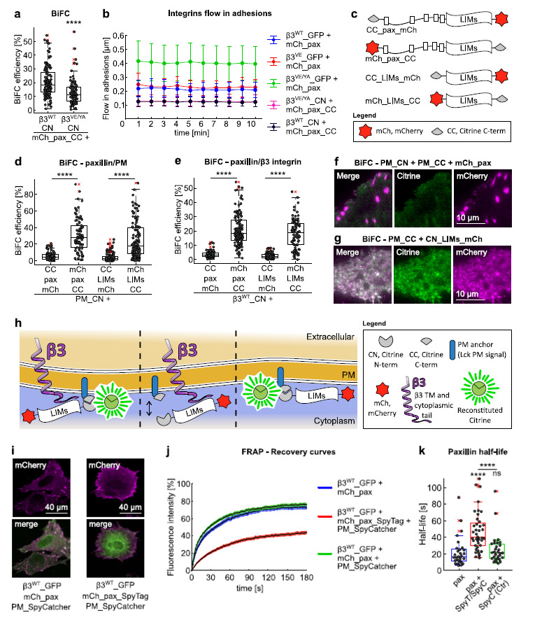
BiFC and SpyTag/SpyCatcher assays to study focal adhesions organization and function. (a) Quantification of the BiFC efficiency upon co-expression of mCherry_paxillin_CC and β3WT_CN or β3VE/YA_CN in NIH-3T3 cells. Statistical analysis is provided in Supplementary Table 1. (b) Quantification of the β3 integrins flow in adhesions, expressed as displacement over time. Error bar ±SD. Statistical analysis is provided in Supplementary Table 2. (c) Paxillin and LIMs fusion proteins tagged with mCherry and the citrine C-terminal fragment, tested
in BiFC assays with alternatively PM_CN or β3WT_CN. (d,e) Quantification of the BiFC signal generated by the co-expression of each of the paxillin constructs shown in figure c with (d) the plasma membrane-localized CN fragment, in Swiss-3T3 or with (e) the β3WT integrin C-terminally tagged with CN, in NIH-3T3 cells. Statistical analysis is provided in Supplementary Table 1. (f) Representative TIRF images of a triple positive Swiss-3T3 fibroblast, co-expressing mCherry_paxillin and the two complementary citrine fragments, each one fused to the PM-targeting peptide. While the BiFC signal is localized throughout to the plasma membrane, mCherry_paxillin only appears in the discreates spots of FAs. Brightness and contrast automatically optimized. (g) Example of a Swiss-3T3 fibroblast in which the co-expression of CN_LIMs_mCherry and PM_CC led to substantial BiFC and concomitant mis-localization of LIMs. Brightness and contrast automatically optimized. (h) Schematic representation of the possible scenarios, in terms of BiFC generation, upon co-expression of CN-tagged LIMs recombinant proteins and the PM-localized complementary citrine fragment. Left: the CN fragment at the C-terminus of LIMs leads to BiFC in adhesions. Middle: the CN fragment in front of LIMs is not compatible with BiFC in adhesions. Right: the CN fragment in front of LIMs can complement PM-localized CC outside adhesions. (i) Differential paxillin distribution among FAs, cytosol and PM, in the absence (left) and in the presence (right) of the irreversible fusion of the C-terminus to the PM_SpyCatcher. (j) Florescence Recovery after Photobleaching (FRAP) of paxillin localized to β3WT_GFP-positive FAs, in the absence (mCherry_paxillin and mCherry_paxillin + PM_SpyCatcher, control) and in the presence (mCherry_paxillin_SpyTag + PM_SpyCatcher) of the irreversible fusion of the C-terminus to the PM_SpyCatcher. (k) Box plot of the half-lives of paxillin in β3WT_GFP-positive FAs. Statistical analysis is provided in Supplementary Table 3.