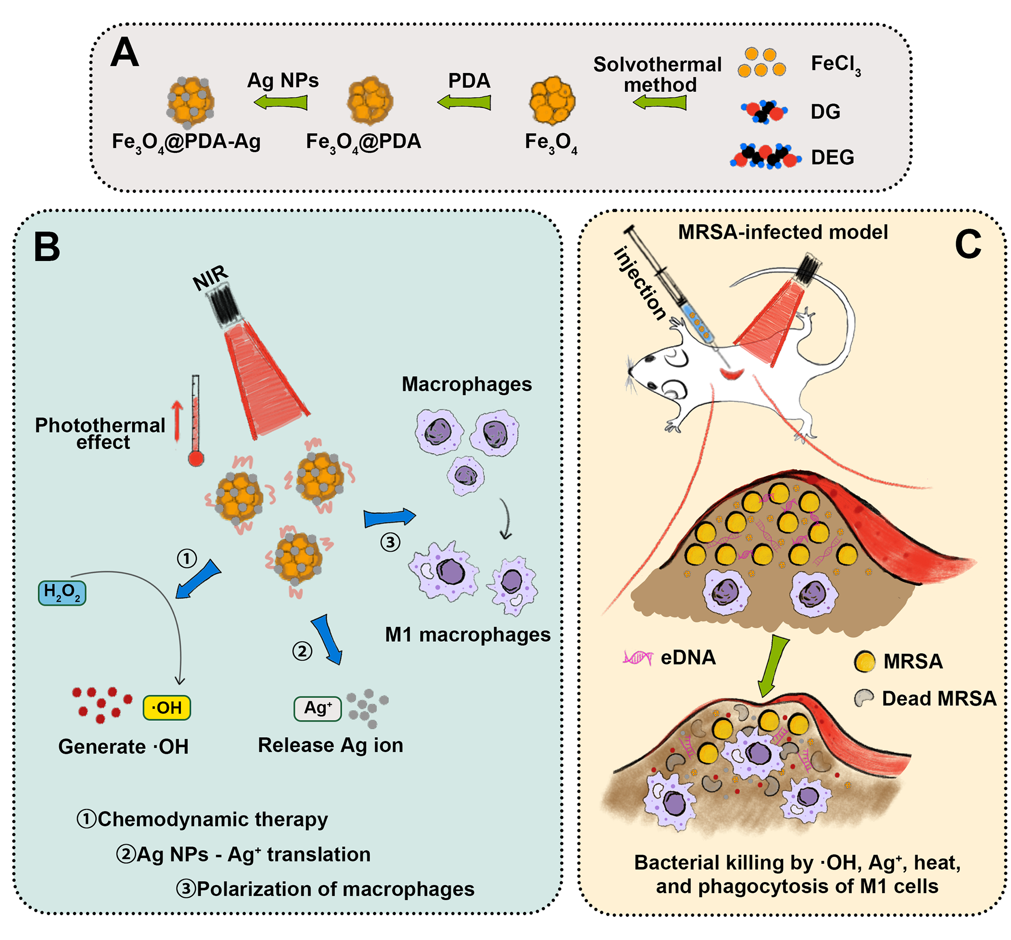
Synthesis and Characterization of Ag nanoparticles loaded core-shell structure Fe3O4 nanoparticles
According to transmission electron microscopy (TEM) and scanning electron microscopy (SEM) results (Fig. 1A), Fe3O4 is a spherical particle with a rough surface, whereas Fe3O4@PDA has a typical core-shell structure with a smooth surface, Fe3O4 as the core and 8 nm thick PDA layer as the shell. Because of the high adhesion of PDA, partial bonding was observed between the particles. Globular Ag NPs approximately 50 nm in size were loaded onto the Fe3O4@PDA surface. The elemental composition of Fe3O4@PDA-Ag was determined using energy-dispersive spectrometry (EDS) with the mass ratio of Ag being approximately 32.15% (Figure S1). Size analysis of the nanoparticles (Fig. 1B) revealed a larger size than that observed by electron microscopy, which may be due to the hydrated layer caused by hydrophilic hydroxyl groups on the surfaces of the Fe3O4 and PDA layers. For Fe3O4@PDA-Ag, a small peak at approximately 60 nm was observed, which may be caused by the shedding of Ag NPs after sonication. The zeta potentials of each nanoparticles were − 16.0 mV (Fe3O4), -33.3 mV (Fe3O4@PDA), and − 30.9 mV (Fe3O4@PDA-Ag), respectively (Fig. 1C).
In results of X-ray diffraction (XRD) (Figure S2A) of Fe3O4, the diffraction peaks of 2θ angles at 30.2°, 35.5°, 43.1°, 53.5°, 57.1° and 62.6° were observed, corresponding to (220), (311), (400), (422), (511) and (440) of Fe3O4 crystal. The Fe3O4@PDA spectra is consistent with that of Fe3O4 owing to the amorphous structure of PDA. When the Ag NPs were loaded, a characteristic diffraction peak was observed at 38.3° corresponding to the (111) plane of Ag. In the X-ray photoelectron spectroscopy (XPS) spectrum (Figure S2B), the Fe 2p peak of Fe3O4 appeared at a binding energy of 720 eV, whose peak intensity in both Fe3O4@PDA and Fe3O4@PDA-Ag was reduced by the PDA layer, with an absorption peak of N 1s appearing at 400 eV simultaneously. In addition, an Ag 3d peak appeared at 370 eV in the Fe3O4@PDA-Ag spectrum, confirming the presence of Ag NPs.
In the Fourier-transform infrared (FTIR) spectrum (Figure S2C), the characteristic absorption peak of Fe3O4 at 536 cm− 1 corresponds to the stretching vibration of the Fe-O bond. In the Fe3O4@PDA spectrum, the characteristic peaks of PDA appeared at 3233 cm− 1 (O-H and N-H), 1580 cm− 1 (C = C), 1409 cm− 1 (N-H), and 1282 cm− 1 (C-OH). After loading Ag NPs, no new characteristic peaks were observed. As shown in Figure S2D of Raman spectroscopy, for bare Fe3O4, the Raman peak at 705 cm− 1 is attributed to the vibration mode of (Fe-O). For Fe3O4@PDA, two peaks were observed at 1347 cm− 1 and 1570 cm− 1 owing to the stretching and deformation of the benzene ring in PDA, which was further enhanced by Ag in Fe3O4@PDA-Ag.
Photothermal Property Assay
We tested the photothermal conversion ability of the nanoparticles using 808 nm near infrared radiation (NIR). The temperatures of each nanoparticles rose continuously for the first 200 s, and finally stabilized at 41℃ (Fe3O4), 58℃ (Fe3O4@PDA) and 52℃ (Fe3O4@PDA-Ag), respectively, whereas the temperatures of H2O only increased by 1.5℃ (Figure S3). Owing to the excellent photothermal properties of PDA, the final temperature of Fe3O4@PDA was significantly higher than of Fe3O4. However, as the shielding effect of the Ag NPs, the temperature of Fe3O4@PDA-Ag decreased slightly. Meanwhile, Fe3O4@PDA-Ag not only exhibited concentration-dependent (Fig. 1D) and laser density-dependent photothermal (Fig. 1E) conversion characteristics, but also had high photostability, which was reflected in the consistent increase in temperature after five cycles of irradiation (Fig. 1G). In addition, the time heat transfer constant τs was obtained, being 105.27s, and the photothermal conversion efficiency was approximately 30.15% (Fig. 1F and Table S1). In summary, Fe3O4@PDA-Ag exhibited a good photothermal conversion effect.
ROS Production Assay
Under acidic conditions, Fe2+ and H2O2 undergo the Fenton reaction and eventually produce ·OH. [26] Herein we used degradation of methyl orange (MO) to detect the generation of ROS (·OH) in a simulated bacterial infected tissues that is weakly acidic and presence of H2O2 in vitro. [39] As shown in Fig. 1H, a few MO were degraded in the presence of only 0.2 wt% H2O2 under acidic conditions (pH5.5). In contrast, after the addition of Fe3O4 or Fe3O4@PDA-Ag, a significant degradation of MO occurred, apparently due to the generation of ·OH by the reaction between H2O2 and the nanoparticles. It was also found that the contact between the inner-core Fe3O4 and H2O2 may be hindered by the encapsulation of the PDA layer and Ag NPs, resulting in Fe3O4@PDA-Ag being less able to promote ROS generation than Fe3O4. In addition, we found that at pH 7.4, 0.2 wt% concentration of H2O2 barely degraded MO, whereas the same significant degradation of MO occurred after the addition of nanoparticles, but to a lesser extent than under acidic conditions, mainly because H+ can promote the Fenton reaction and generate more ROS.
However, lower pH conditions cannot be achieved in bacterial infection microenvironment, thus limiting the rate of ROS production, which can only be mediated by external factors. [40] Given the excellent photothermal conversion of our nanoparticles, this is coincidentally an external factor that can be best controlled. Therefore, we again tested the effects of these nanoparticles on ROS production at 37°C (physiological temperature) and 50°C (photothermal temperature). As expected, increasing temperature promoted the Fenton reaction, producing more ROS (Fig. 1I).
Ion Release Assay
The concentration of Fe ions can affect Fenton reaction, [41] therefore we continuously measured the amount of Fe ions in the Fe3O4@PDA-Ag reaction system. Figure 1J shows that Fe3O4 totally released up to 8.5 wt% Fe ions over a period of 7 d under acidic conditions (pH 5.5) and in the presence of 0.2 wt% H2O2, demonstrating that the reaction was ongoing. The release of Ag+ from Ag NPs during this process is now accepted as an antibacterial mechanism. Therefore, at pH 5.5 and 0.2 wt% H2O2, the Ag+ content of the reaction system was measured. As shown in Fig. 1K, the release of Ag+ progressed from fast to slow, eventually releasing approximately 9.4 wt% of the total amount of Ag+ within 7 days. This indicates that our nanoparticles can continuously release Ag+ under acidic conditions.
Biocompatibility Assay in Vitro
3T3 cells were employed here to evaluate the biocompatibility of nanoparticles. The three groups of nanoparticles (Fe3O4, Fe3O4@PDA, Fe3O4@PDA-Ag) showed the same trend that more than 70% of the cells remained viable when the concentration was kept below 100 µg mL− 1, but as the concentration increased, cytotoxicity started to show, especially above 200 µg mL− 1 (Figure S4). This may be owing to a decreased dispersion in high concentration, and particles accumulating on the cell surface affected the ability of the cells. The increase in Ag+ concentration in the case of Fe3O4@PDA-Ag also is to blame for the decline in cell viability. We assessed the hemolytic activity of the nanoparticles (Figure S5), and the Fe3O4 and Fe3O4@PDA did not show hemolysis in the concentration range tested. Although we found that Fe3O4@PDA-Ag induced hemolysis, it should be noted that this phenomenon only occurred in the high concentration range (> 300µg mL− 1). We further performed live/dead and skeletal staining of the cells after 24 h of co-culture with the nanoparticles. No significant cell death was observed in cells co-cultured with the nanoparticles compared to the control group (Figure S6). Similarly, cytoskeletal staining showed the cells in all groups were well expanded and did not undergo any significant morphological changes or shrinkage owing to the addition of the nanoparticles (Figure S7). Overall, these results demonstrate the biosafety of nanobiomaterial at a range of concentrations and support their subsequent application in vivo.
Anti-Bacterial Assay in Vitro
To obtain a preliminary idea of the antimicrobial properties of the nanoparticles, we performed minimal inhibitory concentration (MIC) measurements. Under general conditions (pH 7.4 without H2O2), methicillin-resistant Staphylococcus aureus (MRSA) was efficiently suppressed by Fe3O4@PDA-Ag at a concentration of approximately 64 µg mL− 1 (Figure S8A,C). Considering that the nanoparticles was also an excellent Fenton reactant, we tested the MIC values under acidic conditions (pH 5.5) and in the presence of H2O2 (200 µM). Unsurprisingly, the bactericidal ability of Fe3O4@PDA-Ag was increased by the dual antibacterial effect, which exhibited bactericidal ability at a concentration of 4 µg mL− 1 and completely inhibited the growth of MRSA at 8 µg mL− 1 (Figure S8B,D). In contrast, MRSA was not significantly inhibited only within H2O2 or without Ag NPs, which can laterally indicate that Fe3O4@PDA-Ag has a synergistic effect with the ROS generated by the Fenton reaction in terms of antibacterial activity. Notably, this concentration was well below the cytotoxic concentration (200 µg mL− 1).
Anti-Bacterial Assay with NIR in Vitro
We investigated the enhanced antibacterial activity of nanoparticles under NIR (Fig. 2D). The results demonstrated no difference in bacterial viability in the FP group with or without NIR, it was concluded the photothermal effect of 10 min alone did not effectively inhibit bacterial growth. Contrary, in the presence of Ag NP, the photothermal effect of Fe3O4@PDA-Ag enhanced its bactericidal ability, suggesting synergistic in terms of antibacterial activity.
The spread-plate method was employed to assess the antibacterial effectiveness in vitro. (Fig. 2B,E). In both the H2O2 and FP groups, the number of colonies was not significantly reduced compared to the control group. However, the quantity of bacteria surviving in the FPA and FPA + H2O2 groups was much lower. It is noteworthy that the temperature generated by the photothermal action of the nanoparticles further reduced bacterial survival; however, considering the synergistic effect, this phenomenon was only observed in the FP + H2O2, FPA and FPA + H2O2 groups. The live/dead fluorescence staining experiment (Fig. 2C,F) supported the findings that FPA, FPA + H2O2, and FP + H2O2 treatments, with or without NIR, significantly inhibited bacterial growth or even killed bacteria directly within a short period of time, as evidenced by the fact that these groups possessed not only fewer green fluorescent particles (live bacteria), but also a larger number and more pronounced red fluorescent particles (dead bacteria).
Figure 2A shows the destructive effect of the nanoparticles on the cell membrane or cell wall of the bacteria. The bacteria exposed to H2O2 or Fe3O4@PDA, regardless of NIR, retained their normal morphology like control group (PBS), that is, a smooth and intact spherical surface without the appearance of damage. In contrast, after simultaneous treatment with H2O2, Fe3O4@PDA, and NIR, the bacteria showed a certain degree of deformation and stretching with irregular contraction and depression of the cell membrane (wall). In both the FPA and FPA + H2O2 groups, damage to bacterial cells was more obvious (this phenomenon was more obvious after NIR treatment).
Influence on Differentially Expressed Genes in MRSA
We performed bacterial transcriptome sequencing to explore the possible antimicrobial mechanisms. Sequencing identified 732 differentially expressed genes (DEGs), of which 331 had their expression levels increased, while 401 had it decreased (Fig. 3A). GO functional enrichment analysis revealed that genes associated with MRSA resistance to heat shock, oxidative stress, and energy metabolism functions were all upregulated (Fig. 3C,D), whereas GO functional annotation analysis revealed that the treated bacteria had a significant impact on biological processes, cell components, and molecular functions associated with DEG (Fig. 3E,F).
Previous studies have shown that Ag+ primarily targets glycolysis and ATP synthesis in Staphylococcus aureus. [42] Notably, among the numerous downregulated genes identified, the DEGs focused mainly on energy metabolism-related aspects (Fig. 3C), further confirming the antibacterial role of Fe3O4@PDA-Ag via the Ag+ pathway. Heat shock proteins (HSPs) are an important class of defense proteins that maintain cellular activity when bacteria are exposed to high environmental temperatures. The sequencing results showed that MRSA produced a heat-resistant response after nanoparticles treatment, with significant upregulation of HSP-related genes, confirming that Fe3O4@PDA-Ag can be bactericidal via PTT (Fig. 3B). We also observed an upregulation in the expression of dps, rclA, and other related genes encoding antioxidative stress-related proteins that scavenge free radicals and counteract oxidative stress products (Fig. 3B), further indicating that Fe3O4@PDA-Ag exerts antimicrobial effects through ROS production. In addition, we observed the downregulation of the expression of genes associated with MRSA virulence factors (crt), cell membrane integrity (cmt), and quorum sensing (Fig. 3B), which have rarely been reported in the mechanism of action of other conventional antimicrobials.
Anti-Biofilm Assay in Vitro
Biofilms are important barriers for bacteria against adverse external environments, and extracellular DNA (eDNA) is an important component for stabilizing the biofilm structure. [43] To our knowledge, ROS have a strong ability to damage DNA. [44, 45] As shown in Fig. 4C, live/dead bacterial fluorescence staining suggested that nanoparticles treatment alone was not effective in disrupting biofilms; however, this effect was slightly enhanced by the addition of H2O2. Considering that the green fluorescence signal of the H2O2-only group was almost identical to that of the control group, it was confirmed that the ROS was the main factor in the disruption of the bacterial biofilms. It was found that the nanoparticles were more destructive to the biofilm after treatment with NIR than without irradiation. We observed a similar phenomenon in the crystal violet staining results (Fig. 4A,B), where only biofilms treated with both nanoparticles and H2O2 were significantly disrupted. NIR further enhanced this effect, may suggesting the synergistic role of photothermal effect in anti-biofilm activity.
Immunomodulatory Effects to Macrophages
Macrophages are critical in the clearance of infections. In particular, M1 phenotype macrophages exhibit enhanced phagocytic and bactericidal capacities under infectious conditions. [46] Therefore, directing more macrophages towards the M1 phenotype during pathogen invasion is an important means of preventing the development of bacterial infections. It has been reported that nano-silver can initiate M1-like polarization of macrophages for antitumor effects via the toll-like receptor 4 signaling pathway. [25] We investigated whether Fe3O4@PDA-Ag could drive macrophage polarization to the M1 phenotype.
We first performed Immunofluorescence (IF) experiments to analyze the expression of inducible nitric oxide synthase (iNOS, highly expressed by M1-type macrophages) and the mannose receptor (CD 206, highly expressed by M2-type macrophages). As shown (Fig. 5A), cells in the FPA and FPA + H2O2 groups expressed more iNOS than those in the control and H2O2 groups, as evidenced by the fact that they possessed denser and brighter green fluorescence, indicating that macrophages in these two groups were more susceptible to polarization towards M1. In contrast, there was little difference in red fluorescence among the four groups of cells.
In addition, we performed cell flow experiments and obtained similar results (Fig. 5B and Figure S9A). Specifically, 16.0% of the cells in the FPA + H2O2 group expressed CD 86, higher than those in the FPA, H2O2, and control groups (13.8%, 8.92%, and 7.34%, respectively). In contrast, the percentage of cells expressing CD 206 in these four groups was significantly lower than that of cells expressing CD 86. Our results suggest that Fe3O4@PDA-Ag can induce macrophages to polarize towards the M1 pro-inflammatory phenotype, and this effect becomes more pronounced after the addition of H2O2.
Finally, we determined the pro-inflammatory cytokines and cellular phenotypic proteins expression level using RT-PCR (Fig. 5C). The expression of intracellular pro-inflammatory-related genes (TNF-β) and genes that mark M1 polarization (CD 86) was upregulated, but there was little difference in the expression of genes related to anti-inflammatory and M2 markers (TGF-β and Arg). Combining these multiple findings, Fe3O4@PDA-Ag can indeed influence the differentiation of macrophages towards the M1 phenotype to fight against bacteria.
Macrophage-Mediated Bactericidal Assay
We evaluated the phagocytic activity of macrophages treated with nanoparticles. Although macrophages in all groups exhibited some phagocytic activity, macrophages stimulated with FPA + H2O2 showed greater phagocytosis of MRSA compared to those stimulated with H2O2 or FPA alone, owing to the enhanced pro-inflammatory effect of ⋅OH, as evidenced by the fluorescence results (Fig. 5D), where the red circle (macrophage cell membrane) was wrapped around more green fluorescent dots (bacteria). In addition, we performed bacterial colony-forming units (CFU) of macrophage lysates and obtained the same results (Fig. 5E and Figure S9B), that is, more bacteria were phagocytosed by macrophages in the FPA + H2O2 treated group. In summary, we verified that Fe3O4@PDA-Ag has a direct antibacterial effect as well as an indirect bactericidal effect by modulating the activity of immune cells.
Antibacterial Properties Assay in MRSA-Infected Mouse Model
We constructed a mouse skin bacterial infection model to evaluate the antibacterial efficacy of the nanoparticles in vivo,and evaluated the temperature to which the skin could rise after NIR (Fig. 6A and Figure S10). Figure 6B shows the changes in the infected tissue from 1–7 days after treatment, and the comparison the abscessed tissue volume in the isolated skin on day 7 (Fig. 6C,E). The mice in both control and FP groups had obvious skin inflammatory oedema and the abscess kept getting larger during the 7 days. In contrast, mice injected with Fe3O4@PDA-Ag significantly inhibited bacterial growth (little change in abscess within 7 days) and possessed a smaller abscess at 7 days post-treatment. It was noted that the antimicrobial performance of the nanoparticles in vivo was greatly enhanced when it exerted both Fenton and photothermal effects, as the infected tissue in the FPA + NIR group not only possessed minimal volume, but even showed signs of healing with surface crusting off at the end point of the experimental observation. Then we assessed the number of bacteria remaining in infected skin tissues. The results (Fig. 6D,F) again validate the inhibition of bacteria by Fe3O4@PDA-Ag. The antibacterial effect of the FPA was significant, as compared to the control group, but still inferior to the FPA + NIR group.
Subsequently, pathological staining was performed to analyze the infected tissue and the severity of inflammation. The H&E-staining (Fig. 7A) showed that both the PBS and FP groups exhibited a marked inflammatory response, as evidenced by the prominent edematous zone around the abscess, heavily infiltrated with inflammatory cells. In both the FP + NIR and FPA groups, the abscess area was relatively small; therefore, the inflammatory response was relatively mild. In the FPA + NIR group, a synergistic bactericidal effect was exerted, such that almost no abscess tissue was visible, while infection-related inflammation was significantly reduced, and even epidermal tissue undergoing repair could be seen. Giemsa staining (Fig. 7B) revealed significant bacterial residues within the subcutaneous tissue of the control group; however, few bacteria were sporadically visible in the FPA + NIR group.
In addition, we assessed IL-6 expression in infected tissues. In the subcutaneous tissues, IL-6 is an important marker of inflammation, and its higher expression indicates a more severe infection. The staining results (Fig. 7C) further supported these previous findings. The control group, in which infection was not controlled, showed more inflammatory IL-6 positive regions. The next most numerous were in the FP, FP + NIR, and FPA groups, indicating that the infection was somewhat controlled. However, it was not as large as that in the FPA + NIR group, as it possessed the smallest positive area for IL-6. We also assessed the biosafety of the nanoparticles in vivo and did not observe significant pathological changes in the major organs of these mice (Figure S11), as well as significant hepatic or renal impairment (Figure S12).
Polarization and Modulation of Macrophages in MRSA-Infected Mouse Model
Macrophages play a key role in innate immunity as a defense mechanism against pathogens. Among these, M1 phenotype macrophages are important proinflammatory cells during the early phase of infection that secrete inflammatory factors to kill microorganisms, whereas M2 phenotype macrophages are involved in the anti-inflammatory response and promotion of wound healing during the late phase of infection. [47] In particular, the relative high temperature can increase the activity of immune cells to enhance host immune defense against bacteria. [48, 49] We label different macrophage subtypes at the site of infection in mice to assess the inflammatory response on days 3 and 7 after treatment. According to the results (Fig. 7D), more M1 phenotype cells were present near the infected area in the FPA + NIR group during the progression of infection (day 3), indicating the organism was able to provoke stronger innate immunity to exert an antibacterial effect. In contrast, there were fewer M1 and more M2 macrophages in this group on day 7, suggesting the local inflammatory response was almost controlled, and the organism entered the process of tissue repair. However, the control group did not have sufficient numbers of M1 phenotype cells infiltrating the infected area by day 3, but instead showed more M1 phenotype cells after day 7. The response in the FPA group was intermediate between these two groups. We speculate that in addition to the ability of ROS to regulate cell polarization towards the M1 phenotype, there may be the fact ROS and NIR together provoke stronger immunogenicity of the pathogen.
Influence on DEGs of Cells Around Infection Site
We further performed DEGs analysis of infected tissues at the transcriptome level to search for other possible roles of Fe3O4@PDA-Ag in resistance to bacterial infection in vivo. In these two groups, we identified 1375 DEGs (Fig. 8A,B), of which 671 were upregulated and 704 were downregulated. GO enrichment analysis (Fig. 8C,D) revealed the top 20 most enriched functions and pathways. We noted that the DEGs were enriched in phagocytic recognition and engulfment, which supports our previous finding that Fe3O4@PDA-Ag promote macrophage polarization towards the M1 phenotype. In addition, functional enrichment was observed in other immune regulation pathways, including the positive regulation of leukocyte activation, immune response-regulating signaling pathway, B cell receptor signaling pathways, regulation of B cell activation, and antigen receptor-mediated signaling pathways. This indicates that Fe3O4@PDA-Ag also positively regulates humoral immunity against bacterial infections.