Multiple sclerosis (MS) is an inflammatory, autoimmune demyelinating central nervous system (CNS) disease. A neuroprotection model is suggested to find novel treatment approaches with oxidative markers interplay in MS. Current study is aimed to find the role of specific neuroinflammatory and oxidative stress biomarkers involved in the progression of EAE-murine-model of MS and to evaluate the neuromodulatory effects of Olea Europaea (Olive oil), and Arachis Hypogaea (peanut oil). Olea Europaea was used for the environmentally friendly synthesis of silver nanoparticles. Marked suppression of leukocyte counts and oxidative-stress markers such as Superoxide dismutase (SOD) (0.66IU/ml), catalase (CAT), (3.89 and 5.56µmol/ml) and Glutathione (GSH) (6.88 µmol/ml) in the cocktail of Olive and Peanut oil extract treated group's serum was noticed. The expression level of interleukin -6 (IL-6) (9.63 ± 0.43) and Tumor necrosis factor- α (TNF-α) was significantly (P≤0.001) increased (7.89±0.24) in the diseased rat group treated with LPS as compared with the control group (1.000±0.00). Olive oil and peanut oil extracts alleviated expression levels of IL-6, TNF-α, INF-α, GAPDH, β-actin and MMP-8. Similarly, standard drµg Nimodipine, Interferon-α, and Dimethyl fumarate also ameliorated pro-inflammatory cytokine production. As per findings, a significant neuroprotective effect with remyelinated axonal-terminal and oligodendrocytes migration, minimal number of lymphocytic infiltrations, and necrosis of Purkinje-cells was observed after treatment with a cocktail of olive and peanut oils by upregulation of Nitric oxide (NO), Matrix metalloprotease-8 (MMP-8) and 8-hydroxy guanosine (8-OHdG) expression levels.
Research Article
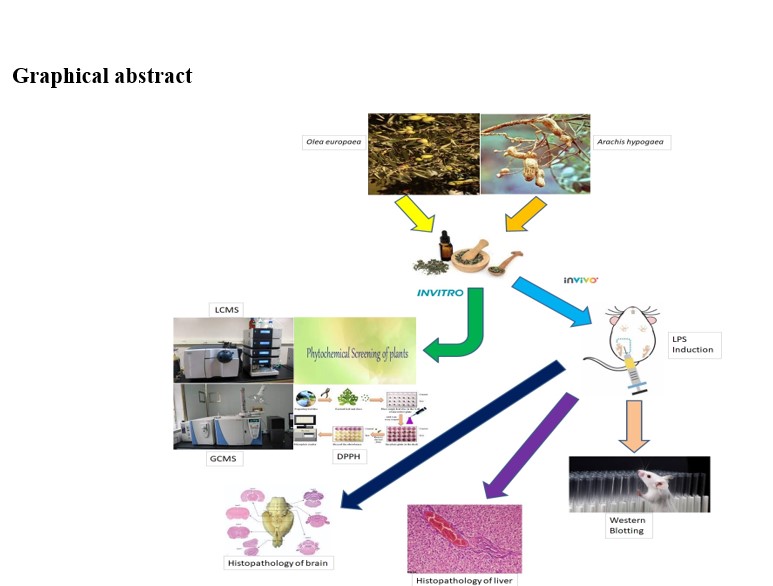
Remyelinating potential of Olea Europaea and Arachis Hypogaea on Experimental autoimmune Encephalomyelitic model of Multiple Sclerosis by downregulating the pro-inflammatory cytokines
https://doi.org/10.21203/rs.3.rs-4358501/v1
This work is licensed under a CC BY 4.0 License
Version 1
posted
You are reading this latest preprint version
DMF
MDA
GSH
INF- α
EAE
8-OhDG
IL-6
TNF-α
Multiple Sclerosis is characterized by the demyelination of neurons in the central nervous system (CNS) and ultimately leads to chronic inflammation of the brain. Etiologically it is an autoimmune disease affecting young adults mostly (Magliozzi et al. 2019). Four clinical types of multiple sclerosis exist in the community and the most prevalent is relapsing-remitting sclerosis and 87% of individuals suffer acute attacks following incomplete recovery. Medications have been developed for therapeutic aspects of disease in individuals via relapsing procedures of sclerosis (Andhavarapu et al. 2019). Medications control disease primarily by depressing the inflammatory cells insinuated by immune cells which can treat the neurologic deficit before diagnostic application. Multiple neurodegenerative illnesses, such as multiple sclerosis (MS) and Alzheimer's disease (AD), have been linked to oxidative stress caused by poor mitochondrial function (Steinman, 2014). Experimental autoimmune encephalomyelitis (EAE) is an inflammatory disease of the central nervous system (CNS) that is induced in Sprague-Dawley (SD) rats via the generation of an autoimmune response against the myelin sheath, an insulating covering around nerve fibers. The typical clinical course is an ascending paralysis that correlates with inflammation and tissue damage in the lumbosacral regions of the spinal cord, although the optic nerves and brain (particularly the subpial white matter and brain stem) are also frequently affected. In recent years, long-term polyphenol-rich olive oil dietary administration in rats has been shown to alleviate age-related motor coordination dysfunctions (Muscoli et al. 2014). Demyelination or the loss of myelin can be induced due to severe injury or disease. Sclerosis is characterized by inflammatory demyelination probably induced by macrophages, monocytes and T-cells. Macrophage-consequential oxidative species cause damage to axonal transmission & results in myelinosis. Demyelination slows axonal conduction which fails to propagate the signal past the demyelinated segment (Chen et al. 2020). Remyelination is an example of spontaneous repair in the CNS where new myelin sheaths are generated around demyelinated terminals. The occurrence of remyelinated areas was documented in early morphological descriptions of the brain of MS patients. While such observations were attributed initially to incomplete demyelination, there had been earlier suggestions that a process of repair could follow damage to myelin (Stadelmann et al. 2019) Neuroprotection is a debatable paradox that highlights various anatomical and immunological differences among scientists on neurodegeneration, synaptic potentials and GABAergic transmissions. Most importantly, functional pathology is the most crucial dilemma supporting the fact that novel neuroprotective strategies surpass devastations created by different enzymes, proteins and oxidative stress markers. Conversely, neurodegeneration cannot be overlooked. Neurodegeneration is characterized by inflammation episodes, demyelination of neurons and gliosis, with characteristic neuronal loss; neurodegenerative episodes can be relapsing, remitting or sometimes can be progressive (Baecher-Allan et al. 2018). Neurodegeneration typically affects 325,000 individuals in the United States and 7.5 million worldwide A neurodegenerative condition known as multiple sclerosis is a very complex disease that attacks CNS from female to male. Almost, 2.5 million population suffered from this disease globally. The frequency of disease is higher in females because they suffered twice the males (Erkkinen et al. 2018). Oxidative stress is a condition in which the balance between reactive oxygen species (ROS) production and antioxidants is significantly disturbed and results in damage to cells by excessive ROS production. ROS contribute to the development of neurodegeneration by modulating the function of biomolecules (Verhaegen et al. 2022). Even though there is no confirmed therapy available in the case of primary disease progression, rarely do numerous drugs exist to improve the secondary progression condition the of disease and when it is controlled through relapsing and remitting way constructively alters the action of the disease. Cereal grains, fruits and vegetables, and plant-based drinks like vine and tea contain phytochemicals(Correale et al. 2017). Consumption of phytochemicals decreases the risk of many long-lasting diseases because they contain antioxidants that protect from the harmful effects of free radicals. Some recent studies have emphasized the potential role of phytochemicals in improving endothelial function and vascular blood flow (Harasym & Oledzki 2014). More than 250 different chemical compounds have been identified in olive oil, including triterpene alcohols, steroids, terpenoids and phenolic compounds. Phenolic compounds are also primary antioxidants found in O. Europaea (olive) and A. hypogaea (peanuts) and contain both lipophilic and hydrophilic phenols (Kontogianni & Gerothanassis 2012). Herbal medication is a cost-effective alternative to conventional medicine. These plant-based medicines have many benefits against several diseases. Some phytochemicals are used to treat multiple sclerosis; including the Parkinsonia plant which can cross the blood-brain barrier and reach the brain. It has antioxidant properties that protect the brain from the oxidative stress and is useful in Parkinson's disease and the relapsing course of sclerosis (Tanaka & Vécsei et al. 2020).
Studies have been conducted on remyelination models in animals. Remyelination occurred early in young animals as compared to older animals. This age-related mechanism is related to transcripts of proteolipid protein & Myelin Basic Protein (PLP and MBP) inside animal lesions (Blakemore & Franklin 2008). The current research is aimed to investigate the role of specific neuroinflammatory and oxidative stress biomarkers involved in the progression of experimental autoimmune encephalomyelitis (EAE) murine model of neurodegeneration and to evaluate the neuromodulatory effects of phytoconstituents alcoholic extracts from O. Europaea and A. hypogaea in treating neurodegeneration.
Organic extraction process for preparation of alcoholic oil extracts
Fresh O. Europaea and A. hypogaea were purchased from a local herbal supplier in Lahore Pakistan and were authenticated by a certified taxonomist and its consent was taken before starting the protocol. Fresh fruits of O. Europaea and A. hypogaea were rinsed with fresh water to remove the extraneous matter and fruits were dried under shade for 7 days. Size reduction was done using a knife and pulverized using an electrical grinder and blender. 100g powder was soaked for 7 days in HPLC- graded ethanol (500 ml) or n-hexane (500 ml) and incubated at room temperature with occasional agitation and then macerated for seven days thrice separately. Mixtures were strained through muslin fabric followed by Whatman filter paper Grade 42, 2.5µm size. Filtrates were then concentrated at 37°C in a rotary evaporator (IKA Germany RV 10B S99) and pressure of 200mmHg in a water bath connected with a vacuum gauge and vacuum pump (Jones & Kinghorn, 2012).
Rotary evaporator procedure for preparation of alcoholic extracts
The round bottom flask was removed from the base of the condenser and was inspected to ensure that it is clean. Then, the sample was loaded into the cleaned round bottom flask which was then, attached to the condenser. Vacuum grease was used to create a vacuum seal between the condenser and the round bottom flask. The collection flask was inspected. Cabinet doors were opened. The chiller was turned sideways tubing was securely connected to the back of the chiller and condenser. The chiller was turned ON using a power switch and set at 20°C. To change the temperature, the knob was rotated and the vacuum pump was turned ON. A hot bath was filled with a round bottom flask to fit in it. UP and DOWN keys were used to raise or lower the setup. A freeze-dried extract was obtained from the liquid extract obtained at 50oC by using a 100% alcoholic mixture (Raaman, 2006).
Freeze drying method for preparation of alcoholic oil extracts
Before freeze-drying, alcohol was removed by a rotary evaporator, and later, the leftover over extract was submitted to freeze-drying. Initially, samples were frozen with liquid nitrogen (-196°C), equilibrated at -80oC, for 2 hrs and then submitted to freeze-drying in a Labconco Freeze Dry System (Labconco Corporation, U.S.A.) at 1.5×10⁻⁴ mbar during 72 h. The moisture content of the freeze-dried particles were determined gravimetrically by weighing small amounts of dried particles i.e. 0.5 g before & after drying in an oven at 110°C. Thereafter, extracts were dried in an incubator. The percentage yield was calculated as 15% for ethanolic oil extract of O. Europaea and 7% for n-hexane extract. A dose of 200 mg/kg body weight was used The weight of rats is between 250-300g. The extract yield of the fresh fruits of O. Europaea and A. hypogaea was calculated using the formula:
Table 1: Percentage yield when solvents were used (mg/100 g crude extract).
|
Medicinal Plant |
Ethanolic |
n-Hexane |
|
Olea Europaea |
2.98 ± 0.86 |
5.74 ± 0.36 |
|
Arachis Hypogaea |
3.87 ± 0.56 |
4.74 ± 0.26 |
Separation funnel method for collection of oil
When four different solvents (n-hexane, ethanol) are selected, fractionation begins by moistening or complete dissolution of crude extract with 250mL of water. This is followed by transfer into a separating funnel, shaken, and allowed to settle. Furthermore, to 250mL of n-hexane, the least polar solvent was added and shaken. The content can settle, and the bottom of the separating funnel opened to remove the aqueous layer. The remaining content in the separating funnel was poured into a clean container to get n-hexane fraction. Equal volume of n-hexane was added again, shaken, and separated. The addition continued until after adding n-hexane and shaken no reasonable quantity of extract appeared to move into the n-hexane portion. Similar cycle was performed for ethanol, methanol, petroleum ether and diethyl acetate fractions (Scopetani et al. 2020).
Quantitative analysis of alcoholic extracts of O. Europaea and A. Hypogaea extracts
Determination of total phenolic content
Folin-Ciocalteu (FC reagent) method was used to determine total phenolic content in aerial parts (50 mg/ml in ethanol) of O. Europaea and Gallic acid (100 µg/ml) is used as standard in concentration range of 2-10 µg/ml. The absorbance was measured at wavelength range of 650-760 nm within Shimadzu double beam UV/Visible spectrophotometer. The standard calibration curve was plotted with Gallic acid as standard and expression of results were done by Gallic acid equivalent (GAE) per gram of dry weight of aerial parts of O. Europaea and A. hypogaea oils (Cao et al., 2020).
Determination of total flavonoid content
The total flavonoid content in aerial parts of O. Europaea and A. hypogaea oils (0.5 ml of extract, 50 mg/ml in methanol) was determined by UV colorimetric method containing aluminum chloride reported by Chia-Chi Chang (2002). Quercetin was used as a standard solution in range 20-200ug/ml which is used to calculate the flavonoid content. The absorbance of the reaction mixture is to be measured at 415 nm with a Shimadzu double beam UV/Visible spectrophotometer (Chang et al., 2002). The Total flavonoid content was calculated from the standard curve and reported as quercetin equivalent in the literature (% w/w).
Determination of total tannins content
Folin-Ciocalteau method is used to determine total tannins content. Different aliquot parts were used which included 100 µl extract plus 750 µl of deionized water plus 30% of sodium carbonate and 500 µl of FC reagent. Dilution was done with 10 milliliters of deionized water and was shaken vigorously with contained mixture extract. 30 minutes’ incubation time was required at room temperature and read at 725 nm UV vis Shimadzu double beam spectrophotometer. The blank contained deionized water whereas Gallic acid was used as standard solution. The GAE were expressed as Gallic acid equivalent (Ogawa & Yazaki 2018).
Determination of total alkaloids content
As far as determination of phytochemical method of O. Europaea and A. hypogaea oils is concerned, 1/2 g of O. Europaea and A. hypogaea oils extracts specimens were added supplementary, in particular test tube and 4 milliliter of n-hexane fraction were blended in it, with vigorous shaking and filtration. At that instance take 10 milliliters of 5% HCl along with pouring in a test tube having the mixture of plant extract and hexane. Plant extract macerate was heated in test tube; filtration was done with pouring of certain drops of picric acid in plant extract’s macerate. Formation of yellow colored precipitates indicates the presence of alkaloids (Ajanal et al. 2012).
Determinations of total carotenoids content
Preparation of Standard Beta-Carotene solution was done. Approximately 5.0 ml of n-hexane was dissolved in 1 mg of β-carotene which gives an equivalent amount of 500 micrograms/milliliter. From this solution, 20 microliters were diluted to 10 ml to have a solution equivalent to 1 mg/ml. The absorbance of this solution was taken at 450 nm in a spectrophotometer using extinction coefficient value of beta-carotene then the solution was made 1 mg/ml. The absorbance of this solution was recorded. Carotenoids show characteristics absorbance spectra, for example, Beta-carotene has an absorption maximum of 450 nm in n-hexane with a molecular extinction coefficient of 2590. Therefore, to estimate total carotenoid in the eluent, absorbance of it was read on a spectrophotometer at 450 nm using a 1 cm cell (Barba et al. 2006).
The total carotenoid concentrated is determined by using the formula below:
Total carotenoid content (µg/g): A = Absorbance at 450 nm,
Volume = Total volume of extract (25 or 50 ml) A1%-cm = Absorption coefficient of β-carotene in petroleum ether
Determination of total steroids content
Steroid solution containing 1 ml of each fraction of plant extracts was transferred in the volumetric flask of quantity 10 ml. 2ml of 4 normal solutions of sulphuric acid prepared plus 2 ml of the 0.5% weight/volume ferric chloride solution, in addition to 0.5 % weight/volume potassium hexa-cyanoferrate solution is added. The temperature required for maintaining water bath stays within range of 75±25ºC for 35 minutes with vigorous shaking and diluted up to the mark with deionized water. The absorbance was measured at 780 nm against the blank using spectrophotometer (Görög 2012).
Antioxidant evaluation of O. Europaea and A. Hypogaea oils
Herbals affluently rich in secondary metabolites, in conjunction to antioxidant phenolics, flavonoids and carotenoids have antioxidant activity, by virtue of their redox potential and chemical structures. The crude ethanolic and n-hexane extracts of O. Europaea and A. hypogaea oils had robust antioxidant potential, contrary to entire free radicals computed. The 2,2-diphenyl-1-picrylhydrazyl (DPPH) radical is generally used in assessing free radical scavenging activity (Munteanu & Apetrei 2021). The stock solution containing plant sample of concentration (1.0 mg/ ml) was diluted to final concentrations of 10 µg/ml, 20 µg/ml, 50 µg/ml ,100 µg/ml, 250 µg/ml, 500 µg/ml in either ethanol or n-hexane. An average of 1 ml of a 0.5 millimolar DPPH ethanol solution/n-hexane solution was added to 3.0 ml of the sample solution, with various dilutions shaking in an orbital shaker and was permitted to function at optimum temperature. Subsequently, after 45 minutes absorbance values were recorded at 518 nm as well as converted into percentage anti-oxidative potential employing the following mathematical statement described below:
Afterwards, a curve of % DPPH scavenging capacity versus concentration was plotted and IC50 values were calculated. IC50 denotes the concentration of sample required to scavenge 50% of DPPH free radicals with reference to bioactive modulation of crude extracts is concerned, flavonoids and antioxidant phenolics are responsible (Marinova & Batchvarov, 2011). In regards to vastly scavenging efficacious nature of flavonoids is concerned, predominantly oxidizing molecular species, including singlet oxygen, and various other free radicals implicated in several diseases. Flavonoids suppress reactive oxygen formation, chelate trace elements involved in free-radical production, scavenge reactive species and upregulate and protect antioxidant defenses. Similarly, phenolics conferring oxidative stress tolerance on plants.
Elemental analysis of O. Europaea and A. Hypogaea oil extracts
The O. Europaea and A. hypogaea oil extract were subjected to analysis of major elements such as calcium, potassium, sulfur and trace elements (iron, copper, and zinc) according to the method of camri.
Table 2: Elemental Analysis of the Ethanol and n-hexane Extracts of O. europaea and A. hypogaea
|
|
Minerals |
Olea Europaea |
Arachis Hypogaea |
|
1 |
Calcium |
36.3003 ±1.23 |
3.108 ± 0.17 |
|
2 |
Iron |
0.694 ± 0.12 |
4.500 ± 0.25 |
|
3 |
Potassium |
22.332 ± 1.12 |
3.049 ± 0.20 |
|
4 |
Magnesium |
4.708 ± 0.21 |
0.408 ± 0.03 |
|
5 |
Sodium |
3.149 ± 0.19 |
22.332 ± 1.16 |
|
6 |
Phosphorus |
0.408 ± 0.05 |
0.694 ± 0.15 |
|
7 |
Sulphur |
1.467 ± 1.00 |
0.033 ± 0.004 |
|
8 |
Zinc |
0.033 ± 0.002 |
1.032 ± 1.16 |
The mineral content was determined using inductively coupled plasma atomic emission spectrophotometer (Hitachi, USA, ZA3300). It is an analytical method which uses flame with atomic absorption and flame photometry. Optical model with Double beam (Polarized Zeeman method). Sample chamber switching is not required in case of Atomic absorption spectrophotometer. Diffraction grating for ZA3300 model turns out to be Czerny-Turner mount, 1800 Lines /mm, Blazed at 200nm. Wavelength range setting was 190 to 900 nm with automatic spectral peak setting. Focal length, reciprocal dispersion is 400 mm, 1.3 nm /mm. Slit width is in 4 steps i.e. 0.2, 0.4, 1.3, 2.6 nm. Photomultiplier detector was with 2 pieces. Number of lamps turn-on current were 8 Lamps (turret), 2 Lamps with simultaneous lightning, 1.0 to 20 mA (average amperage). The Pre-mix fish tail type burner is installed in ZA 3300 model atomic absorption spectrophotometer (Van Loon. 2012). Number of sample containers are 60 pcs of 1.5 mL container each with 96 micro plate option available. Contiguous dispensing with sample injection volume up to 1-100ul. The concentration of each element in the sample was calculated as the percentage of dry matter. 10 mg of the O. Europaea and A. hypogaea oils were extracted successively with petroleum ether at 42-62°C B.p., ethanol and n- hexane, and using rotary evaporator apparatus. The separation with each solvent system, was carried out prior to the solvent was achromatic for 20 hours. Decisively, the mark left was separated with deionized water digesting on a warming water bath. The separation was continued until a few drops of the last portion of the extract left no residue on drying. The extracts were taken in a tared porcelain dish and evaporated to dryness on a water bath and dried at 105°C to a constant weight (Hart & Fisher. 2012). The percentage extractives were calculated with reference to air-dried drug.
Gas chromatography mass spectrometric analysis of O. Europaea and A. Hypogaea oils
GC-MS analysis of O. Europaea and A. hypogaea oils was performed using the equipment Agilent Technologies Ultra Version: 6.0, MS Model 5975C GC model 7890°A equipped with a fused silica column. Control Information (Sample inlet: GC, Injection Source: PAL sampler, Mass Spectrometer: Enabled, Injection Volume: 1ul, Overlap Injection Mode: No Overlap) PAL Method Information (Syringe: 10ul, Cycle: MACRO GC_ Liq4-V2). Parameters of the PAL cycle {(Air Volume (ul): 0, Pre Clean with Solvent 1: 2, Pre clean with Solvent 2: 0, % Syringe Fill for Cleaning (%): 40, Pre Clean with Sample: 0, Sample Amount for Cleaning (ul): 0, Filling Speed (ul/s): 2, Filling Strokes:1, Inject to: GC Inj 1, Injection Speed: (ul/s): 50, Pre inject Delay (MS):500, Post Inject Delay (MS): 500, Post Clean with Solvent 1: 0, Post Clean with Solvent 2 :2, Oven, Equilibrium Time:0.5min, Max Temperature: 350 degrees C, Slow Fan: Disabled, Oven Program : ON, 50° C: for 3 min : # 1 then 7 °C/min to 180 °C for 20 min, # 2 then 7 °C/min to 300 °C for 30 min, Run Time : 88.714min, QQQ Collision Cell EPC : He Quench gas : Off, N2 Collision Gas: Off, Sample Overlap : Sample Overlap is not enabled, Front SS Inlet He, Mode: Split, Heater: On 260°C, Pressure : On 9.05 Psi, Total Flow : On 26.709mL/min, Septum Purge Flow : On 3mL/min, Gas Saver : On 20mL/min after 2min, Split Ratio: 20:1, Split Flow : 22.58ml/min, Thermal Aux 2 ( MSD Transfer Line) : Heater : ON, Temperature Program : On 260°C for 0 min, Run Time : 88.714 min, Column # 1: OPTIMA –OPTIMA 5MS ; 340 °C: 30m × 250um × 0.25 um, In : Front SS Inlet He, Out: Vacuum, (Initial ) : 50°C , Pressure : 9.05 Psi, Flow : 1.129mL/min, Average Velocity : 38.724 cm/sec, Hold Up Time : 1.2912 min, Flow Program : On 1.129mL/min for 0 min, Run Time : 88.714 min, Signals : Signal # 1 : Test Plot : Save Off 50Hz, Signal # 2 : Test Plot : Save Off 50Hz, Signal # 3 : Test Plot : Save Off 50Hz, Signal # 4 : Test Plot : Save Off 50Hz}. Many phytoconstituents were obtained from GC-MS spectra Oleuropein, Fagarasterol etc (Karasek & Clement, 2012).
Liquid chromatography–mass spectrometric analysis O. Europaea and A. Hypogaea oils
An Agilent 1100 HPLC Series system (Agilent, USA) equipped with a degasser, binary gradient pump, column thermostat, auto sampler, and UV detector, was used. The LC system was coupled with an Agilent 1100 mass spectrometer (LC/MS). For the separation, a reverse-phase analytical column was employed (Zorbax SB- C18 column of 1.8ùm 2.1 × 50 mm particle); the work temperature was 48°C. The detection of compounds was performed on both UV and MS mode (MS Model 5975C). The UV detector was set at 350 nm until 17.5 min, then at 380 nm. The MS system operated using an electrospray ion source in negative mode. The chromatographic data were processed using ChemStation and Data Analysis software from Agilent, USA. The mobile phase was a binary gradient: acetonitrile and formic acid 0.05% (v/v). The elution started with a linear gradient, beginning with 5% acetonitrile and ending at 42% acetonitrile, for 35 minutes; then 42% acetonitrile for the next 3 minutes (López-Fernández et al. 2020)
In vivo studies
Sample collection
A total of 8 groups of 4-6 months old Sprague-Dawley albino rats were bred at Animal house, Institute of Molecular Biology and Biotechnology, The University of Lahore, Lahore, Pakistan after fulfilling all ethical considerations. Serum samples of lipopolysaccharide-induced rats (n=64) were collected. O. Europaea and A. hypogaea were purchased from local herbal market, Lahore, Pakistan.
Induction of neurotoxicity
Lipopolysaccharide (LPS) with concentration 0.1µg/µl prepared in normal saline (N/S). The Lumbosacral (LS) injection of Lipopolysaccharide was injected at 15µg/rat (7.5µg in 5µl/site) using Hamilton micro syringe; for 28 days to induce neurotoxicity. Induced neurotoxicity was later confirmed by Complete Blood Count (CBC), and histopathology. LS injection of Lipopolysaccharide dissolved in normal saline 15 µg/rat (7.5 µg in 5 µl/site), bilaterally into the lumbosacral region using Hamilton micro syringe, Lipopolysaccharide (LPS)+ N/S @ (0.3ml/kg B. wt. /day) for 14 days. Olive Oil Extract (OE), Peanut oil Extract (PE) @ (200 mg/Kg B. wt. /day) for 14 days. Nimodipine® @ (30 mg/Kg B. wt. /day) for 14 days, -Interferon alpha @ 1.5ug/kg for 14 days, Dimethyl fumarate 120mg/kg for 14 days.
Table 3: Experimental Design Related To Induction of Neurotoxicity
|
Groups (n=8) |
Treatments |
|
|
LPS (15 µg, icv) |
Phytochemicals (200 mg/kg BW per oral) |
|
|
A |
No (Sham Control) |
Nil |
|
B |
Yes (Control) |
Nil |
|
C |
Yes |
LPS+NIM® |
|
D |
Yes |
LPS+INF® |
|
E |
Yes |
LPS+DMF® |
|
F |
Yes |
LPS+OE |
|
G |
Yes |
LPS+PE |
|
H |
Yes |
LPS+OE+PE |
Development of lipopolysaccharide induced experimental autoimmune encephalomyelitis (EAE) model
The SD rats were treated with intracerbroventricualr (icv) injection of Lipopolysaccharide (Batista et al., 2019). Briefly, the rats were anaesthetized with pentobarbital 45 mg/kg body weight) and placed in stereotaxic apparatus. Through a midline sagittal incision, the scalp was reflected and two drill holes made in the skull for placement of the injection canula in the lateral cerebral ventricle. The animals were given post-operative antibiotic (safinamide 5 mg/kg, intraperitoneally) to ward off sepsis. Rats were then administered rotenone (15 µg dissolved in 5 µl of artificial cerebrospinal fluid) using Hamilton micro syringe. To facilitate drµg diffusion, the canula was left in place for 2–3 min after the injection. The wound was then sealed with sterile wax and Neosporin powder sprayed externally as an additional antiseptic measure.
Determination of antioxidants and reactive oxygen species (ROS) in the serum of albino rats
Lipid peroxidation in blood samples was estimated calorimetrically by measuring Thiobarbituric acid reactive substances (TBARS) (De Leon & Borges, 2020). In cell culture medium and biological fluids, nitrogen oxide level was estimated by analyzing stable nitrogen oxide derived oxidation products; NO2-and NO3-. The concentration of nitrite was measured by Calorimetric Griess assay (Romitelli et al. 2007). Superoxide dismutase (SOD) activity determined by the method of Weydert and Cullen (2010). Meanwhile anti-8-hydroxydeoxyguanosine Antibody kit (catalog number AB5830) Sigma-Aldrich, was used to determine level of 8-hydroxy-2'–deoxyguanosine (8-OHdG). The levels of matrix metalloproteinases-8 (MMP-8) were also measured by human available diagnostic ELISA kit (ABCAM).
Quantitative real-time PCR
Total RNA at various time points was isolated using TRizol reagent (Invitrogen, Carlsbad, CA, USA) and reverse transcribed into cDNA and an oligo (dT)18 primer (TaKaRa Biotechnology, Dalian, China). Real-time quantitative reverse transcription PCR (qRT-PCR) was performed using an SYBR Green PCR Kit (Roche, Basel, Switzerland) by equipment (Applied Biosystems, Foster City, CA, USA). A 20 µl reaction mixture contained 10µl of SYBR Green buffer, 0.5µl of forward primer, 0.5 µl of reverse primer, 1 µl of H2O and 8 µl of cDNA. GAPDH was used as the internal control. Specific primers for the target genes were designed using Primer 5.0 based on the corresponding gene sequences of the proteins. The data was analyzed using VII-A7 software (Applied Biosystems, Foster City, CA, USA) in a relative quantification study model.
Table 4: PCR reaction mixture (20 μl)
|
PCR master mix component |
Volume per reaction |
|
cDNA template |
8 μl |
|
SYBR green master mix with ROX |
10 μl |
|
Primer pair mix |
1.5 μl from 10μM working solution |
|
Nuclease free water |
6.5 μl |
|
Total Volume |
20 μl |
Table 5: Primer sequences of IL-6, INF-α, TNF- α, GAPDH, β-actin and MMP-8
|
Gene |
Direction |
Sequence |
|
IL-6 |
Forward |
5΄AGGAGACTTGCCTGGTGAAA 3΄ |
|
|
Reverse |
5΄CAGGGGTGGTTATTGCATCT 3΄ |
|
INF-α |
Forward |
5΄GCCTCGCCCTTTGCTTTACT 3΄ |
|
|
Reverse |
5΄CTGTGGGTCTCAGGGAGATCA 3 |
|
TNF-α |
Forward |
5΄GAGGCCAAGCCCTGGTATG 3΄ |
|
|
Reverse |
5΄CGGGCCGATTGATCTCAGC 3΄ |
|
GAPDH |
Forward |
5΄GCACCGTCAAGGCTGAGAAC 3΄ |
|
|
Reverse |
5΄TGGTGAAGACGCCAGTGGA 3΄ |
|
β-actin |
Forward |
5΄CACCTTCTACAATGAGCTGC 3΄ |
|
|
Reverse |
5΄AGGCAGCTCGTAGCTCTTCT 3΄ |
|
MMP-8 |
Forward |
5΄ATGGACCAACACCTCCGCAA 3΄ |
|
|
Reverse |
5΄GTCAATTGCTTGGACGCTGC 3΄ |
HISTOPATHOLOGICAL EXAMINATION
The histopathology of the tissue samples (brain, liver, and kidneys) was performed according to the procedures described by Bancroft (2019). The tissue slides were stained using hematoxylin and eosin (H&E) staining procedure.
Table 6: Steps for Tissue Dehydration, Fixation and Embedding
|
Name of Step |
Reagent used |
Duration |
|
|
Fixation |
10% neutral buffered formalin |
24-72 hours |
|
|
Washing |
Running tap water |
2-4 hours |
|
|
Dehydration |
Graded alcohol |
70% |
1 hour |
|
80% |
1 hour |
||
|
95% |
1 hour |
||
|
100% |
1 hour |
||
|
Clearing |
Xylene |
Xylene 1 |
30 minutes |
|
Xylene 2 |
30 minutes |
||
|
Xylene 3 |
45 minutes |
||
|
Embedding |
Paraffin wax at 58ºC |
Paraffin wax 1 |
30 minutes |
|
Paraffin wax 2 |
30 minutes |
||
|
Paraffin wax 3 |
30 minutes |
||
|
Paraffin wax 4 |
45 minutes |
||
Histopathological examination
Sample for analysis was collected from affected tissue regions and preserved in freshly prepared formalin at a volume ratio o 1:10 of tissue: formalin. The same process was carried out for all the samples.
Steps of tissue fixation and processing
Tissue fixation involves dehydrating the tissues using different alcohol solutions and xylene and then fixing the tissues in preheated paraffin for cutting and sectioning. For dehydration different alcohol concentrations are used mentioned.
Block preparation
Block preparation was carried out with steel molds using paraffin. Tissues from 60 ºC were embedded in blocks and placed immediately in refrigerator at 4 ºC for a day or at -20 ºC for long term preservation.
Sectioning
Microtome was used for sectioning and cutting of block. Microtome was adjusted to cut sections with 4-6 μm. Sections were immediately placed in cold water for five seconds and then transferred to water bath preheated to 45ºC before placing them onto slides.
Section mounting on glass slides
Adherent slides were prepared using protocol by (Mayer’s egg albumin). Slides were labelled properly. Tissues floating on the surface of warm water embedded in paraffin will be picked on the slides. These slides were allowed to cool in an incubator for 10-15 minutes at 37ºC (table 12).
Hematoxylin and eosin staining
Hematoxylin and eosin staining was carried for histological analysis. Different alcoholic reagents were also used for staining along with stains. The protocol of technique and role of the reagents is listed in table 13. All the steps are carried out in darkness. After completion of staining process, mounting step is carried out. Post staining the slides are immediately covered with slide cover slips to prevent any contact with air. DPX mounting media was used in this process. Mounted slides were placed at an angle of 45ºC for drying. Slides were then observed under compound microscope and images were taken at resolution of 10X and 40X for analysis of damage at tissue level.
Table 7: Protocol For Staining Process of Hematoxylin and Eosin
|
Reagents |
Time |
Role |
|
Xylene I |
15 minutes |
To remove the paraffin wax from the tissues |
|
Xylene II |
15 minutes |
|
|
Xylene III |
15 minutes |
|
|
Ab. Alcohol I |
3 minutes |
To remove the xylene from the tissues |
|
Ab. Alcohol II |
3 minutes |
|
|
90% Alcohol |
3 minutes |
|
|
70% Alcohol |
3 minutes |
|
|
D. Water |
3 minutes |
To remove alcohol from the tissues |
|
Hematoxylin |
15 minutes |
For staining of nucleus and basophilic parts of the cell |
|
D. Water |
5 minutes |
For washing |
|
Acid Alcohol |
3-5 dips |
To remove the stain from the background of the tissue section |
|
D. Water |
3 minutes |
For washing |
|
Ammonia Water |
3 minutes |
For restoration of nucleus blue colour (Blueing) |
|
D. Water |
3 minutes |
For washing |
|
70% Alcohol |
3 minutes |
|
|
Eosin |
15 minutes |
For staining of eosinophilic parts of the cell |
|
D. Water |
1 minutes |
For washing |
|
70% Alcohol |
3 minutes |
The mounting material does not miscible with the water found in the tissues. Therefore, water was eliminated by placing slides in ethyl alcohol in ascending order |
|
Ab. Alcohol I |
3 minutes |
|
|
Ab. Alcohol II |
3 minutes |
|
|
Xylene I |
15 minutes |
Permanent tissue sections must be translucent. Therefore, slides were dipped in the xylene as xylene is miscible with mounting medium |
|
Xylene II |
15 minutes |
|
|
Xylene III |
15 minutes |
STATISTICAL ANALYSIS
The correlation analysis of the raw data for all attributes was computed using coSTAT computer package. Comparison of means was done using Duncan’s Multiple Range (DMR) tests.
Quantitative analysis of alcoholic extracts of O. Europaea and A. Hypogaea extracts
Total phenolic content
In the first phase, results showed that ethanol extract was evaluated to estimate the highest percentage of total phenolic content in the O. Europaea and A. hypogaea extract. The maximum phenols 11.64 ± 0.65 µgof GAE/g of extract were recorded in ethanolic extract of A. hypogaea followed by O. Europaea 11.08 ± 1.76 µg of GAE/g extract
Total tannins content
Total condensed tannins present in a given plant extract contains polyphenolic biomolecules and the maximum proanthocyanidins are present in the ethanolic extract O. Europaea (27.87 ± 2.54µgof GAE/g of extract) followed by A. hypogaea extract (15.39 ± 1.71µgof GAE/g of extract)
Total alkaloids content
The maximum alkaloids were recorded 20.84 ± 2.69 µg of GAE/g of A. Hypogaea extract followed by 17.87 ± 2.76 µg of GAE/g of O. Europaea and extract. Total tannins were in minute quantities in n-hexane extract and a water fraction
Total flavonoid content
The ethanolic extract of O. Europaea showed maximum radical scavenging activity as maximum flavonoids were recorded in this extract (56.87 ± 3.98 of QE/g of extract) followed by ethanolic extract (32.62 ± 2.31 µg of QE /g) of A. Hypogaea extract. Their anti-carcinogenic, anti-mutagenic, anti-inflammatory, and anti-oxidative properties coupled with their capacity to modulate key enzyme functions.
Total carotenoids content
Quantitative analysis results showed that O. Europaea ethanolic extract has 24.09 ± 3.87 µg of GAE of antioxidant tetraterpenoid which are potent scavengers of free radicals present in the plant extract followed by 14.27 ± 1.56 µgof GAE/g of A. Hypogaea extract. They play significant roles as antioxidants in promoting human health and reducing the risk of heart diseases due to their vitamin A activity.
Total steroids content
Quantitative Analysis of Bioactive steroids showed that n-hexane extract of O. Europaea has more concentration of steroids i.e. 10.78 ± 0.67 µg/g of extract CAE/g as compared to ethanolic fraction 5.34 ± 0.86 µg/g of extract CAE/g. Cycloartenol is a working standard in case of steroids. They are involved in the metabolism of proteins, carbohydrates and lipids; help to maintain immune system, homeostasis, maintain also control renal excretion of electrolytes & blood volume.
| Medicinal Plant | Phenols (GAE) | Tannins (GAE) | Alkaloids (GAE) | Flavonoids (QAE) | Carotenoids (GAE) | Steroids (CAE) |
|---|---|---|---|---|---|---|
| Olea Europaea | 11.08 ± 1.76 | 27.87 ± 2.54 | 17.87 ± 2.76 | 56.87 ± 3.98 | 24.09 ± 3.87 | 10.78 ± 0.67 |
| Arachis Hypogaea | 11.64 ± 0.65 | 15.39 ± 1.71 | 20.84 ± 2.69 | 32.62 ± 2.31 | 14.27 ± 1.56 | 5.34 ± 0.86 |
DETERMINATION OF THE FREE RADICAL SCAVENGING ACTIVITY OF O. EUROPAEA & A. HYPOGAEA PLANT EXTRACTS
The antioxidant activity of the various solvent fractions of O. Europaea and A. Hypogaea was further assessed by the DPPH radical scavenging method, Ascorbic acid was used as standard control. The highest radical scavenging activity was observed for A. hypogaea n-hexane extract (IC50 = 285.74 ± 1.03 µg/ml), while lowest for O. Europaea ethanolic extract (105 ± 1.76 µgmL1) The IC50 (DPPH) values of the extracts increased in the following order: A. hypogaea n-hexane extract ≤ A. hypogaea ethanolic extract ≤ O. Europaea n-hexane extract ≤ O. Europaea ethanolic extract. This is in good settlement with the TPC values listed in Table 9. Therefore, it is likely that the phenolic & flavonoid phytoconstituents present in the A. Hypogaea and O. Europaea are responsible for the antioxidant and free radical scavenging activities. Compared to the reference compound, ascorbic acid (IC50 = 120.05 ± 0.54 µg/ml), the extracts of both plants showed higher antioxidant capacity, at the same concentration (25 µg samples/ml). The result of the present study suggests that these plant materials especially O. Europaea and A. Hypogaea can be used as a natural source of antioxidants.
| Samples (25 µg samples/ml) | DPPH Radical Scavenging Activity (%) | IC50 (µg/ml) |
|---|---|---|
| Olea Europaea ethanolic extract | 25.41 ± 0.59 | 105.74 ± 1.76 |
| Olea Europaea n-hexane extract | 15.49 ± 1.01 | 150.05 ± 1.95 |
| Arachis Hypogaea ethanolic Extract | 9.89 ± 0.14 | 170.86 ± 0.14 |
| Arachis Hypogaea n-hexane Extract | 13.54 ± 0.38 | 285.61 ± 1.03 |
| Ascorbic Acid | 91.60 ± 1.4 | 120.05 ± 0.54 |
Gas chromatography–mass spectrometric analysis of O. Europaea and A. Hypogaea plant extracts
The extracts were subjected to gas chromatography and mass spectrometric analysis essential oils which indicates the presence of secondary metabolites. The cocktail extract contains high concentrations of Oleuropein (35.03%), Oleocanthal (22.21%), Stigmasterol (7.23%), Luteolin (4.0%), Hexadecane (3.65%), Phytol (3.45%), Spinacene (3.21%), Isopropyl (9Z,12Z,15Z)-9,12,15-octadecatrienoate (3.00%), Isophytol (2.03%), Stearic Acid (2.18%), Pinoresinol (1.92%), n-hexadecanoic acid (1.78%), 1-Monolinoleoylglycerol Trimethylsilyl Ether (1.76%), Clionasterol (1.34%), Fagarasterol (1.03%), Isopropyl palmitate (1.00%), Caryophyllene oxide ( 0.99%), Diisooctyl phthalate (0.94%), Heptadecane (0.77%), Vitamin E (0.75%) Hentriacontane (0.71%), 3-Ethyl-5-(2'-ethylbutyl) octadecane (0.60%), Mangiferic Acid (0.57%), Heptacosane (0.34%), Isopropyl linoleate (0.16%) as shown in Table 10
Table 10:
Liquid chromatography-mass spectrometric analysis of O. Europaea and A. Hypogaea plant extract
Liquid chromatography and mass spectrometric analysis tentatively identified the presence of phytoconstituents as secondary metabolites including Epigallocatechin gallate, Helioamplexine N Oxide, Isomammein, 1-[(Fluoromethyl)hydroxyaminomethyl]-2-nitro-1H-imidazole, Rutin, Vicenin-2, Geranate, Hirsutidin, Mellitic acid, Anisomelic acid, Cerebrodiene, Sodium;2-[(E)-tetradec-1-enyl]butanedioic acid, Zeaxanthin, Sodium;7-(5-hexyl-2-hydroxy-6-oxabicyclo[3.1.0]hexan-1-yl)heptanoic acid/14deoxyandrographiside, Sodium ricinoleate, 2-[Bis(2-dec-9-enoxyethyl)amino]ethyl dec-9-enoate, Sodium dioctyl benzene-1,2-dicarboxylate based on Retention time, m/z [M + H] + (experimental), m/z [M + H]+ (calculated) and Error in ppm
| Retention time (Rt) | m/z [M + H]+ (experimental) | m/z [M + H]+ (calculated) | Error in ppm | Elemental Formula | Tentative identification |
|---|---|---|---|---|---|
| 0.30–0.43 | 365.1055 | 365.1134 | 0.83 | C22H18O11 | Epigallocatechin gallate |
| 3.08–3.66 | 330.1908 | 330.2100 | 0.53 | C16H27NO6 | Helioamplexine N Oxide |
| 3.78–3.99 | 372.2015 | 372.2121 | 0.31 | C22H28O5 | Isomammein |
| 4.24–4.71 | 438.2385 | 438.2411 | 2.35 | C5H7FN4O3 | 1[(Fluoromethyl)hydroxyaminomethyl]-2-nitro-1H-imidazole |
| 5.14–5.45 | 611.1607 | 611.2399 | 1.06 | C27H30O16 | Rutin |
| 5.53–5.98 | 595.1659 | 595.1000 | -0.27 | C27H30O15 | Vicenin- 2 |
| 5.96–6.36 | 167.1066 | 167.1119 | 0.18 | C10H15O2− | Geranate |
| 6.62–7.16 | 345.1073 | 345.2543 | 3.18 | C18H17O7 | Hirsutidin |
| 7.22–7.48 | 375.1073 | 375.6574 | -2.85 | C12H6O12 | Mellitic acid |
| 7.51–8.22 | 359.1124 | 359.1020 | -0.51 | C20H26O4 | Anisomelic acid |
| 7.91–8.03 | 239.1620 | 239.1999 | -0.04 | C18H35NO | Cerebrodiene |
| 8.41–8.73 | 335.2193 | 335.2190 | 0.52 | C18H32NaO4 | Sodium;2-[(E)-tetradec-1-enyl]butanedioic acid |
| 8.75–9.10 | 301.1416 | 301.1417 | 0.66 | C40H56O2 | Zeaxanthin |
| 8.79–9.20 | 317.2087 | 317.2099 | -1.07 | C18H30O3Na+ | Sodium;7-(5-hexyl-2-hydroxy-6-oxabicyclo[3.1.0]hexan-1-yl)heptanoic acid/14deoxyandrographiside |
| 9.20–9.62 | 352.3393 | 352.4657 | -0.98 | C18H34O3Na | Sodium ricinoleate |
| 9.97–10.15 | 280.2634 | 280.2655 | -0.98 | C36H67NO4 | 2-[Bis(2-dec-9-enoxyethyl)amino]ethyl dec-9-enoate |
| 11.09–11.46 | 413.2655 | 413.2650 | 1.78 | C24H38NaO4+ | Sodium;dioctyl benzene-1,2-dicarboxylate |
Figure 2: LCMS Spectra of various Phytoconstituents reported by NIST MS Phytoconstituents Library. Tentative identification of Detected Phytoconstituents according to retention time and m/z. Epigallocatechin gallate, Helioamplexine N Oxide, Isomammein, 1-[(Fluoromethyl)hydroxyaminomethyl]-2-nitro-1H-imidazole, Rutin, Vicenin-2, Geranate, Hirsutidin, Mellitic acid, Anisomelic acid, Cerebrodiene, Sodium;2-[(E)-tetradec-1-enyl]butanedioic acid, Zeaxanthin, Sodium;7-(5-hexyl-2-hydroxy-6-oxabicyclo[3.1.0]hexan-1-yl)heptanoicacid/14deoxyandrographiside,Sodiumricinoleate,2-[Bis(2-dec-9enoxyethyl )amino]ethyl dec-9-enoate, Sodium;dioctyl benzene-1,2-dicarboxylate
Effect of standard drugs and alcoholic oil extracts on MDA, GSH, NO, SOD, CAT concentrations in LPS treated rats
All seven treatments had significant effects on MDA concentration than control/healthy rats as shown in Fig. 3a and Table 7. MDA concentration for Control rats was found to be 0.93nmol/ml while 12.35nmol/ml in diseased rats. MDA value for drugs treated respectively Dimethyl fumarate treated group was (8.48 nmol/ml), Nimodipine treated group (10.29 nmol/ml), and MDA levels after olive and peanut oils treated groups were7.88 6.58 nmol/ml, respectively. This synergic application of both olive and peanut oils treated group reduced MDA level to 4.19 nmol/ml, causing maximum decline among 7 diseased rat groups. The mean value for the concentration of GSH was 8.64 µmol/ml in healthy rats and 2.34 µmol/ml in diseased rats treated with LPS. GSH concentration for the Dimethyl fumarate treated group was 4.07 µmol/ml, Interferon-α treated group is 3.16 µmol/ml GSH, Nimodipine treated group is 3.34 µmol/ml. GSH concentrations in olive and peanut oils extract treated group was 4.99 and 5.16 µmol/ml, respectively. The synergic application of both drugs enhanced the GSH concentrations to 6.88 µmol/ml.
The mean value for the concentration of NO was 11.55 µmol/L in the healthy rats and 16.26 µmol/L in diseased rats treated with LPS. Interferon-α treated group was 12.59 µmol/L, Nimodipine treated group was 14.29 µmol/L, Dimethyl fumarate treated group was 13.35 µmol/L. Olive and peanut oils treated groups reduced NO level in serum 11.57 and 11.29 µmol/L, respectively. The synergic application of both olive and peanut oils brought down the NO level to 11.58 nmol/ml. The mean value of SOD was 1.09 IU/ml in healthy rats, and 0.46 IU/ml in diseased rats treated with LPS. Among the drugs used for treatment, SOD concentration for the Dimethyl fumarate treated group and Nimodipine treated group was 0.93 IU/ml, Interferon-α treated group was 0.87 IU/ml, olive and peanut oils treated groups were 0.77 and 0.83 IU/ml, respectively. The synergic application of both olive and peanut oils treated groups enhanced the SOD concentration to 0.66 IU/ml. The mean value for the CAT concentration was 6.39 µmol/ml in healthy rats, 2.18 µmol/ml in diseased rats treated with LPS. Among Dimethyl fumarate CAT concentration was 2.11 µmol/ml, respectively the other two medicines, i.e., Interferon-α and Nimodipine treated groups were 2.51 and 2.88 µmol/ml, respectively. Oil fractions enhanced the CAT concentration in serum to 3.89 and 5.56 µmol/ml, respectively. The synergic application of olive and peanut oils treatment enhanced the level in diseased rats to an almost comparable level with control/healthy rats.
| Groups | Mean ± SD (n = 8) | |||||||
|---|---|---|---|---|---|---|---|---|
| MDA (nmol/ml) | GSH (µmol/ml) | NO (µmol/L | SOD (IU/ml) | CAT (µmol/ml) | IL-6 (ng/ml) | 8-OHdG (pg/ml) | MMP-8 (ng/ml) | |
| A | 0.93 ± 0.063 | 8.64 ± 2.23 | 11.55 ± 3.55 | 1.09 ± 0.08 | 6.39 ± 1.77 | 3.20 ± 1.13 | 2.09 ± 0.16 | 21.25 ± 2.19 |
| B | 12.35 ± 3.59 | 2.34 ± 0.93 | 16.26 ± 3.29 | 0.46 ± 0.017 | 2.18 ± 0.09 | 10.13 ± 2.0 | 21.29 ± 3.29 | 181.26 ± 5.26 |
| C | 10.29 ± 3.17 | 3.34 ± 0.99 | 14.29 ± 5.26 | 0.93 ± 0.038 | 2.88 ± 0.89 | 9.08 ± 3.05 | 17.19 ± 2.28 | 102.26 ± 7.28 |
| D | 9.68 ± 1.88 | 3.16 ± 1.22 | 12.59 ± 5.19 | 0.87 ± 0.081 | 2.51 ± 0.65 | 8.16 ± 2.00 | 15.29 ± 3.29 | 98.26 ± 6.25 |
| E | 8.48 ± 2.25 | 4.07 ± 2.00 | 13.356 ± 1.59 | 0.93 ± 0.045 | 2.11 ± 0.56 | 8.05 ± 1.98 | 13.29 ± 2.18 | 78.26 ± 7.26 |
| F | 7.88 ± 1.19 | 4.99 ± 1.33 | 11.57 ± 1.47 | 0.77 ± 0.019 | 2.01 ± 0.19 | 6.95 ± 2.01 | 8.89 ± 1.28 | 47.26 ± 5.26 |
| G | 6.58 ± 2.47 | 5.16 ± 0.96 | 11.29 ± 2.23 | 0.83 ± 0.037 | 3.89 ± 0.99 | 6.06 ± 1.34 | 14.26 ± 3.29 | 68.26 ± 4.44 |
| I | 1.56 ± 0.32 | 2.06 ± 1.03 | 14.26 ± 2.26 | 14.26 ± 2.26 | 1.06 ± 0.076 | 1.09 ± 0.04 | 4.29 ± 1.90 | 25.26 ± 4.23 |
| LSD (0.05) | 2.07 | 1.75 | 0.99 | 0.316 | 1.09 | 1.08 | 6.29 | 8.16 |
| P-value | 0.011 | 0.008 | 0.013 | 0.001 | 0.038 | 0.011 | 0.014 | 0.012 |
| Variables | MDA | GSH | CAT | SOD | NO | IL-6 | 8-OHdG | MMP-8 |
|---|---|---|---|---|---|---|---|---|
| MDA | 1.000 | 0.423 | 0.519 | 0.399 | 0.435 | 0.512* | 0.423 | 0.823** |
| GSH | 1.000 | 0.645* | 0.715** | 0.619** | 0.774** | 0.684** | 0.659* | |
| CAT | 1.000 | 0.648** | 0.671** | 0.719** | 0.589* | 0.726** | ||
| SOD | 1.000 | 0.746* | 0.659** | 0.589** | 0.865** | |||
| NO | 1.000 | 0.614** | 0.741** | 0.665 | ||||
| IL-6 | 1.000 | 0.621* | 0.596** | |||||
| 8-OHdG | 1.000 | 0.619* | ||||||
| MMP-8 | 1.000 |
**Correlation is significant at the 0.01 level (two-tailed). *p < 0.05, **p < 0.01, ***p < 0.001.
Effect of drugs and alcoholic oil extracts on MMP-8 and 8-OXO-2'-deoxyguanosine (8-OHDG) concentration in LPS treated rats
The mean value for the concentration of MMP-8 was 3.2 ng/ml in healthy and 10.13 ng/ml in diseased rats treated with LPS. MMP-8 concentration of all three drugs Dimethyl fumarate, Interferon-α and Nimodipine were was 8.05, 8.16 and 9.08 ng/ml, respectively. Olive and peanut oils fractions MMP-8 levels in serum 6.05 ng/ml, respectively. The synergic application of both olive and peanut oils brought down the MMP-8 level to 5.19 ng/ml (Fig. 3 & Table 12). The 8-OHdG concentration of 0.76 pg/ml in the healthy rats and 31.88 pg/ml in diseased rats treated with LPS. 8-OHdG concentration for Dimethyl fumarate, Interferon-α and Nimodipine, slightly reduced the 8-OHdG concentration to 27.35, 30.29, and 30.89 pg/ml, respectively. Olive and peanut oils fractions reduced the 8-OHdG level in serum to 18.59 and 18.07 pg/ml, respectively. The synergic application of both olive and peanut oils brought down the 8-OHdG level to 18.79 pg/ml.
mRNA expression of IL-6, TNF-α, INF- α, MMP-8, β-Actin and GAPDH genes in RT-PCR
IL-6 gene was significantly (P ≤ 0.001) increased (10.13 ± 2.00) in the diseased rat group treated with LPS as compared with the healthy rat or control group (3.20 ± 1.13). Treatment of diseased rats with Dimethyl fumarate, Interferon-α and Nimodipine drugs reduced mRNA expression of the IL-6 gene (7.36 ± 0.58, 8.45 ± 1.12, and 8.84 ± 0.37) recorded respectively. The diseased rats that were treated with olive and Peanut oil extracts and their synergic application showed significantly (P ≤ 0.001), lower IL-6 expression (3.95 ± 0.89, 3.13 ± 1.29, 2.17 ± 0.87 respectively).
TNF-α gene was significantly (P ≤ 0.001) increased (7.89 ± 0.24) in the diseased rat group treated with LPS as compared with the healthy rat or control group (1.000 ± 0.00). Treatment of diseased rats with Dimethyl fumarate, Interferon- α and Nimodipine drugs reduced mRNA expression of the TNF-α gene (4.71 ± 0.22, 5.95 ± 0.43 and 6.36 ± 0.19) respectively recorded. The diseased rats that were treated with olive and peanut oils fractions and their synergic application showed significantly (P ≤ 0.001), lower TNF-α expression (2.52 ± 1.45, 2.37 ± 1.15, 1.91 ± 0.49 respectively).
INF-α gene was significantly (P ≤ 0.001) increased (5.98 ± 1.43) in the diseased rat group treated with LPS as compared with the healthy rat or control group (1.000 ± 0.00). Treatment of diseased rats with Dimethyl fumarate, Interferon- α, Nimodipine, olive oil extract, Peanut oil extract, and olive and peanut oils synergic application reduced mRNA expression of INF-α gene (4.78 ± 0.62, 5.31 ± 0.38, 5.53 ± 0.34, 3.76 ± 0.35, 2.49 ± 0.19, 2.14 ± 0.21 respectively).
MMP-8 gene was significantly (P ≤ 0.001) increased (5.42 ± 0.11) in the diseased rat group treated with LPS as compared with the healthy rat or control group (1.000 ± 0.00). Treatment of diseased rats with Dimethyl fumarate, Interferon- α and Nimodipine drugs reduced mRNA expression of the MMP-8 gene (4.33 ± 0.36, 4.86 ± 0.30 and 5.06 ± 0.17 respectively). The diseased rats that were treated with olive and peanut oils fractions and their synergic application showed significantly (P ≤ 0.001), lower MMP-8 expression (3.11 ± 0.45, 2.76 ± 0.39, 2.05 ± 0.14) respectively.
β-actin gene was slightly increased (1.23 ± 0.08) in the diseased rat group treated with LPS as compared with the healthy rat or control group (1.000 ± 0.00). Treatment of diseased rat with Dimethyl fumarate, Interferon- α, Nimodipine, olive oil extract, Peanut oil extract and olive and peanut oils synergic application did not affect mRNA expression of the β-actin gene (1.14 ± 0.13, 1.21 ± 0.13, 1.12 ± 0.0.5, 1.09 ± 0.11, 1.39 ± 0.21 and 1.05 ± 0.03) respectively. Similarly, the expression level of the GAPDH gene was slightly increased (1.31 ± 0.08) in the diseased rat group treated with LPS compared with the healthy rats or control group (1.000 ± 0.00). Treatment of diseased rats with Dimethyl fumarate, Interferon- α, Nimodipine, olive oil extract, Peanut oil extract and olive and peanut oils synergic application did not affect mRNA expression of the GAPDH gene (1.05 ± 0.33, 1.14 ± 0.23, 1.39 ± 0.19, 1.21 ± 0.14, 1.15 ± 0.21, 1.21 ± 0.21 respectively) (Fig. 4).
Expression of IL-6 and TNF- α Proteins
Western blotting showed persistent expression of the IL-6 and TNF-α protein in control or healthy rats, diseased rats treated with LPS, diseased rats treated with Dimethyl fumarate, Interferon- α, Nimodipine, olive oil extract, Peanut oil extract and olive and peanut oils synergic application. The expression level of IL-6 protein was significantly (P ≤ 0.001) increased (18.36 times) in the diseased rat group treated with LPS as compared with the healthy rat or control group (1.00 ± 0.00). Treatment of diseased rats with Dimethyl fumarate, Interferon- α and Nimodipine drugs reduced IL-6 protein expression (8.46, 11.31, and 13.78 times, respectively) compared with LPS-treated diseased rat. The diseased rats were treated with olive and peanut oils fractions and their synergic application showed significant (P ≤ 0.001), lower IL-6 protein expression (5.79, 3.21, and 2.11 times, respectively) as compared with LPS-treated diseased rats. The expression level of TNF-α protein was significantly (P ≤ 0.001) increased (19.72 times) in the diseased rat group treated with LPS as compared with the healthy rat or control group (1.00 ± 0.00). Treatment of diseased rats with Dimethyl fumarate, Interferon-α and Nimodipine drugs reduced mRNA expression of the TNF-α gene (7.71, 6.79, and 14.79 times, respectively) as compared with LPS-treated diseased rats. The diseased rats that were treated with olive and peanut oils fractions and their synergic application showed significantly (P ≤ 0.001), lower TNF-α protein expression (7.85, 5.73, 4.71 times, respectively) as compared with LPS-treated diseased rats (Fig. 4).
Histopathological examination
Histopathology of brain
Photomicrographs show (H&E 40X) (A) No microscopic lesions were observed with intact convoluted cerebellar folia, sulci, and meninges. (C) Heterogeneous lymphocyte proliferation was evident in the granular layer Endothelial lining of the central vein was disrupted along with an influx of inflammatory cells in sinusoids. (E) Mild to moderate necrosis of the molecular layer and Purkinje cells were noticed along with RBCs' seepage from blood vessels to white matter. Pia mater was sloughed off in most of the cerebellar folia. (G) Chromatolysis were pronounced in neurons in molecular and granular layers. Moderate necrosis of Purkinje cells was evident with vacuolation of granular and Purkinje cell layers; moreover, multifocal coalescing foci of hemorrhage were also observed in the cerebellum. (I) Microscopic lesions showed lymphocytic cuffing surrounding the molecular layer of the cerebellum. Mild vacuolation in the granular layer and mild hemorrhages in Purkinje cell layers were evident. (K) The astrocytes were scattered around the profound oligodendrocytes; no specific lesions were present in the cerebellum; only a few areas with pronounced vacuolation along with fibrous molecular and granular layers. There was atrophy of the granular layer associated with necrosis of neurons and Purkinje cells. (M) Photomicrograph (H&E 40×) showed thickening of meninges due to lymphocytic infiltration in pia mater accompanied by extravasation of red blood cells (O) Degenerative changes in molecular and granular layers were pronounced, along with the white matter's demyelination. Focal necrosis of Purkinje cells was evident in Purkinje cell layers. There was evidence of inflammatory cells in granular cell layers of encephalomyelitic rat brain (Fig. 5).
Histopathology of liver
Photomicrographs show (H&E 40×) (B) No evidence of microscopic lesions as observed in the hepatocellular pattern. (D) Fatty changes in the hepatocytes were pronounced with visible pyknotic nuclei in hepatocytes. (F) The focal areas of hepatic hemorrhages and binucleated hepatocytes were visible in some areas. Moderate degenerative changes in hepatocytes with abnormal hepatic cords were evident in hepatic parenchyma. (H) Vacuolation in hepatocytes is developed away from the central vein. Focal areas of mild hemorrhages with Karyolysis were detected in some hepatocytes. (J) There was mild hepatic congestion with disrupted endothelial lining of the central vein. The hepatic triad was normal with intact hepatic artery, portal vein, and bile duct. No histologic changes in hepatocytes and sinusoids. (L) Moderate hepatic congestion accompanied by mild to moderate hemorrhages in sinusoids was detectable. Perivascular infiltration of mononuclear cells is evident with mild hydropic degenerative changes in the hepatic parenchyma. (N) Hepatic congestion in the central vein with severe degenerative changes in hepatocytes. Fragmentation in the nucleus of hepatocytes was pronounced. (P) In most of the areas, the architecture of hepatocytes was recovered completely. The endothelial lining of the central vein was disrupted (Fig. 5).
An episode of neurological malfunction that lasts for several weeks and either a full or partial recovery marks the beginning of neurodegeneration. Evidence suggested that neurotoxicity is caused by immune system-mediated inflammation and neurodegenerative progressions (Mottahedin et al. 2017). Anti-oxidants stop the oxidation processes initiated by atmospheric oxygen species. The mechanism of an organism against diseases brought on by free radicals depends heavily on these species. To determine phytochemicals and antioxidant potential, O. Europaea and A. hypogaea extracts were prepared via maceration technique in ethanol, hexane, petroleum ether, and methanol. The phytochemical examination was done according to international standards listed in the Phytochemistry manual (Tzanova et al. 2020). Due to their potential health benefits, the secondary metabolites flavonoid and phenolic, have drawn significant attention from researchers. The presence of flavonoids and phenolics with encephalomyelitis and remyelinating potential in the extracts of O. Europaea & A. hypogaea might contribute to therapeutic potential (Yan et al. 2022). In the current settings, total phenolic levels were observed in O. Europaea & A. hypogaea extract (11.08 ± 1.76 GAE/g) and flavonoid contents (56.87 ± 3.98GAE/g) in dry extract. These higher values may be attributed to the higher polyphenols content in Olives as compared to Peanuts. The O. Europaea & A. hypogaea were identified to have significant antioxidant potential through DPPH free radical scavenging activity as shown in Table 3. Quantitative analysis of various solvent fractions of O. Europaea and A. hypogaea showed a higher % of inhibition of both antioxidants when compared to their respective standard, i.e., the inhibition of Antiencephalomyelitic activity was evaluated against the percentage of demyelinating nerve fibers (Montero Torres, 2020). The Olive oil extract showed an inhibition ranging from (15.49 ± 1.01) to (25.41 ± 0.59), and the Peanut Oil extract showed an inhibition ranging from (9.89 ± 0.14) to (13.54 ± 0.38) compared to the standard Ascorbic Acid (91.60 ± 1.40). The cocktail of O. Europaea & A. hypogaea was subjected to (GCMS). 25 compounds and metabolites were tentatively identified by the NIST library including Oleuropein, Luteolin, Pinoresinol, Diisooctyl phthalate, Hentriacontane, Stigmasterol, Oleocanthal, Stearic acid, Heptacosane, Isopropyl palmitate, Fagarasterol, n-hexadecanoic acid, Caryophyllene oxide, Clionasterol, Vitamin E, 3-Ethyl-5-(2'-ethyl butyl) octadecane, Isophytol, Mangiferic Acid, Phytol, Heptadecane, Isopropyl (9Z,12Z,15Z)-9,12,15-octadecatrienoate, Hexadecane, Spinacene, Isopropyl linoleate,1-MonolinoleoylglycerolTrimethylsilylEther. Similarly, 18 compounds and metabolites were identified when a cocktail of Olea Europaea & Arachis Hypogaea was also subjected to (LCMS). Major identified compounds among those were Epigallocatechin gallate, HelioamplexineNOxide, Isomammein, 1-[(Fluoro-methyl)-hydroxy-amino-methyl]-2-nitro-1H-imidazole, Geranate, Hirsutidin, Morphine, Mellitic acid, Anisomelic acid, Cerebrodiene, Sodium;2-[(E)-tetradec-1-enyl]-butanedioicacid, Zeaxanthin, Sodium-7-(5-hex yl-2-hydroxy-6-oxabicyclo[3.1.0]-hexanyl)-6-oxabicyclo-[3.1.0]-hexanyl)heptanoiacid/deoxyndrographisideacid / 14deoxyandrographiside, Sodium ricinoleate, 2-[Bis(2-dec-9-enoxyethyl)-amino]-ethyl-dec-9-enoate, Sodium dioctyl-benzene-1,2-dicarboxylate. Inflammation in demyelinating lesions can generate oxygen and nitrogen free radicals as well as pro-inflammatory cytokines, which contribute to the onset and progression of neurodegenerative episodes during pathogenies of neurotoxicity (Kamma et al. 2022). Activated immune cells are linked with the production of reactive nitrogen and oxygen species which take part in phagocytosis, myelin breakdown, axonal degeneration, and oligodendrocyte destruction (Vodjgani et al. 2020). The results of this investigation indicated a multifactorial autoimmune remyelinating reaction linked to the central nervous system (CNS). The balance of biomarker quantities including carbonyl protein content, lipid peroxidation, non-proteinous chemicals, vitamin E, SOD, vitamin C, CAT activity, and DNA damage were also assumed (Boucelha et al. 2021). Peroxy-nitrite, one type of free radical found in neural tissue, can increase the inflammatory response that leads to neurotoxicity. Many mediators like reactive substances of thiobarbituric acid, end products of oxidation proteins like fructosamine, and activated levels of α-2-macroglobulin (Gorecki et al. 2018). Malondialdehyde (MDA), nitric oxide (NO), Matrix metalloproteinase-8 (MMP-8) or Neutrophil collagenase, and 8-Oxo-2'-deoxyguanosine (8-OHdG) are important biomarkers for oxidative stress in animals. MDA, NO, MMP-8, and 8-OHdG concentrations increased up to 42 times in the serum of diseased rats (compared with healthy control rats) treated with LPS endotoxin superoxide’s oxidative stress in the neuronal cells in the brain and spinal cord (Davies, 2021). This increase showed the development of oxidative stress. These biomarkers are produced in response to oxidative stress. O. Europaea & A. hypogaea reduced the production of MDA, NO, MMP-8, and 8-OHdG contents significantly by reducing oxidative stress and highlighting the neuroprotective role of these extracts. In most cases, these alcoholic oil extracts showed higher effectiveness than traditional drugs, while their cocktail showed a synergic effect after their application (Chagas et al. 2022). These findings are in line with the previous finding on the subject that has been linked with the regulation of ERK/CREB/BDNF signaling pathways by these oil extracts providing neuroprotection in the rat. GSH, CAT, and SOD are essential antioxidants against oxidative stress in animal cells (Sharifi-Rad et al. 2020). GSH, CAT, and SOD concentrations decreased almost 2.5 to 4 times in the serum of diseased rats (compared with healthy control rats) treated with LPS, an endotoxin inducing super oxides oxidative stress in the brain and other cells. This reduction showed the development of oxidative stress, and GSH, CAT, and SOD are produced in fewer amounts than required in response to oxidative stress. GSH is a delicate indicator of oxidative stress in many neurodegenerative diseases (Demirci-Çekiç et al. 2022). GSH, CAT, and SOD contents were produced in significantly higher quantities, thus reducing the oxidative load of free radicals when LPS-induced diseased rats were treated with standard drugs like Dimethyl fumarate, Nimodipine, and Interferon α, O. Europaea & A. hypogaea, and a cocktail of O. Europaea & A. hypogaea (Sarker & Oba, 2018). These findings support the previous finding on the subject, thus providing neuroprotection in the rat. The qPCR analysis showed that the expression level of mRNA of IL-6, TNF-α, INF-α, and MMP-8 genes expression was significantly increased in the diseased rat group treated with LPS compared with the healthy rats in the control group. Treatment of diseased rats with standard drugs such as Dimethyl fumarate, Interferon and Nimodipine, olive oil extract, and Peanut oil extracts, and their synergic application reduced mRNA expression of these genes (You et al. 2011). In most cases, these oil extracts showed higher effectiveness in reducing gene expression. However, the current study showed their synergistic effects if applied together. The neuroprotection and disease mediations involving oxidative stress have also been shown to be done by the downregulation of these genes in previous studies (Dumont & Beal, 2011). The mRNA expression of β-actin and GAPDH genes did not show any significant change in different conditions. Some evidence proposed the role of oxidative stress in the demyelinating pathways. Oxidative stress, energy failure, and mitochondrial injury could be linked with neurodegeneration as well as plaque formation in lesions of gray and white matter (Mao et al. 2013). Neurodegeneration in the sclerosis is linked with oxidative stress, hemoglobin's long-lasting extravasation in the lesions, and the disability of many cellular protective procedures against hemoglobin reactivity. Some studies revealed that the change in antioxidant and oxidant balance contributes to disease pathophysiology ant this can be applied to levels of many substances in the body like contents of carbonyl protein, lipid peroxidation, nonproteins compounds, vitamin E, SOD, vitamin C, CAT activities as well as DNA damage (Dani & Buonocore).
The study deduces that formation of oxidative stress triggers the activation of NADPH oxidase, inflammatory cytokines such as IL-6, IL-12, IL-17, and caspases that ultimately causes DNA damage. Phytochemical analysis of the crude oils of O. Europaea and A. hypogaea results in the number of secondary metabolites, i.e., phenols, flavonoids, alkaloids, coumarins, tannins, terpenes, etc. that are significant in overcoming the oxidative damage caused by the inflammatory triggers. Hence, treatment with the O. Europaea, A. hypogaea, their cocktail and standard drugs Nimodipine, INF-α, and DMF blocks the calcium flux and cellular leakage across the blood-brain barrier and treat the demyelination in the axonal and neuronal terminals. However, the extracts of olive oil and peanut oil showed higher effectiveness than traditional drugs and showed higher synergistic activities.
Ethical statement
This Research is approved from Animal Research Ethical Committee IMBB- The University of Lahore reference letter (IMBB/UOL/20/149) dated (December 20, 2020).
Acknowledgement
The Authors are highly thankful for the valuable contribution of students of Lab 313, Institute of Molecular Biology & Biotechnology, The University of Lahore-Pakistan and Department of Environmental Design, Health & Nutritional Sciences, Allama Iqbal Open University Islamabad
- Ajanal M, Gundkalle M. B, & Nayak, SU (2012). Estimation of total alkaloid in Chitrakadivati by UV-Spectrophotometer. The ancient science of life 31(4):198.
- Andhavarapu S, Mubariz F, Arvas M, Bever Jr, C & Makar T. K (2019). Interplay between ER stress and autophagy: a possible mechanism in multiple sclerosis pathology. Experimental and molecular pathology, 108:183-190.
- Baecher-Allan C, Kaskow B. J, & Weiner HL (2018). Multiple sclerosis: mechanisms and immunotherapy. Neuron 97(4):742-768.
- Barba A.O, Hurtado M.C, Mata M.S, Ruiz V. F & De Tejada M. L. S. (2006). Application of a UV–vis detection-HPLC method for a rapid determination of lycopene and β-carotene in vegetables. Food chemistry 95(2): 328-336.
- Batista C. R. A, Gomes G. F, Candelario-Jalil E, Fiebich B. L, & De Oliveira A. C. P (2019). Lipopolysaccharide-induced neuroinflammation as a bridge to understand neurodegeneration. International Journal of Molecular Sciences 20(9): 2293.
- Blakemore W & Franklin R (2008). Remyelination in experimental models of toxin-induced demyelination. Advances in multiple sclerosis and experimental demyelinating diseases 193-212.
- Boucelha L, Abrous-Belbachir O, & Djebbar R (2021). Is protein carbonylation a biomarker of seed priming and ageing? Functional Plant Biology 48(6): 611-623.
- Cao W, Zhang J, Liu C, Bai W & Cheng N (2020). A modified Folin-Ciocalteu method for the microdetermination of total phenolic content in honey. International Food Research Journal 27(3).
- Chagas M. d. S. S, Behrens M. D, Moragas-Tellis C. J, Penedo G. X, Silva A. R & Gonçalves-de-Albuquerque C. F (2022). Flavonols and Flavones as Potential anti-Inflammatory, Antioxidant, and Antibacterial Compounds. Oxidative Medicine and Cellular Longevity 20-22.
- Chang C C, Yang M H, Wen H M & Chern JC (2002). Estimation of total flavonoid content in propolis by two complementary colorimetric methods. Journal of food and drug analysis 10(3).
- Chen D, Huang Y, Shi Z, Li J, Zhang Y, Wang K, Gao Y (2020). Demyelinating processes in aging and stroke in the central nervous system and the prospect of treatment strategy. CNS Neuroscience & Therapeutics 26(12): 1219-1229.
- Correale J, Gaitán M. I, Ysrraelit M. C & Fiol M. P (2017). Progressive multiple sclerosis: from pathogenic mechanisms to treatment. Brain 140(3):527-546.
- Dani C & Buonocore G. Role of Oxidative Stress in Maternal and Neonatal Diseases.
- Davies M. J (2021). Myeloperoxidase: Mechanisms, reactions and inhibition as a therapeutic strategy in inflammatory diseases. Pharmacology & Therapeutics 218:1076-85.
- De Leon J. A. D & Borges C. R (2020). Evaluation of oxidative stress in biological samples using the thiobarbituric acid reactive substances assay. JoVE (Journal of Visualized Experiments) (159): 611-22.
- Demirci-Çekiç S, Özkan G, Avan A. N, Uzunboy S, Çapanoğlu E & Apak R (2022). Biomarkers of oxidative stress and antioxidant defense. Journal of pharmaceutical and biomedical analysis 209: 1144-77.
- Dumont M & Beal M. F (2011). Neuroprotective strategies involving ROS in Alzheimer disease. Free radical biology and medicine 51(5): 1014-1026.
- Erkkinen M. G, Kim M O, & Geschwind M. D (2018). Clinical neurology and epidemiology of the major neurodegenerative diseases. Cold Spring Harbor perspectives in biology 10(4): 0331-18.
- Fan Y, Meng H., Hu, G.-R., & Li F L(2018). Biosynthesis of nervonic acid and perspectives for its production by microalgae and other microorganisms. Applied microbiology and biotechnolog, 102(7): 3027-3035.
- Gorecki G, Rusu E, Moldovan H & Tudorache I. S (2018). Reactive nitrogen species and cardiovascular diseases. Hormones 5:6.
- Görög S (2012). Quantitative analysis of steroids: Elsevier.
- Harasym J & Oledzki R (2014). Effect of fruit and vegetable antioxidants on total antioxidant capacity of blood plasma. Nutrition 30(5): 511-517.
- Hart F. L, & Fisher H. J (2012). Modern food analysis: Springer Science & Business Media.
- Jones W. P, & Kinghorn A. D (2012). Extraction of plant secondary metabolites. Natural products isolation 341-366.
- Kamma E, Lasisi W, Libner C, Ng H. S, & Plemel J. R (2022). Central nervous system macrophages in progressive multiple sclerosis: relationship to neurodegeneration and therapeutics. Journal of Neuroinflammation 19(1): 1-27.
- Karasek F. W & Clement R. E (2012). Basic gas chromatography-mass spectrometry: principles and techniques: Elsevier.
- Kontogianni V. G & Gerothanassis I. P (2012). Phenolic compounds and antioxidant activity of olive leaf extracts. Natural Product Research 26(2):186-189.
- López-Fernández O, Domínguez R, Pateiro M, Munekata P. E, Rocchetti G, & Lorenzo J. M (2020). Determination of polyphenols using liquid chromatography–tandem mass spectrometry technique (LC–MS/MS): A review. Antioxidants 9(6): 479.
- Magliozzi R, Howell O. W, Durrenberger P, Aricò E, James R, Cruciani C, Reynolds R (2019). Meningeal inflammation changes the balance of TNF signalling in cortical grey matter in multiple sclerosis. Journal of neuroinflammation 16(1): 1-16.
- Mao P, Manczak M, Shirendeb U. P & Reddy P. H (2013). MitoQ, a mitochondria-targeted antioxidant, delays disease progression and alleviates pathogenesis in an experimental autoimmune encephalomyelitis mouse model of multiple sclerosis. Biochimica et Biophysica Acta (BBA)-Molecular Basis of Disease 1832(12): 2322-2331.
- Marinova G & Batchvarov V (2011). Evaluation of the methods for determination of the free radical scavenging activity by DPPH. Bulgarian Journal of Agricultural Science 17(1): 11-24.
- Montero Torres J (2020). Importancia nutricional y económica del maní (Arachis hypogaea L.). Revista de Investigación e Innovación Agropecuaria y de Recursos Naturales 7(2): 112-125.
- Mottahedin A, Ardalan M, Chumak T, Riebe I, Ek J, & Mallard C (2017). Effect of neuroinflammation on synaptic organization and function in the developing brain: implications for neurodevelopmental and neurodegenerative disorders. Frontiers in cellular neuroscience 11: 190.
- Munteanu I.G & Apetrei C (2021). Analytical methods used in determining antioxidant activity: A review. International Journal of Molecular Sciences 22(7): 3380.
- Muscoli C, Lauro F, Dagostino C, Ilar S, Giancotti L, Gliozzi M, Casale F (2014). Olea Europea-derived phenolic products attenuate antinociceptive morphine tolerance: an innovative strategic approach to treat cancer pain. J. Biol. Regul. Homeost. Agents 28(1):105-116.
- Ogawa S, & Yazaki Y (2018). Tannins from Acacia mearnsii De Wild. Bark: Tannin determination and biological activities. Molecules 23(4): 837.
- Raaman N (2006). Phytochemical techniques: New India Publishing.
- Romitelli F, Santini S. A, Chierici E, Pitocco D, Tavazzi B, Amorini A. M, Di Stasio E (2007). Comparison of nitrite/nitrate concentration in human plasma and serum samples measured by the enzymatic batch Griess assay, ion-pairing HPLC and ion-trap GC–MS: the importance of a correct removal of proteins in the Griess assay. Journal of Chromatography B 851(1-2): 257-267.
- Sarker U, & Oba S (2018). Catalase, superoxide dismutase and ascorbate-glutathione cycle enzymes confer drought tolerance of Amaranthus tricolor. Scientific reports 8(1): 1-12.
- Scopetani C, Chelazzi D, Mikola J, Leiniö V, Heikkinen R, Cincinelli A & Pellinen J (2020). Olive oil-based method for the extraction, quantification and identification of microplastics in soil and compost samples. Science of The Total Environmen, 733: 139-338.
- Sharifi-Rad M, Lankatillake C, Dias D. A, Docea A. O, Mahomoodally M. F, Lobine D, Catarina Moreira A (2020). Impact of natural compounds on neurodegenerative disorders: from preclinical to pharmacotherapeutics. Journal of Clinical Medicine 9(4): 1061.
- Stadelmann C, Timmler S, Barrantes-Freer A, & Simons M (2019). Myelin in the central nervous system: structure, function, and pathology. Physiological reviews 99(3): 1381-1431.
- Steinman L (2014). Immunology of relapse and remission in multiple sclerosis. Annual review of immunology 32: 257-281.
- Tanaka M & Vécsei L (2020). Monitoring the redox status in multiple sclerosis. Biomedicines 8(10): 406.
- Tzanova M, Atanasov V, Yaneva, Ivanova D, & Dinev T (2020). Selectivity of current extraction techniques for flavonoids from plant materials. Processes 8(10): 1222.
- Van Loon A. T (2012). Analytical atomic absorption spectroscopy: selected methods: Elsevier.
- Verhaegen D, Smits K, Osório N, & Caseiro A (2022). Oxidative Stress in Relation to Aging and Exercise. Encyclopedia 2(3): 1545-1558.
- Vodjgani M, Salehi Z & Izad M (2020). The influence of reactive oxygen species in the immune system and pathogenesis of multiple sclerosis. Autoimmune diseases 20-20.
- Yan L, Guo M.-S, Zhang Y, Yu L, Wu JM, Tang Y, Chen, Q (2022). Dietary Plant Polyphenols as the Potential Drugs in Neurodegenerative Diseases: Current Evidence, Advances, and Opportunities. Oxidative Medicine and Cellular Longevity, 20-22.
- You Z, Luo C, Zhang W, Chen Y, He J, Zhao Q, Wu Y (2011). Pro-and anti-inflammatory cytokines expression in rat's brain and spleen exposed to chronic mild stress: involvement in depression. Behavioural brain research 225(1):135-141.
Table 10 is not available with this version.