All methods used in this study are in compliance with the procedures and regulations set down by the University of Medical Sciences, Ondo, Ondo State, Nigeria for experimental research.
Collection of Plant Materials
The leaves of Ocimum gratissimum were obtained at Iya-Laje Market, Ondo City, Ondo State, Nigeria. The plant was then authenticated by the Department of Pharmacognosy, University of Ibadan, Oyo State with authentication number 15409.
Preparation of Extract
The leaves were washed, air dried and pulverized using mortar and pestle. Thereafter, the pulverized leaves were soaked in 100% methanol for 72 hours and the filtrate was obtained using muslin cloth. The filtrate was concentrated using rotary evaporator. The concentrate was then partitioned successively between hexane, chloroform, ethyl acetate and methanol using vacuum liquid chromatography (VLC) in order to obtain various fractions.
Animals
Animals weighing 100-150g were used for this study.The experimental animals used for this study were locally bred male Wistar rats from Animal House Holding of the University. The animals were acclimatized for one week and maintained under the standard environmental conditions on 12hr day/night cycle and were given feed and water ad libitum. After which they were intubated with ethyl acetate fraction of Ocimum gratissimum leaf extract and honey for one week.
Ethical approval
Approval for this research was obtained from Animal Care and Use Research Ethics Committee with reference number: UNIMED-AREC/Apv/2023/015.
Drugs and Chemicals
Indomethacin was purchased from Uche-Care pharmaceuticals, Ondo. All reagents were of analytic grade obtained from Sigma-Aldrich Ltd, USA.
Induction of Ulcer in rats
Gastric ulceration was induced in the animals according to the procedure described by Sayanti et al., (2007). Twenty-four hours before the experiments the animals were deprived of food and water to clear the stomach of all food materials. The rats were induced with indomethacin (30 mg/kg) dissolved in 0.9% phosphate buffer by oral gavaging with five minutes interval between each animal. The animals were allowed a period of four hours after the administration of indomethacin so as to exert its ulcerative effect.
Experimental Design
Twenty rats were divided into five equal groups as follows:
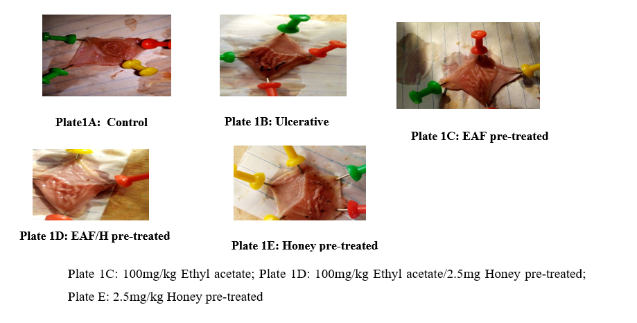
Group 1: Control (2.5 mL Olive oil as vehicle)
Group 2: Ulcerative untreated (rats administered with 30 mg/kg indomethacin)
Group 3: Ulcerogenic rats pre-treated with 100 mg/kg body weight ethyl acetate fraction of Ocimum gratissimum leaf extract.
Group 4: Ulcerogenic rats pre-treated with 100 mg/kg body weight ethyl acetate fraction of Ocimum gratissimum leaf extract and 2.5 g/kg body honey.
Group 5: Ulcerogenic rats pre-treated with 2.5 g/kg body weight honey.
Assessment of Ulceration
Macroscopic Scoring
After the experiment, the stomach of each rat was excised and opened through the lesser curvature and examined macroscopically. A magnifying lens was used to examine the degree of ulceration. The ulcers were scored using the Alphin and Ward method (Alphin and Ward, 1967).
Histopathological Study
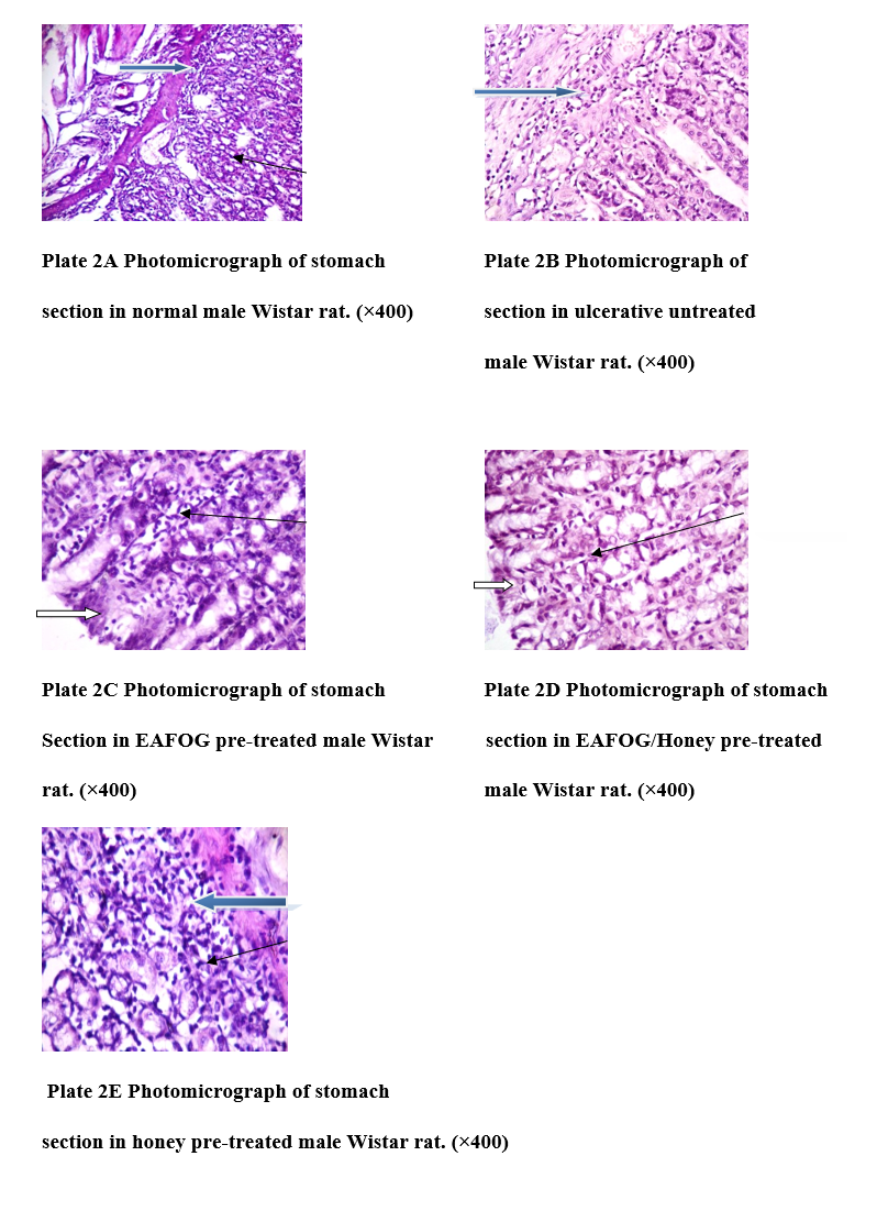
Samples were fixed in 10% formal saline after which they were embedded in paraffin, sections were stained with hematoxylin and eosin for histological examination of gastric damage by light microscopy.
Preparation of homogenate
The stomach was excised, homogenized and centrifuged at 10,000 rpm for 15 minutes to obtain the post-mitochondrial fraction stored at 4oC.
Measurement of Gastric Acidity
1 mL of gastric supernatant was titrated against 0.1 M NaOH to get a faint pink coloration. The total gastric acid secretion was determined using:
CAVA=CBVB
Where, CA=Concentration of Acid
CB=Concentration of NaOH
VA=Volume of acid
VB=Titre Value
Determination of Total Protein
The protein concentration of the various homogenates was determined by means of the Biuret method as described by Gornal et al., (1949) with some modifications: the addition of potassium iodide to prevent precipitation of Cu2+ ions as cuprous oxide.
Determination of Catalase Activity
Catalase activity was determined according to the method of Claiborne (1985). The method is based on the loss of absorbance observed at 240 nm as catalase splits hydrogen peroxide. Despite the fact that hydrogen peroxide has no absorbance maximum at this wavelength, its absorbance correlates well enough with concentration to allow its use for a quantitative assay. An extinction coefficient of 0.0436 mM-1cm-1 (Noble and Gibson, 1970) was used.
Assessment of Lipid Peroxidation
Lipid peroxidation was determined by measuring the formation of thiobarbituric acid reactive substances (TBARS) present in the test sample according to the method of Varshney and Kale (1990). Under acidic conditions, malondialdehyde (MDA) produced from the peroxidation of fatty acids reacts with the chromogenic reagent 2-thiobarbituric acid to yield a pink coloured complex with maximum absorbance at 532 nm.
Determination of Superoxide Dismutase (SOD) Activity
The activity of SOD was determined by the method of Misra and Fridovich (1972). The ability of SOD to inhibit the autoxidation of epinephrine at pH 10.2 makes this reaction a basis for a simple assay for this dismutase. Superoxide radical causes the oxidation of epinephrine to adrenochrome and the yield of adrenochrome produced per superoxide radical introduced increases with increasing pH and concentration of epinephrine.
Estimation of Glutathione-S-Transferase Activity
Glutathione S-transferase activity was determined according to Habig, Pabst and Jacoby (1974). The assay is based on the principle that all known glutathione S-transferase isotypes demonstrate a relatively high activity with 1-chloro-2, 4-dinitrobenzene (CDNB) as the second substrate. When CDNB is conjugated to reduced glutathione, its absorption maximum shifts to a longer wavelength and the absorption increase at the new wavelength of 340 nm provides a direct measurement of the enzymatic reaction.
Assay for Glutathione Peroxidase Activity
Glutathione peroxidase (GPx) activity was measured according to the procedure of Rotruck et al., (1973) with some modifications. Glutathione peroxidase is allowed to conjugate hydrogen peroxide to glutathione for a fixed period of time after which the reaction is quenched. The remaining glutathione is reacted with Ellman’s reagent and the GSH consumed is then used as a measure of enzyme activity.
Estimation of Reduced Glutathione (GSH) Level
The method of Beutler et al., (1963) was followed in estimating the level of reduced glutathione (GSH). This method is based upon the development of a relatively stable yellow coloured product when 5,5′–dithiobis-2-nitrobenzoic acid (DTNB; Ellman’s reagent) is added to sulfhydryl compounds of which glutathione comprises the bulk in tissues. The resulting chromophoric product possesses maximum absorbance at 412 nm.
Statistical Analysis
All data were represented as Mean ± Standard Deviation. Statistical significant difference was accessed by one-way analysis of variance (ANOVA) with Duncan’s multiple range test. They were carried out using graph pad prism 8 (GraphPad Software, San Diego, USA). P<0.05 was considered as statistically significant.