2.1 Reagents
Baicalin (25 + B00521EANO) was purchased from Heown Biotechnology (Tianjin, China). Carbomer 940 (J0601A) was purchased from Meilun Bio (Dalian, China). Zinc hyaluronate (22071302) was obtained from Bloomage Biotechnology Co., Ltd. (Beijing, China). Trolamine (20140811) was purchased from Yonghua Chemical Co. Ltd. (Suzhou, China). Glycerol (20220930) was purchased from Lingfeng (Shanghai, China). 5% Imiquimod (IMQ) (40230616) was obtained from Sichuan Mingxin Pharmaceutical Co. Ltd. Carpotriol ointment was purchased from LEO Laboratories, Ltd. IL-17A, TNF-α, and IL-1β were obtained from Multisciences (Nanjing, China). FreeZol Reagent, HiScript II Q RT SuperMix for qPCR, and ChamQ Universal SYBR qPCR Master Mix were purchased from Vazyme (Nanjing, China).
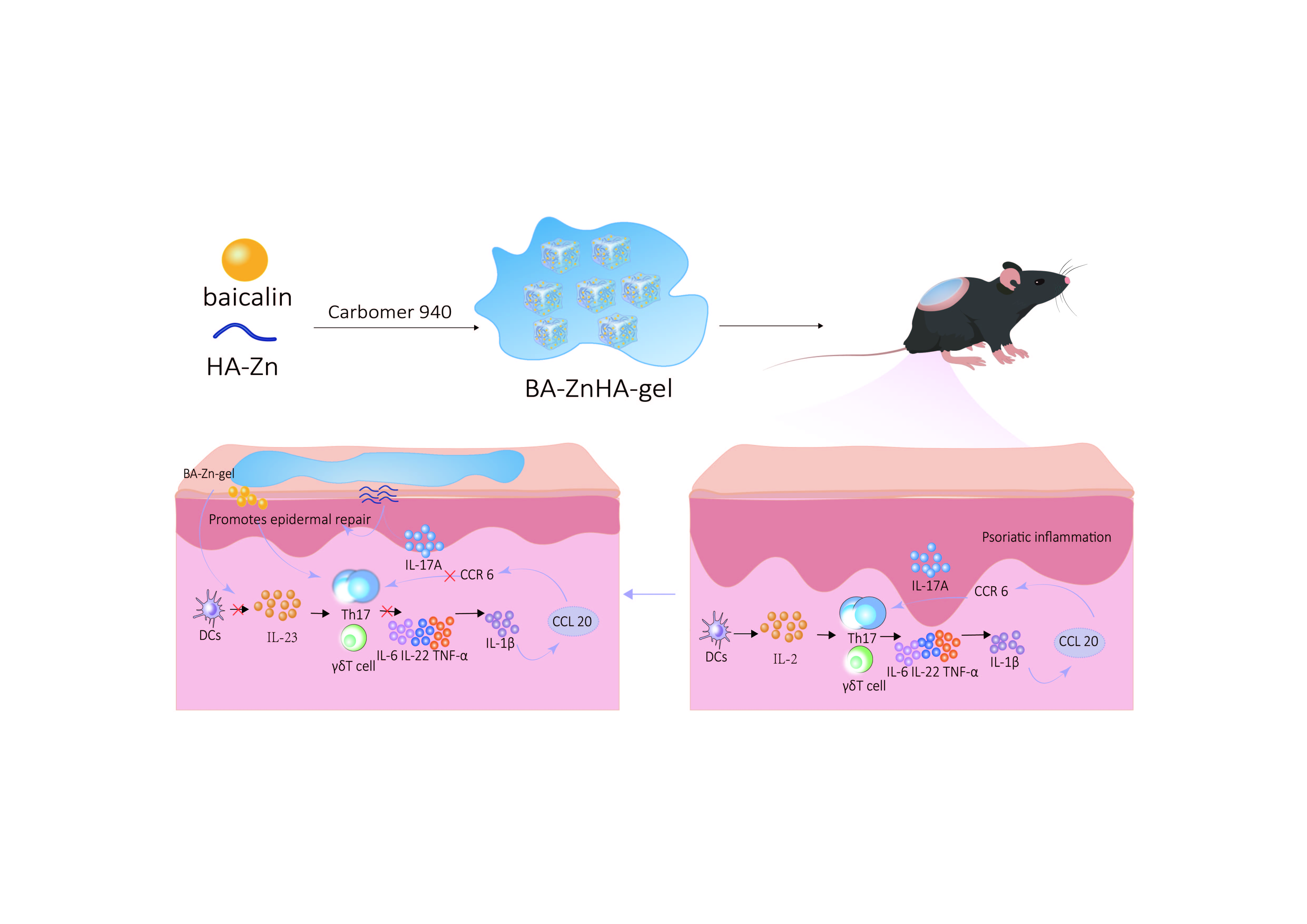
2.2 Preparing baicalin-zinc hyaluronate hydrogel
Preparing baicalin-low-dose zinc hyaluronate hydrogels: Baicalin (40 mg) was added to 20 mL of pure water by stirring and mixing. Triethanolamine was used to adjust the pH to dissolve baicalin, and zinc hyaluronate (100 mg) was added. After it was fully dissolved, 0.3 g of carbomer 940 was added, and 1.5 mL of propanetriol was added after dissolution and stirred well. The pH was adjusted to 6.5 with triethanolamine to obtain a baicalein-low-dose zinc hyaluronate hydrogel [21].
Preparing baicalin-high-dose zinc hyaluronate hydrogels: Baicalin (40 mg) was added to 20 mL of pure water by stirring and mixing. Triethanolamine was used to adjust the pH to dissolve baicalin, and zinc hyaluronate (200 mg) was added. After it was fully dissolved, 0.2 g of carbomer 940 was added, and 1.5 mL of propanetriol was added after dissolution and stirred well. The pH was adjusted to 6 with triethanolamine to obtain baicalein-high-dose zinc hyaluronate hydrogel.
Preparing zinc hyaluronate hydrogel: Zinc hyaluronate (100 mg) was weighed, added to 20 mL of pure water, dissolved, and 0.2 g of carbomer 940 was added. After dissolution, 1.5 mL of propanetriol was added and stirred, and the pH was adjusted by adding triethanolamine to obtain a zinc hyaluronate hydrogel.
Preparing blank gels: Blank gels were obtained by adding 0.2 g of carbomer 940 to 20 mL of pure water, leaving it to dissolve, adding 1.5 mL of propanetriol, and mixing well. The pH was adjusted using triethanolamine.
2.3 Characterizing baicalin-hyaluronic acid zinc gels
pH test: Five grams of baicalin-low-dose zinc hyaluronate hydrogel and baicalin-high-dose zinc hyaluronate hydrogel were placed in a beaker, dispersed by adding 20 mL of ultrapure water, and subjected to pH testing using a pH meter.
Viscosity test: Appropriate amounts of baicalin-low-dose zinc hyaluronate hydrogel and baicalin-high-dose zinc hyaluronate hydrogel were placed in a beaker, and the viscosity of the gels was measured at 25 ℃ using a viscometer.
Centrifugal stability study: The two hydrogels were added to 1.5 mL centrifuge tubes and centrifuged in a centrifuge at 3,000 rpm for 30 min, and the gel properties were observed to determine the presence of delamination and liquefaction.
2.4 Animals
Forty-eight specific pathogen-free (SPF)-grade healthy male C57BL/6J mice with an average weight of approximately 20 ± 1 g were purchased from SPF Biotechnology Co., Ltd. (Suzhou, China), and they were housed in the Animal Experiment Center of the Jiangsu Institute of Traditional Chinese Medicine. All animals were housed in conventional cages at a temperature of 24 ℃ ± 2 ℃, with a relative humidity of 60% ± 10%, and under a 12-h light/12 h dark cycle. Each cage contained six mice per cage, all of which had ad libitum access to food and water.
2.5 Experimental design
Healthy C57BL/6J mice were acclimatized and fed for 1 week and randomly divided into the following groups: normal control, model control, carpotriol ointment group (CB), blank gel group (CA-gel), baicalin gel group (BA-gel), zinc hyaluronate gel group (ZnHA-gel), baicalin low-dose zinc hyaluronate hydrogel group (BA-Zn-L), and baicalin high-dose zinc hyaluronate hydrogel group (BA-Zn-H), with six animals in each group. A rectangular hairless area of approximately 2 cm × 3 cm in length and width was shaved on the backs of all mice using an electric shaver the day before the experiment; the normal control group was coated with 62.5 mg of white petroleum jelly every morning, while the remaining seven groups were induced with psoriasis phenotypes in the hairless area of the back every morning using an equal amount of IMQ for 7 days [22]. The positive drug group was treated daily in the afternoon with carpotriol ointment, the blank gel group with drug-free blank gel, the baicalin gel group with baicalin gel, the zinc hyaluronate gel group with zinc hyaluronate gel, the baicalin-low-dose zinc hyaluronate gel, and the baicalin-high-dose zinc hyaluronate gel groups were treated with gels loaded with low and high doses of baicalin-zinc hyaluronate, respectively, for 7 days, while the blank and model groups were not administered. On day 8, the mice were sacrificed, and blood samples were collected using an anticoagulant-free blood collection tube to obtain serum. The supernatants were centrifuged at 3000 rpm for 10 min and frozen at − 80°C for determining inflammatory factor expression levels. Spleens were collected to record the spleen weight and calculate the spleen index. Skin was taken from the shaved area on the back of mice, and one part of the skin was stored at − 80°C and the other part was stored in 4% paraformaldehyde.
2.5 Skin lesion status and psoriasis area and severity index (PASI) score in mice
During the animal experiments, the skin lesions of the mice were photographed, recorded, and scored using the PASI. The clinical PASI scoring system was used to assess the overall condition of the skin lesions in the mice, which were scored from mild (0) to severe (4) according to the severity of the lesions in terms of erythema, infiltration, and epidermal scaling.
2.6 Assessing spleen indices
The spleen mass of each group of mice was weighed, and the spleen index was calculated (spleen index = spleen weight/body weight × 100%).
2.7 Histopathological examination
Mouse skin tissues fixed in 4% paraformaldehyde were washed, dehydrated, embedded, sectioned, deparaffinized, and stained using H&E.
2.8 Enzyme-linked immunosorbent assay
Serum levels of IL-1β, IL-17A, and TNF-α expression were detected using an enzyme-linked immunosorbent assay kit using collected mouse serum.
2.9 Quantitative real-time polymerase chain reaction (qRT-PCR)
Total RNA was extracted from mouse skin samples and isolated using the FreeZol Reagent kit (Lot No. 7G770J3) according to the manufacturer’s instructions. cDNA was synthesized using the Vazyme two-step qRT-PCR kit, according to the manufacturer’s instructions. cDNA was analyzed by qRT-PCR using the StepOnePlus™ Real-Time Fluorescence Quantitative PCR System. The SYBR method for qRT-PCR and the relative gene expression levels were calculated using the ΔΔCT method. The primer sequences used in this study (Table 1) were custom-synthesized by Sangon Biotech (Shanghai, China).
Table 1
| Primer name | Primer sequence |
| IL-23 F | ACCAGCGGGACATATGAATCTAC |
| IL-23 R | CTGGCTGTTGTCCTTGAGTC |
| IL-17A F | CTCAGACTACCTCAACCGTTCC |
| IL-17A R | ATGTGGTGGTCCAGCTTTCC |
| IL-22 F | CTGAGAAATGCTTGCGTCTG |
| IL-22 R | CGTTAGCTTCTCACTTTCCTTTAG |
| TNF-α F | GAGTGACAAGCCTGTAGCC |
| TNF-α R | CTCCTGGTATGAGATAGCAAA |
| GAPDH-F | AAGAAGGTGGTGAAGCAGG |
| GAPDH-R | GAAGGTGGAAGAGTGGGAGT |
2.10 Western blotting
An appropriate amount of mouse skin tissue was lysed in a pre-cooled protein extraction reagent containing inhibitors. The total protein was separated by 30% acrylamide gel electrophoresis and transferred to a polyvinylidene difluoride (PVDF) membrane, which was incubated with 5% skimmed milk powder dissolved in Tris-buffered saline and 0.1% Tween 20 (TBST) for 1 h at room temperature. The sample was incubated with primary antibodies against IL-23 (DF13760, 1:2000, affinity), IL-17 (66148-1-IG, 1:2000, Proteintech), CCL20 (DF2238, 1:2000, affinity), CCR6 (DF10207, 1: 2000, affinity), β-actin (GB11001, 1: 15000, Servicebio), and β-actin (GB11001, 1: 15000, Servicebio). affinity), β-actin (GB11001, 1: 15000, Servicebio) and incubated at 4°C overnight. It was rinsed five times for 5 min each using TBST and then incubated with secondary antibodies, goat anti-rabbit IgG (H + L) (G1213, 1: 15000, Servicebio) diluted in 5% skim milk powder, and goat anti-mouse IgG (H + L) (G1214, 1: 15000, Servicebio, Wuhan, China) (1: 15000), at room temperature for 1 h. The blots were visualized using a chemiluminescent ECL substrate (Pinofide Bio, Wuhan, China), and the results were analyzed using ImageJ software.
2.11 Statistical analysis
The experimental data were processed using IBM SPSS Statistics 26 (IBM SPSS Inc., Chicago, USA), and one-way analysis of variance (ANOVA) and least significant difference (LSD) were used to analyze and compare the data of all experimental groups. A p < 0.05 indicates a significant difference.