Preparation and Characterization of MPDA
Owing to their characteristics of surface functionalization, targeting, good degradability and biocompatibility, nanoparticles have been widely used in modern biomedicine as interventions of anti-tumor, antioxidation, drug delivery and tissue regeneration, et.al. Recently, the application of nanoparticles in the field of wound healing has gradually attracted extensive attention, for example, the nanofibrous matrix based on biomimetic elastomeric peptide has been developed to overcome the multidrug-resistant bacterial and promote the skin regeneration[27], in addition, grape seed-inspired smart hydrogel scaffolds has also been reported to significantly accelerate the healing of wound[28]. MPDA nanoparticles shows unique characteristics featured by simple preparation protocol, strong adhesive properties, easy and straightforward functionalization, and biocompatibility, and thus has been widely used in the fields of biomedicine, sensing, catalysis, environment and energy[29]. However, for such an important nanoparticle, there are few reports on its application in accelerating the regeneration of skin wounds.
In the current research, MPDA nanoparticles were synthesized according to previously reported procedures.[24] As described in the Experimental section, MPDA nanoparticles were formed in an aqueous solution containing triblock copolymer Pluronic F-127 and 1, 3, 5-trimethylbenzene (TMB) as organic templates. Dopamine self-polymerized into PDA particles through an ammonia-catalyzed approach, and the resultant PDA particles were assembled on Pluronic F-127-stabilized TMB droplets via π-π stacking interactions. The PDA particles were finally obtained as the organic templates were removed. Both FE-SEM (Figure 1A, B) and TEM images (Figures 1C and D) confirmed that the resultant PDA particles showed well-defined spherical morphologies and mesoporous structures. Their average diameter and polydispersity were 205 nm and 3.9%, respectively (see inset in Figure 1A). Close observation of the MPDA nanoparticles at higher magnification (Figure 1B and D) revealed that the cylindrical open channels were exposed on the MPDA nanoparticle surfaces, in which some mesopores had diameters of 10 nm. The elemental mapping patterns revealed a uniform distribution of C, N, and O elements (Figure 1E), further verifying the formation of MPDA nanoparticles. Thus, due to their mesoporous structure, large surface area, and nano-sized spherical morphology, MPDA particles exhibit great potential as versatile platforms for drug delivery, diagnosis, and therapy[30].
Characterization of Nanocomposites of MPDA+RL-QN15
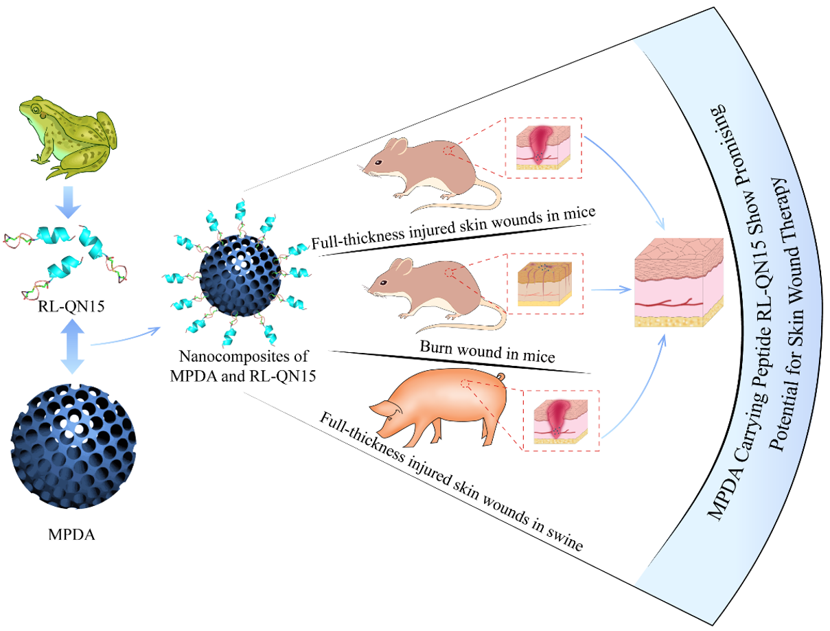
As a molecular tool, peptides have not only contributed substantially to clarify physiological processes in humans but also led to the discovery and development of novel therapeutics, hence it is widely recognized that peptide molecules have made indelible contributions to both basic scientific research and development of new drugs that are represented by insulin, exenatide, hirudin, captopril, et al.[31]. Amphibian skin secretions are rich in a variety of bioactive peptides, such as antimicrobial peptides, antioxidant peptides, bradykinins, neuromodulating peptides, and neurotoxins, et, al, and thus have been considered as a treasure trove of the natural bioactive peptide[16, 32, 33]. In recent years, peptides with obvious potency of accelerating the healing of skin wounds have aroused significant attention and several prohealing peptides have been identified, including RL-QN15, cathelicidin-OA1 and cathelicidin-NV, Ot-WHP, tylotoin, OM-LV20, et al.[21, 34-38]. Our previous research have revealed that RL-QN15 contains an amino acid sequence of QNSYADLWCQFHYMC and an intramolecular disulfide bond located between C9 and C15, when at a relative low concentration of nM scale, more importantly, RL-QN15 significantly promote the healing of full-thickness injured wounds and diabetic skin chronic wounds in mice, and thus is considered as a promising prohealing agent[22]. However, we still hoped that the ability of RL-QN15 to promote repair activity can be improved by some ways, so as to provide new strategies for the development of novel therapeutics for the treatment of wound healing which are still a harsh clinical challenge. One of the available methods was to use the nanoparticles to enhance the prohealing potency of RL-QN15.
In the current research, considering the characteristics of MPDA adhesion to each other, we first dispersed the prepared MPDA in PBS, as shown in Figure 2A, the PBS solution of RL-QN15 is colorless, MPDA and MPDA+RL-QN15 nanocomposites are brown. Then, FT-IR analysis, which is useful in qualitative analysis as no two bioactive compounds have the same FT-IR spectra[39], was carried out to verify whether RL-QN15 was successfully loaded onto the MPDA shell. Figure 2B showed the characteristic peaks of MPDA and MPDA+RL-QN15 nanocomposites. The broad peak at 3440 cm-1 in the PDA spectra was the characteristic adsorption of amine N-H and phenolic O-H stretching vibrations, and the band centered at 1 630 cm-1 was assigned to C=O stretching[40, 41]. Compared with MPDA, the wavenumber of the MPDA+RL-QN15 nanocomposites was lower at these two sites, which was attributed to the intermolecular hydrogen bond between MPDA and RL-QN15. In addition, there was a significant difference in the spectral fingerprint (1800-500 cm-1) between MPDA and MPDA+RL-QN15 nanocomposite. The characteristic absorption peaks of MPDA could be observed in the spectrum at the dotted frames, while the characteristic absorption peaks of MPDA/RL-QN15 nanocomposites were obviously weakened or even disappeared in the two places. These results indicated the successful load of RL-QN15 on to MPDA. In addition, results of SEM also provided evidence of the successful formation of MPDA+RL-QN15 nanocomposite. Compared with the MPDA shell (Figure 1A and B), the surface of the MPDA+RL-QN15 nanocomposites was rough and contained small particles (Figure 2C and D). The diameter of MPDA and nanoparticles of MPDA+RL-QN15 shared similar diameter of about 200 nm.
In summary, we successfully prepared nanocomposite of MPDA+RL-QN15, and then we focused on the loading efficiency and release profile of the composites.
In-Vitro Drug Release and Encapsulation Efficiency of RL-QN15 Loaded MPDA nanocomposites
The loading and releasing of RL-QN15 is based on the porous structure of MPDA microspheres. Here, the amount of RL-QN15 was determined at about 280 nm on the UV spectrophotometer according to UV full-wavelength scanning image (Figure S1). As shown in Figure 3A, the encapsulation rate of MPDA+RL-QN15 nanocomposites slowed down significantly after 6 hours, and the maximum encapsulation was 53.01%. In addition, the slow release of RL-QN15 from MPDA microspheres has also been studied. As shown in Figure 3B, RL-QN15 was continuously released from the microsphere during the period of 0-16 h, and the release rate was 92.14% at 24 hours.
The MPDA microsphere nanoparticles have the characteristics of efficient loading and slow release of RL-QN15 and in the following experimental procedures, the main focus was to verify if the load of MPDA significantly enhanced the prohealing potency of RL-QN15.
The Load of MPDA Significantly Enhanced the Prohealing Potency of RL-QN15 on Full-thickness Injured Wounds in Mice
The full-thickness skin wound model of mice is adopted to preliminarily evaluate whether the load of MPDA could significantly increase the activity of RL-QN15. PBS, PDA, RL-QN15, nanocomposite of MPDA+RL-QN15 was administered topically twice a day to the wounds constructed on the dorsal skin of mice. As shown in Figure 4A and B, compared with PBS, MPDA (0.2 mg/mL) could not whereas RL-QN15 (1 nM) could significantly promote the skin wound healing. On the eighth post-operative, RL-QN15 increased the healing effect by nearly 20% (**p<0.01) compared with the control group, showing excellent potential to accelerate wound healing, which was consistent with the data in our previous report[22]. It was worth pointing out that the healing ability of MPDA loaded with lower concentration of RL-QN15 (1 nM) was similar to that of RL-QN15 (50 nM) alone. In other words, MPDA nanoparticles as the carrier of peptides can significantly enhanced the prohealing potency of RL-QN15 by nearly 50 times. Specifically, on the postoperative days 2, 4, 6, and 8, the wound healing rates in the RL-QN15 was 58.35±5.08%, 67.81±5.14%, 76.39±5.56%, 84.3±2.38%, respectively, however, by means of the load of MPDA, healing rate in nanocomposites of MPDA+RLQN15 increased to 69.69 ±5.1%, 80.24±4.36%, 87.07±5.85%, 98.48±2.46%, respectively, which showed increases of 19.43%, 18.33%,13.98%, 17.14%, respectively. In addition, the repair-promoting activity of nanocomposites of MPDA+RLQN15 was time-dependent (Figure 4B).
Mice were sacrificed for histological analysis at postoperative days 4 and 8, and the effects of MPDA+RL-QN15 on wound healing in vivo were further investigated. As shown in Figure 4C and D, on postoperative day 4, the neoepidermis thickness of mice in PBS, PDA, RL-QN15 groups were 113.29±4.36 μm, 139.84±27.78 μm, 96.68±1.59 μm, respectively, while the neoepidermis thickness of mice in MPDA+RL-QN15 group was only 59.85±3.25 μm (Figure 43C). On the postoperative day 8, there was no significant difference in the thickness of epidermis between PBS and MPDA groups (72.89±2.35 m vs. 83.74±2.52 μm). The thickness of epidermis in RL-QN15 was about 54.70±3.63 μm, which was significantly lower than that in PBS and PDA groups, but still higher than that in MPDA+RL-QN15 group (31.24±3.87 μm). We also evaluated the granulation tissue thickness, as shown in Figure 4F and G, on postoperative day 4, there was almost no difference in granulation tissue thickness between the PBS, PDA and RL-QN15 groups, and MPDA+RL-QN15 was lowest among the four groups. On postoperative day 8, there was no significant in granulation thickness between PBS and PDA groups (858.53±96.13 μm vs. 863.71±89.14 μm). Granulation thickness of RL-QN15 was 672.76±74.43 μm, which was significantly lower than that of PBS and PDA groups, but still higher than that of MPDA+RL-QN15 group (506.60±83.62 μm). In a word, after topical administration of nanocomposites of MPDA+RL-QN15, the regeneration and reconstruction of the epidermis and granulation tissue were significantly enhanced.
In previous studies, there are few reports on the combination of nanomaterials and peptides to promote wound healing. One of available references is that KR-12 peptide combined to the fibrous eggshell membrane to promote angiogenesis and hence accelerate skin re-epithelialization[42]. The report on the binding of MPDA particles to peptides as prohealing therapeutics remains unavailable. Our results showed that, by the load by MPDA, the prohealing potency of RL-QN15 against the full-thickness injured wounds in mice were increased by an average of 17.14%. Hence, for the first time, we provided evidence to indicate that MPDA nanoparticles could be used as a peptide-carrier to achieve better pro-healing potency.
The Prohealing Potency of RL-QN15 Against Burn Skin Wounds Was Significantly Enhanced by the Load of MPDA.
Burn is a common trauma in daily life and the clinically available means to treat burn is the application of skin grafting and wound dressing. However, slow wound healing, infection, pain, and hypertrophic scarring continue to remain a major challenge in burn research and management[43]. In our previous research, we demonstrated that RL-QN15 could promote chronic skin wound healing in diabetic mice, but it is unknown whether RL-QN15 could promote burn wound healing. In this study, we successfully established the mice model of deep-Ⅱ degree burn on the dorsal skins. The macroscopic morphology of the experimentally induced burns was presented in Figure 5A. After inducing the burns on experimental animals, the local area was characterized by a white eschar, the surface skin layers (epidermis and dermis) were damaged. In the first four days, the wound turned brown due to extravasation of injured cells and an increase in the level of inflammation. Eight days after the burn, the skin formed a scab and gradually fell off. PBS, MPDA, RL-QN15, nanocomposite of MPDA+RL-QN15 was administered twice a day to the burns of mice. As shown in Figure 5B, MPDA itself could not promote skin wound healing in mice compared with PBS, while RL-QN15 (1 nM) could significantly promote wound healing. On day 12 post-operation, the wound healing rate in the PBS and MPDA groups was only 73.23±4.25% and 71.16 ± 3.73%, respectively, while the wound healing rate in the RL-QN15 group was 82.47±3.53%, which showed that RL-QN15 has the potential to promote the healing of burn wounds in mice. What is worth mentioning is that the prohealing potency 0f RL-QN15 against chronic skin wounds was significantly enhanced by the load of MPDA. On post-operative day 12, the healing rate of MPDA+RL-QN15 was 98.80±0.80%, which was 19.80% higher than that of RL-QN15 (**p<0.01).
In a parallel experiment, mice were sacrificed for histological analysis at postoperative days 8, and 12, and the effects of nanocomposites of MPDA+RL-QN15 on wound healing in vivo were further investigated. Histological analysis indicated that mice topically treated with nanocomposites of MPDA+RL-QN15 displayed prominently accelerating regeneration of neo-epidermis (neo-epithelial tongue) in the wound compared with the PBS, MPDA or RL-QN15 group (Figure 5C). One of the major systemic damage responses after burn injuries is caused by proinflammatory cytokines released by inflammatory and vascular endothelial cells[44]. During the healing process, the burn wound showed more inflammatory infiltration than the full-thickness wound (Figure 4C vs. Figure. 5C). On the post-injury day 8, all groups were still in the stage of inflammation, while MPDA+RL-QN15-treated group showed the best re-epithelialization and best-formed granulation tissue amongst four groups. On the post-injury day 12, epidermal regeneration and reconstruction of the dermis were complete in the MPDA+RL-QN15 group, which were similar to that in normal mice. Histological evaluation of mice skin tissue sections stained with H&E was also carried out. As illustrated in Figure 5D, E, during the healing process, the level of re-epithelialization in MPDA+RL-QN15 group was always higher than that in other groups. On postoperative day 12, neo-epithelial tongue was completely covered the whole wounded area in the MPDA+RL-QN15-treated mice (re-epithelialization 98.80±1.15%). In contrast, only little partial neo-epithelial tongue was found in the MPDA (re-epithelialization of 50.33±4.64%) and most partial neo-epithelial tongue in the RL-QN15 (re-epithelialization of 82.47±3.67%). In conclusion, histological analysis revealed that mice treated with MPDA+RL-QN15 showed better granulation tissue contraction, and high re-epithelialization.
This is the first time to prove that RL-QN15 showed pro-healing activity on burns in the dorsal skins of mice. Moreover, the load of MPDA significantly enhanced the prohealing potency of RL-QN15. Although RL-QN15 could promote the healing of full-thickness, burns and diabetic wounds in mice, the difference in skin structure between mice and humans made it impossible to provide scientific means for clinical treatment of wound. Considering the clinical application, we next explored the healing activity of RL-QN15 in porcine full-thickness injury.
The Effect of RL-QN15 Against Full-thickness Dorsal Skin Wounds in Swine Was Significantly Accelerated by the Load of MPDA.
Due to the panniculus carnosus, healing in these small animals is largely achieved through wound contraction, as opposed to re-epithelialization in humans[45, 46]. Furthermore, the murine epidermis is only 50 μm thick, so it is technically difficult to create partial-thickness wounds[47]. Porcine models have emerged as promising models to study wound healing. An advantage of using swine is that they are anatomically and physiologically similar to humans[48], and have been used to study many other diseases[49-51]. Like humans, they have a relatively thick epi-dermis, distinct rete pegs, dermal papillae, and dense elastic fibers in the dermis[52, 53]. Swine also have sparse hair rather than fur, although the hair is coarser than human hair. Similarities between swine and human skin also make swine an appropriate choice for the construction of cutaneous wound healing animal models.
A full-thickness dermal skin wound model was established in swine to assess whether the load of MPDA could significantly increase the prohealing activity of RL-QN15. The full-thickness wounds of the dorsal skins were treated with PBS, PDA, RL-QN15, nanocomposite of MPDA+RL-QN15 twice a day. As shown in Figures 6A and B, compared with PBS, MPDA could hardly promote wound healing of swine skin, while RL-QN15 significantly promoted wound healing. For example, on day 28 post-operation, the wound healing rate in PBS and MPDA groups was only 49.01±4.48% and 54.85±4.11% (n=6) respectively, while in RL-QN15 group, the wound healing rate was 65.02±4.70% (n=6). In particular, the effect of RL-QN15 against full-thickness dorsal skin wounds in swine was significantly accelerated by the load of MPDA. For instance, at day 7 and 14, there was almost no difference between the two groups, however, at postoperative days 21 and 28, the wound healing rates in the nanocomposite of MPDA+RL-QN15 group was 57.67±2.98% and 79.35±3.30% (n=6), respectively, which showed increases of 15.43% (*p<0.05), 21.86% (**p<0.01) compared with RL-QN15 group.
Histological analysis was also carried out on skin sections (stained using H&E and Masson’s trichrome) from swine on day 28 post-operation. New blood vessels and hair follicles appeared in the group treated with MPDA+RL-QN15 rather than RL-QN15 groups (Figure 6C). In addition, the thickness of new epidermis in RL-QN15 and MPDA+RL-QN15 groups was 74.25±15.89 μm, 54.46±4.33 μm (n=6) respectively, which was significantly lower than that in PBS group (181.52±17.83 μm) and MPDA group (156.14±14.81 μm) (n=6) (Figure 6D). These results show that RL-QN15 not only accelerated wound healing, but also effectively improved the increase in skin thickness, which is consistent with our previous research[22]. In addition, we also quantified the proportion of collagen. There was no significant difference in the proportion of collagen between PBS and MPDA groups (36.39±6.78% vs. 39.38±2.84% (n=6)) and the proportion of collagen in RL-QN15 was about 63.11±3.22% (n=6), which was significantly higher than that in PBS and PDA groups, but still lower than that in MPDA+RL-QN15 group (71.13±1.78%) (n=6) (Figure 6E). In a word, as a carrier of RL-QN15, MPDA successfully improved the prohealing strength of RL-QN15.
The Construction of Nanocomposite of MPDA and Prohealing Peptide Might Be a Promising Option for the Development of Novel Prohealing Interventions.
At present, the available intervention for wound repair mainly include various growth factors, small molecular compounds, wound dressings and hydrogels, which are far from meeting the clinical needs, thus the development of novel wound healing therapy is urgently highlighted[54]. In our study, the peptide RL-QN15 secreted from the skin of R. limnocharis frogs was loaded onto MPDA particles (Figure 7A). The MPDA particles as drug delivery system loaded with peptides adhere to the wound surface and release RL-QN15 (Figure 7B). Then, according to our previous research, during the inflammatory phase, RL-QN15 stimulates inflammatory cells, especially macrophages, to secrete cytokines such as TGF-β1[22] (Figure 7C). Which is a multifunctional growth factor that exerts pleiotropic effects on wound healing by regulating cell proliferation and migration, differentiation, ECM production, and immune modulation[55]. Next, the wound enters the proliferative phase. TGF-β1 and RL-QN15 promote the proliferation and migration of skin fibroblasts and keratinocytes, resulting in wound closure (Figure 7D). Finally, during the tissue remodeling period, the skin in the wound site is fully restored, including primarily restoration of the epidermal and dermal layers with appendages, such as hair follicles (Figure 7E).
Many nanoparticles have been found to promote wound healing through their intrinsic characteristics (antibacterial, anti-oxidant, promote cell proliferation and migration). For example, cerium oxide nanoparticles accelerate the healing of full-thickness dermal wounds in mice via enhancement of the proliferation and migration of fibroblasts, keratinocytes[56]. MPDA nanoparticles showed no healing ability, but improved the healing effect of RL-QN15. On the one hand, MPDA nanospheres prolonged the effective concentration of RL-QN15 on the wound surface by continuously releasing RL-QN15. On the other hand, the MPDA microsphere shell protected RL-QN15 from modification by endogenous (e.g., tyrosinase, elastase, metalloproteinase) and exogenous (e.g., produced by colonized microorganisms) enzymes, thus enhancing the therapeutic effect. Therefore, the construction of nanocomposite of MPDA and prohealing peptide might be an option for the development of novel prohealing interventions.