Emerging evidences have shown that HSP within children is a systemic disease; however, the pathogenesis of HSP remains unknown. Functional mutations of T cells, such as abnormal cytokine secretion, were reported to be involved in the pathogenesis of HSP(11). Abnormal B cell activation with increased IgA secretion indicated that humoral immunity underlies the manifestations of HSP; however, whether abnormal B cell development and Tfh-dependent B cell responses play a role in the pathogenesis of HSP remain unknown.
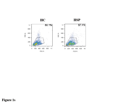
In the present study, we enrolled children diagnosed with HSP, as well as HCs; the percentage of total CD3+CD19− B cells in each group was determined by flow cytometry. We found that the percentage of total B cells was significantly increased in children with HSP compared with HC patients. However, the percentage of CD27++CD38++ plasma cells was significantly reduced in children with HSP.
To the best of our knowledge, no studies have been conducted to investigate the populations of total B cells and plasma cells in association with HSP. Our study is the first to reveal an expansion of total B cells and a reduced frequency of plasma cells in children with HSP. Further study was still needed to discover the detail mechanisms.
Functionally distinct B cell subsets can be divided into different subsets by the phenotypic expression of CD27 and IgD. IgD+CD27− B cells were defined as naïve B cells, whereas CD27 expression by B cells has been considered as a hallmark for somatic hypermutation and memory. CD27+ memory B cells can also be divided into pre-switch (IgD+CD27+) and post-switch (IgD−CD27+) B cell subsets. We sought to determine whether differences in the B cell compartment occur in HSP children and our results showed that the percentage of naïve B cells was significantly reduced, while that of class-switched B cells was significantly increased in children with HSP. Our results indicated that the abnormal development of B cells might be related to abnormal B cell responses in children with HSP.
A large number of studies have shown that Ig production was associated with the frequency of Tfh cells in a variety of autoimmune diseases, such as rheumatoid arthritis and systemic lupus erythematosus. In the current study, the percentage of CD3+, CD4+ and CD8+ T cells was determined. In accordance with previous reports, our results showed that the frequency of CD3+ and CD4+ T cells was significantly decreased in children with HSP compared with HCs. CXCR5, a marker of Tfh cells, mediates the migration of Tfh cells to B cells within follicular areas containing germinal centers. We showed significant increases in the expression of CXCR5 on CD4+ T cells and the total frequency of CXCR5+CD4+ Tfh cells in children with HSP compared with HC patients. PD-1, a functional marker of Tfh cells, exhibited no differences in expression between the two groups. A similar finding reported an expansion of circulating Tfh cells in children with acute HSP; a significant increase in CXCR5+CD4+ cells in children with HSP and no differences in PD-1 expression between two groups were noted(12). Moreover, correlations between Tfh cells and naïve B cells, and class-switched B cell subsets, as well as memory B cells, were found in children with HSP in our study. We speculated that the HSP patients employed in our study may not exhibit an acute form of this disease as shown by the variations in their symptoms. Therefore, increased efforts should be made to classify HSP into different subtypes and the potential relationship between the frequency of CXCR5+CD4+ Tfh cells and IgA-producing B cells should be explored.
This study has some potential limitations. The sample size of this study is small, which may lead to statistical bias. Large scale studies in HSP children are required in order to address this potential limitation.
In summary, our study showed abnormal B cell subsets and a Tfh cell-related abnormal B cell compartment in children with HSP. Our findings may provide a new insight into the pathogenesis of children with HSP.
Table 1
Demographic and Clinical Characteristics of HSP patients and HCs
| | HSP | HCs |
| Number | 14 | 14 |
| Age (years) | 7.5 ± 2.1 | 7.2 ± 1.9 |
| Sex (M/F) | 8/6 | 7/7 |
| WBC (⋅ 109) | 11.5 ± 2.3 | 6.5 ± 1.8 |
| Serum IgA (g/dL) | 1.98 ± 0.92 | 1.77 ± 0.53 |
| Serum IgG (g/dL) | 10.56 ± 3.42 | 9.34 ± 2.52 |
| Serum IgM (g/dL) | 1.45 ± 0.51 | 1.13 ± 0.47 |
|