2.1. Chemicals and kits
Prunetin (CAS Number: 552-59-0, ≥ 98.0 % TLC) was obtained from Sigma-Aldrich. Annexin V-FITC Apoptosis Detection Kit I with 1x Binding Buffer and Propidium Iodide (PI) were purchased from BioVision Inc. Human inducible nitric oxide synthase (iNOS) ELISA Kit was obtained from MyBioSource, Inc., USA. Fetal Bovine Serum (FBS, F2442) was purchased form Sigma-Aldrich. All of the chemicals were of analytical grade.
2.2. Methods
2.3. In vitro Cytotoxicity test
2.3.1. Cell lines and cell culture
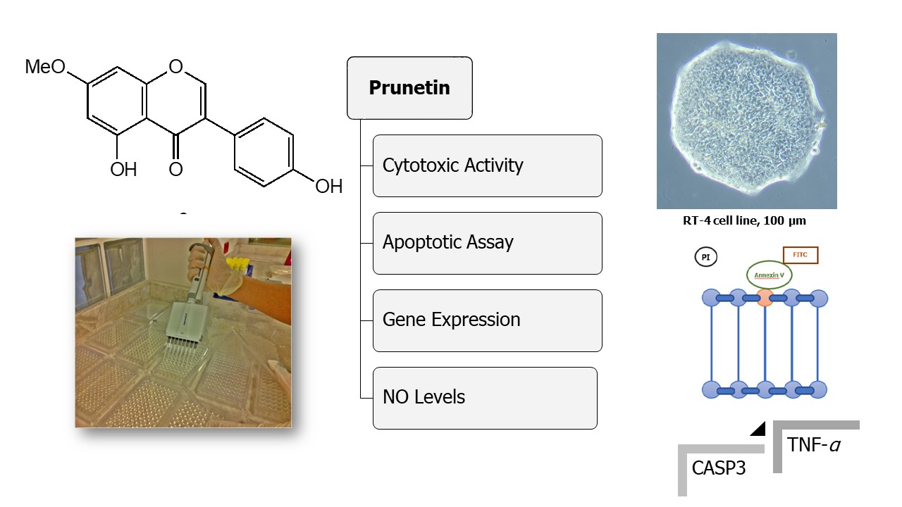
Breast cancer (MDA-MB 231, HTB-26™ MCF-7, HTB-22™), cervical cancer (HeLa, CCL-2™), alveolar adenocarcinoma (A549, CCL-185™), prostate cancer (PC3, CRL-1435™), urinary bladder cancer (RT-4, HTB-2™), colon cancer (Caco-2 HTB-37™) and embryonic kidney cells (HEK293, CRL-1573™ as noncancerous cell line) were obtanied from the American Type Culture Collection. Cells were maintained in different media (DMEM, DMEM/F12, RPMI-Gibco, UK) supplemented with 4% heat inactivated FBS (Sigma-Aldrich, F2442), 1% L-glutamine (Biochrome, Germany) and 1% gentamycine (Invitrogen) at 37 °C in 5% CO₂. Doxorubicin was used individually as positive control (40 μL in each well).
2.3.2. Cell viability assay and determination of IC50
The in vitro cytotoxic effect of prunetin was assessed based on metabolic cell viability procedure. Mitochondrial reductase activity and proliferation of cells were determined according to a modified MTT assay [17]. Cell lines were cultivated at the density of 1x10⁴ cells/mL in V-bottom 96 well plates maintained overnight at 37 °C. After the cultured cells were treated with 0.5, 5, 50 μg/mL of prunetin for 48 h at 37 °C, 150 μL DMSO (5 mg/mL) solution/well was added and incubated at 37 °C for 4 h to dissolve formazan crystals and produce a purple solution that indicates viable cells. Untreated controls were used as comparison of growth inhibition to determine the prunetin concentration that inhibited cell growth by 50% (IC50). The optical density was measured with UV spectroscopic analysis at 560 nm and the percentage of viable cells was calculated. The experiment was done in triplicats and the percentage of cell survival was calculated as follows:
The GraphPad PRISM (version 7) programme was used to calculate the IC50 values which were assessed at ± 95% confidence intervals.
2.4. Apoptotic Assay-Annexin V-FITC/ PI double staining
Annexin V-Fluorescein isothiocyanate conjugate (FITC) and propidium iodide (PI) staining was used to quantify the apoptotic rate of RT-4 and HEK-293 cells according to the manufacturer's directions. Briefly, 5×10⁵ cells were exposed to different concentrations of prunetin (based on the calculated IC50 values) in 6-well plates for 48 h. Then, cells were stained with Annexin V-FITC (5 uL-15 min) and PI (10 uL-5 min) in the dark condition at room temperature. Cellular apoptotic rate was measured using a flow cytometer (BD Biosciences, US) [18].
2.5. Real-time PCR assay
The doses of prunetin used to assess apopotic effects on RT-4 and HEK-293 cells were chosen based on the results of cell viability assays and IC50 values. Total RNA was isolated from RT-4 and HEK-293 cells using the Qiagen RNeasy kit according to the manufacturer’s instructions. Residual genomic DNA was removed from total RNA with DNAseI. Then, cDNA was subsequently synthesized from the total RNA using a EvoScript Universal cDNA Master Kit. The expression of CASP3 and TNF-α mRNA were assessed with Power SYBR Green Master Mix (Applied Biosystems) with RT-PCR assays (Applied Biosystems StepOne Plus-System). As an internal control, β-actin was used to normalize the level of CASP3 and TNF-α mRNA. Primer sequences are listed in Table 1. As a reference point, non-treated cells were chosen and calculation of relative expression levels for genes were conducted with respect to this reference value (set to 1). StepOne Plus software was used for qRT-PCR data analyses.
2.6. Nitric oxide synthase activity assay
RAW 264.7 cells (ATCC® TIB-71™) were cultered (1×10⁵ cells) in LPS and phenol red free RPMI 1640 medium with 10% FBS then incubated for 24 hours. After inducing with 1 μg/mL LPS, 5, 10 and 50 μg/mL concentrations of prunetin were added and incubated under the same conditions for another 24 h. Dose curves were used to calculate IC50 values and positive control was parthenolide. All tests were conducted in triplicate and Griess reagent was used to determine the amount of NO2- in supernatants at 540 nm absorbance [19].
2.7. Statistical analysis
Statistical data are presented as means ± SD from three independent experiments and were analyzed using Student’s t-test or one-way ANOVA to determine statistically significant differences at P <0.05.