Cervical tumors samples and cell cultures
In this study, 30 pairs of cervical cancer and adjacent normal tissues, ten advanced cervical cancer without adjacent normal tissue, and seven normal cervical epithelial tissues of the patients who had a hysterectomy due to myoma were obtained from Liaoning Cancer Hospital. Any cases that received chemotherapy or radiotherapy before collection were excluded. Only squamous cervical cancer cases were collected in order to decrease heterogeneity due to different histological types.
Human cervical cancer cell lines (HeLa and SiHa) were purchased from the Shanghai Institutes for Biological Sciences, China. All cell lines were cultured in Dulbecco’s Modified Eagle’s medium (DMEM) high glucose medium (HyClone, Logan, UT, United States) supplemented with 10% fetal bovine serum (FBS).
circRNAs expression profile analysis
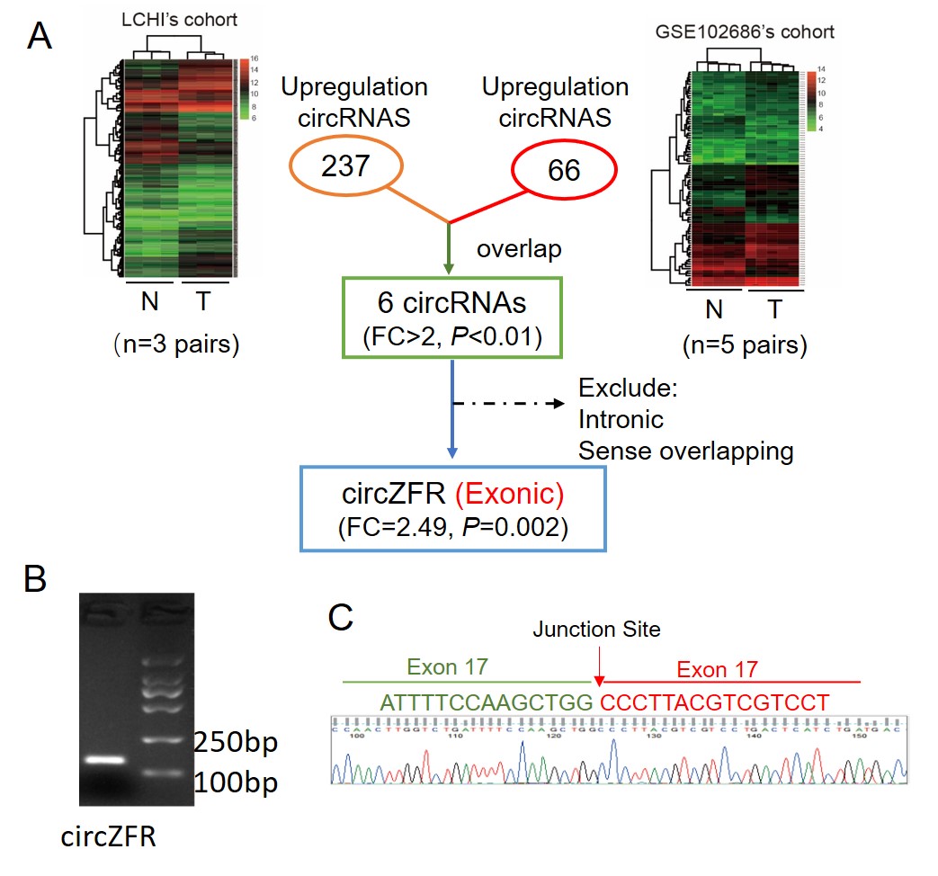
We performed circRNA microarray analysis (human CircRNA microarray V2.0) using three cervical cancer tissues and paired adjacent normal tissues in our Liaoning Cancer Hospital & Institute (LCHI)’s cohort. Another profile using the same microarray (GSE102686) was downloaded from the Gene Expression Omnibus database (GEO, http://www.ncbi.nlm.nih.gov/geo). GSE102686 consisted of 5 cervical cancer tissues and paired adjacent normal tissues. R version 3.6.2 software (https://www.r-project.org/) was used to compare the differentially expressed genes (DEGs) of two profiles separately.
TCGA and GTEx data analyses
A total of 306 cervical cancer and 13 healthy cervical tissues and the corresponding clinical data were obtained from The Cancer Genome Atlas (TCGA, https://cancergenome.nih.gov/) and Genotype-Tissue Expression (GTEx, https://www.gtexportal.org/home/index.html). All the data included in this study are in agreement with the TCGA and GTEx publication guidelines.
Quantitative reverse transcription-polymerase reaction (qRT-PCR)
Total RNA from tissues and cells was isolated using TRIZOL reagent (Invitrogen, CA, USA). For circRNA and mRNA, cDNA was synthesized using the PrimeScript RT reagent Kit with gDNA Eraser (Takara, Otsu, Japan). The quantification of circRNA and mRNA was performed using SYBR Premix Ex Taq II (Takara, Otsu, Japan). CircZFR and mRNA expression were detected using the specific primer pairs (Supplementary Table 1). β-actin was used as the internal reference for the quantification of circRNA and mRNA. qRT-PCR was conducted on the Bio-Rad CFX96 system (Bio-Rad, CA, USA). The relative expression of circRNAs and mRNAs was calculated with the 2-ΔΔCT method.
CircRNA plasmid construction and stable cell lines
Stable cell lines expressing CircZFR or sh-CircZFR and controls (CircCtrl and sh-Ctrl) were performed as previously described[17]. In brief, recombinant lentiviruses were produced in HEK293 cells. The viruses were harvested and purified by centrifugation. Pools of stable transductions were generated by selection using puromycin (1.0 μg/ml HeLa and 5.0 μg/ml for SiHa) for two weeks. The sequences of siRNAs and shRNAs targeting circZFR are listed in Supplementary Table 2.
SSBP1 plasmid construction
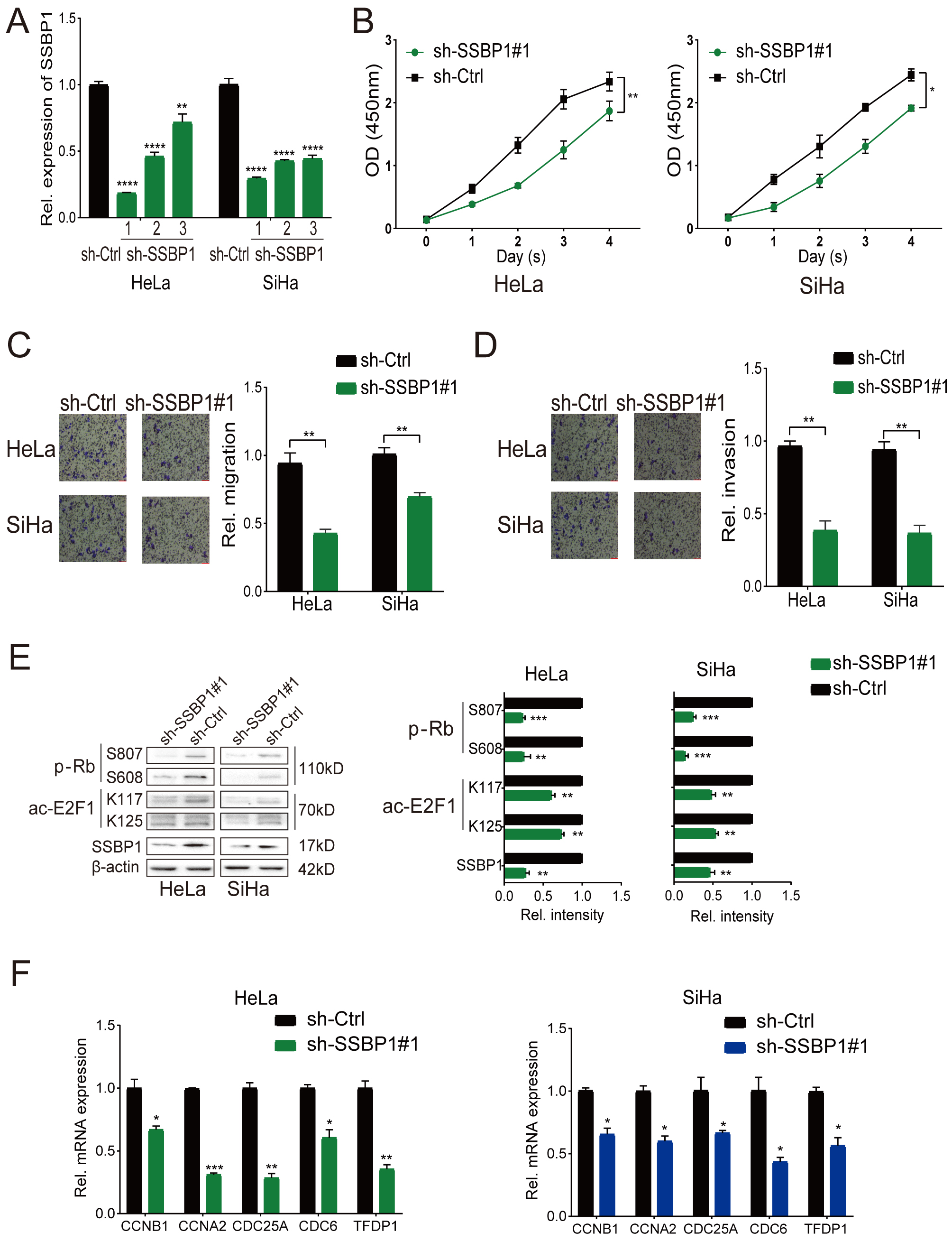
The target sequences of SSBP1 shRNAs are listed in Supplementary Table 3. The shRNA with the most significant knock-down efficiency was selected in this study.
Cell proliferation and cell cycle analyses
We performed cell counting and 5-Ethynyl-20-deoxyuridine (EdU) assays to assess cell proliferation. The cell counting assay was performed using Cell Counting Kit-8 (Dojindo Laboratories, Kumamoto, Japan) as previously described.[17] The stably transfected cells (2.0 x 10³/well) were plated into 96-well plates and incubated at 37℃ for eight hours until adherence and were referred to as day 0. Cell numbers were counted at days 1, 2, 3, and 4. The EdU incorporation assay was performed using a Cell-Light EdU DNA Cell Proliferation Kit (RiboBio, Guangzhou, China) according to the manufacturer’s protocol[18]. The percentage of EdU-positive cells was calculated. We also performed colony formation assay to validate the function of circZFR on cervical cancer cell proliferation. Total 500 stably transfected cells were seeded into six-well plates. The plates were photographed and counted after 14 days.
Cell cycle analyses were performed as previously described[19]. Briefly, all of the cells were first synchronized through serum deprivation for 36 h. The cells were stained using PI/RNase Staining Buffer (BD BioScience) and then analyzed in BD AccuriTM C6 (BD BioScience).
Wound healing, migration and invasion assays
Wound healing, migration and invasion assays were performed as previously described[20]. For wound healing assay, the stably transfected cells were cultured in 6-well plates until 90-100% confluent. After scratching, the cells were washed and maintained in serum-free medium. At 0, 12 and 24h three time points after wounding, the width of wounds was examined in three-independent wound sites per group and normalized to a control group.
Migration and invasion assays were performed using transwell chambers which contain inserts with a pore size of 8μm (Corning Incorporated, Corning, NY, USA). Total 2 x 10³ stably transfected cells were plated into an upper chamber coated with or without Matrigel (Corning Incorporated, Corning, NY, USA) with serum-free medium, and the lower chamber was added 10% fetal bovine serum. After incubation for 24h, the cells passed through the membrane were fixed, stained, and counted in 10 randomly selected fields with a 200x magnification microscope (Leica DMi8, Wetzlar, Germany).
Western blot and immunohistochemistry
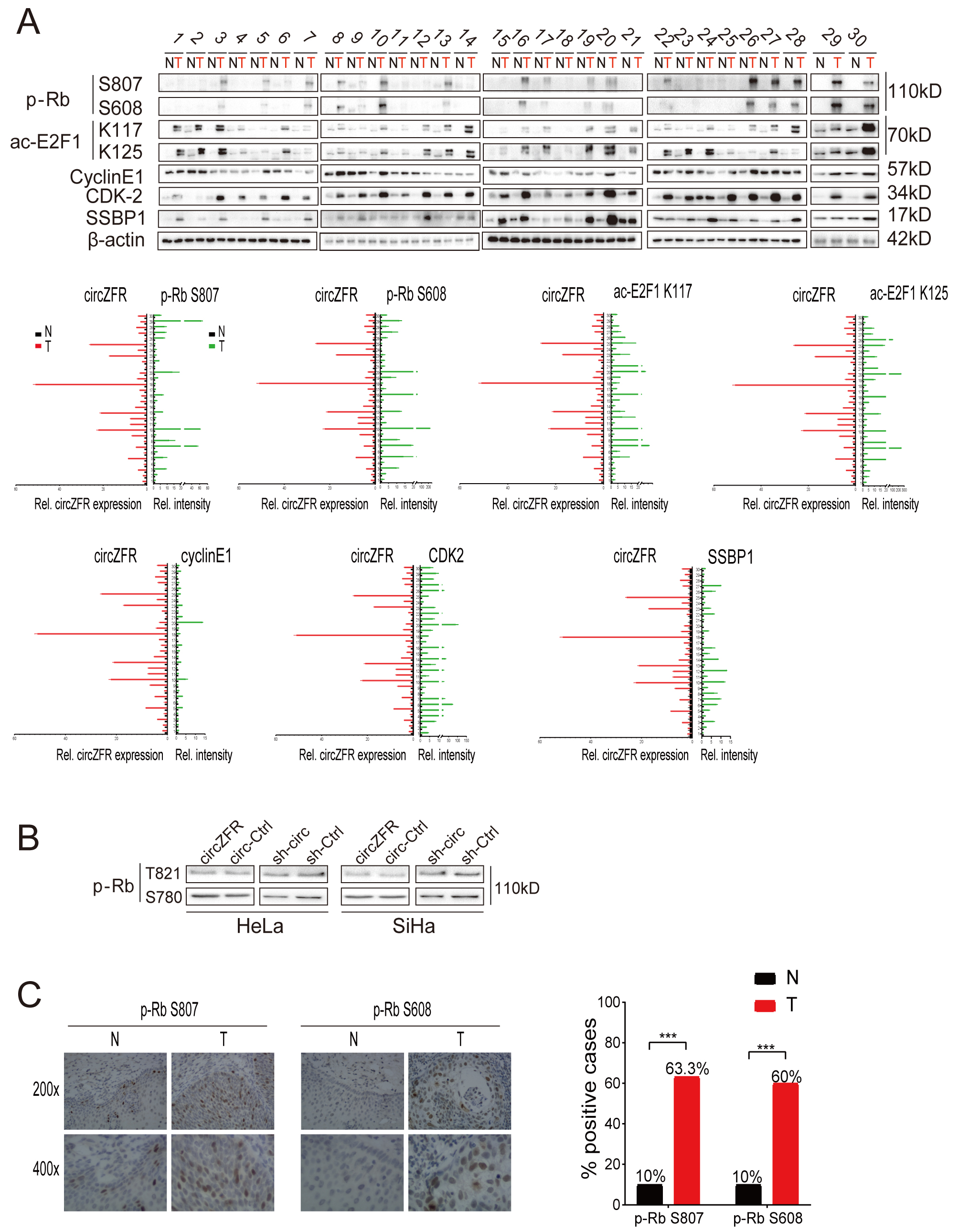
Western Blot analyses were performed according to the published protocol[21]. Cells and tumor tissues were lysed by RIPA lysis buffere with protease and phosphates inhibitor. Then the lysates were resolved by 8-12% SDS-PAGE and transferred on polyvinylidene fluoride membranes (Milipore, Bedford, MA, USA). After blocking, the membranes were immunoblotted with primary antibodies against p-Rb S807 (ab184796, 1:1000, Abcam, Cambridge, UK), p-Rb S608 (ab172975, 1:20000), p-Rb S780 (ab173289, 1:10000), p-Rb T821 (ab32015, 1:5000), p-Rb (ab181616, 1:2000), E2F1 (ab179445, 1:2000), cyclin E1 (ab33911, 1:2000), CDK4 (ab108357, 1:5000), CDK2 (ab32147, 1:5000), ac-E2F1 K117 (YK0087, 1:2000, Immunoway, Texas, USA), ac-E2F1 K125 (YK0088, 1:1000), cyclin D1 (60186-1-Ig, 1:10000, Proteintech, Wuhan, China), SSBP1 (12212-1-AP, 1:1000, Proteintech), and β–actin (60008-1-Ig, 1:20000, Proteintech) overnight at 4℃. After the incubation with secondary antibodies, the signals were developed with chemiluminescent western blotting substrate (Beyotime, Shanghai, China). ImageJ software was used to quantify signal intensity.
For immunohistochemical staining, total 30 pairs of formalin-fixed, paraffin-embedded cervical cancer and adjacent normal tissue specimens were analyzed. Briefly, after dewaxing the paraffin sections of tissues, heat-induced antigen retrieval was conducted. The slides were incubated with primary antibodies against p-Rb S807 (ab184796, 1:200) and p-Rb S608 (ab172975, 1:50) at 4 °C overnight, incubated with the biotinylated secondary antibody, and then subjected to the DAB kit. The evaluation of p-Rb S807 and S608 phosphorylation protein expression levels were performed as described previously[20].
circRNA pull-down and circRNA immunoprecipitation (circRIP) assays
MS2 bacteriophage coat protein (MS2-CP) circRNA pull-down assay was performed using MS2 tagging technique, which is based on the natural binding between a stem-loop structure of MS2 and MS2-CP[22]. In brief, we constructed the plasmid with circZFR and MS2, which was fused with a green fluorescent protein (GFP) (circZFR-MS2GFP). We also constructed the plasmid with MS2-CP-Flag, which was fused with mCherry tag (MS2-CP-FlagmCherry). HeLa cells were transfected with these two plasmids and precipitated circZFR through pulling down using anti-Flag antibodies. As controls, the lysates derived from the cells without the MS2 tagging system were used. The cell lysates were incubated with Protein A/G beads overnight at 4℃. After washing, circZFR-MS2-bound proteins were eluted with urea buffer supplemented with dithiothreitol and trypsin and LysC as previously described[23]. Next, the RNA and bound proteins were eluted with the HiPure Total RNA Mini Kit (MAGEN, Guangzhou, China). RNA was reverse transcribed and analyzed by qPCR, as described above. The bound proteins were analyzed by label-free mass spectrometry (MS).
The circRIP assay was performed with BersinBioTM RNA Immunoprecipitation Kit (BersinBio, Guangzhou, China) according to manufacturer instructions. HeLa and SiHa cells stably overexpressing circ-ZFR or circ-control were used. Briefly, cells were lysed using the complete RNA lysis buffer and then incubated with the RIP buffer containing the magnetic beads conjugated with SSBP1 antibodies (12212-1-AP, 1:1000, Proteintech) or negative control IgG at 4℃, overnight; then, the beads were washed three times. After Proteinase K treatment, the immunoprecipitated RNAs were extracted using phenol-chloroform-isoamylol (25:24:1). Finally, qRT-PCR was performed to identify the expression of circZFR.
Co-immunoprecipitation (Co-IP)
The Co-IP assay involving CDK2 and SSBP1 was performed using Pierce Crosslink Immunoprecipitation Kit (Pierce, Rockford, IL) according to the manufacturer’s instructions. Briefly, cells were serum-starved for 36 h prior to cell lysis. Then antibodies cross-linked Protein A/G Plus-Agarose were added, and the eluted samples were analyzed by western blot, as described above.
Animal Models
Stably-transfected HeLa cells (2 × 10⁷ cells) were subcutaneously injected into 4 to 6-week-old female BALB/c nude mice (Beijing HFK Bioscience, Beijing, China). The length and width of the subcutaneous tumor were measured once 3 three days, and the volume of the subcutaneous tumor was calculated according to this formula: (length×width2)/2. After 21 days, the mice were sacrificed and imaged. And the weight of tumors was measured. The animal procedures were approved by the Institutional Animal Care and Use Committees of China Medical University.
Statistical analysis
Data were presented as the mean ± standard deviation (SD) from three independent experiments. A paired t-test (two-tailed) was used to analyze the differences in circZFR levels between cervical cancer and paired normal cervical tissues. Other differences between the two groups were analyzed using the Student’s t-test (two-tailed) or Chi-square test. Pearson’s correlation coefficient analysis was used to analyze the correlations. P < 0.05 was considered statistically significant. The statistical analyses were conducted with SPSS19.0 (Chicago, IL, USA) or GraphPad Prism 7.0 (La Jolla, CA, USA).