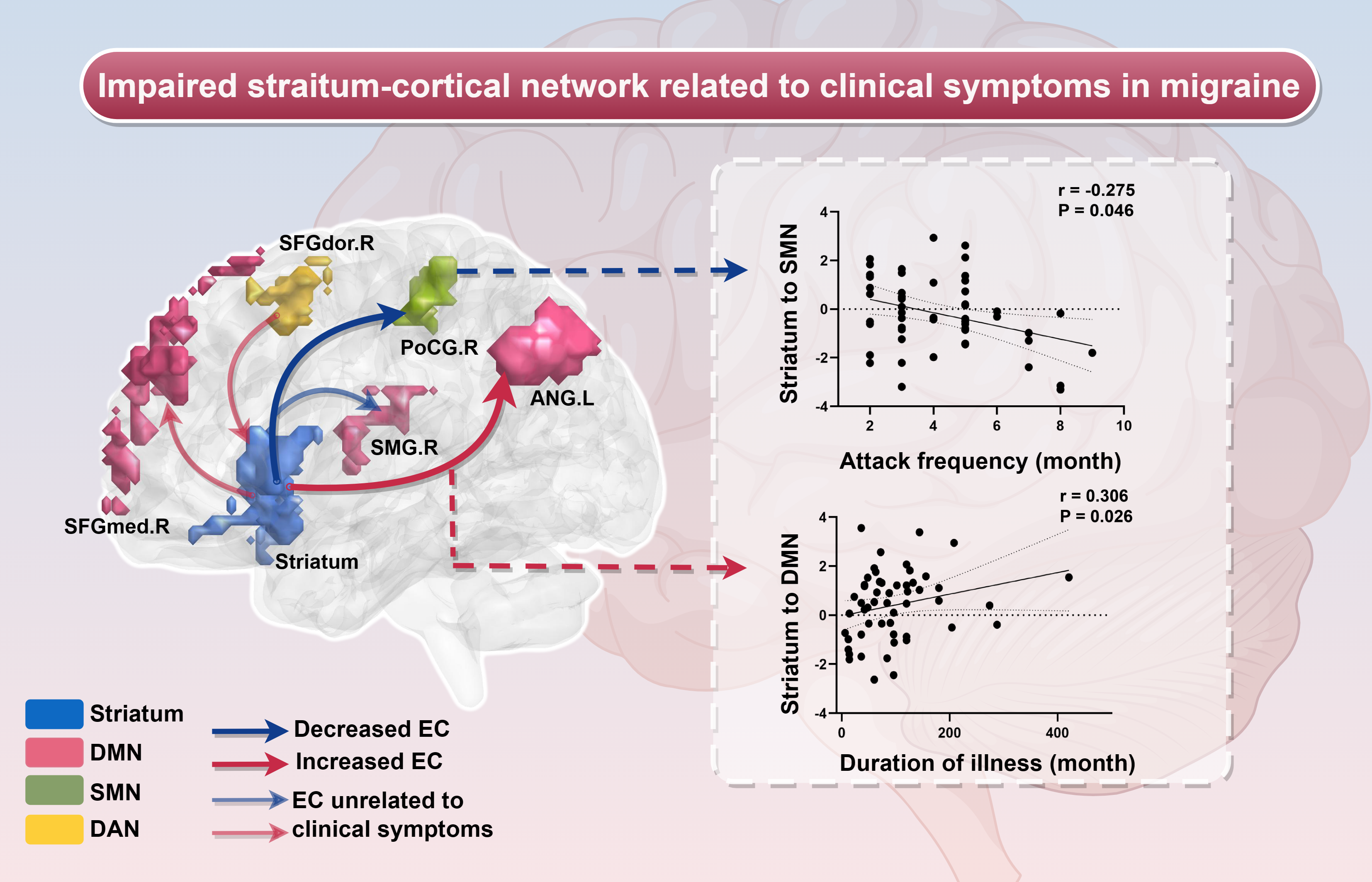
To the best of our knowledge, this study is the first to combine whole brain functional connectivity and EC to characterize abnormal connectivity in MWoA patients compared to HCs. We utilized DC analysis to investigate impaired functional hubs in MWoA patients we found decreased DC value in PUT.L and increased DC value in ANG.L, which respectively belongs to subcortical region (striatum) and cortical area (DMN). To further investigate the directional influence, we employed PUT.L and ANG.L as seeds to evaluate their EC with whole brain applying GCA and the altered EC were mainly in striatum-cortical network. In addition, the EC abnormalities from PUT.L to PoCG.R and CAU.R to ANG.L were significantly correlated with headache attack frequency and duration of illness. Conclusively, these finding confirmed the hypothesis that MWoA exhibited abnormal functional and effective connectivity in subcortical brain regions (striatum) and cortical network including DMN, SMN and attention network, and the dysfunction were related to pain sensory during the occurrence and progression of migraine. Specifically, on the one hand, the pain sensory of migraine is amplified by the weakened modulation of pain signals from the subcortex (striatum) to the cortex network, and on the other hand, the cortical network (attention network) pays too much attention to pain signals from the subcortex (striatum), resulting in excessive convergence of pain signals.
4.1 Impaired whole-brain functional hubs in MWoA
Migraine involves extensive functional abnormalities in the cortex and subcortical brain regions[30]. Our study found that the dysfunctional brain regions belong to cortical (DMN) and subcortical areas (striatum). Degree centrality represents the status and role of voxels in the whole-brain network[24]. We discovered the DC value of left putamen significantly decreased in MWoA patients compared with HCs. The putamen is part of striatum, being activated frequently during pain attacking[31]. Previous study reported that putamen may play a significant role in the mechanisms that convert nociceptive information into pain sensory[32], and a diffusion tensor imaging (DTI) study which could quantify the chance from one brain region to other areas showed that the putamen connected with several regions pertained to the procession of pain sensory such as DMN (middle frontal gyrus), insula and hippocampus[33]. Thus, the decreased DC value indicated weaker functional connectivity with other regions, which may lead to dysregulation of pain sensation.
We also observed increased DC value in left angular in MWoA patients compared with HCs. The angular locates at posterior part of the inferior parietal, a study using diffusion tensor imaging and tractography techniques explored rich structural link between angular with other areas including precuneus, caudate, frontal and temporal gyrus[34], which provided structural support that angular participates in consisting of DMN and its relationship with striatum. In addition, the angular converges multisensory information involving pain[35], increased DC value may result in excessive convergence of pain signal, then amplified the pain sensory.
4.2 Effective connectivity from striatum to cortical network
To further investigate the direction of functional connectivity, we selected PUT.L and ANG.L as seeds to perform the GCA. Our study indicated significantly increased EC from PUT.L to SFGmed.R, and decreased EC from PUT.L to SMG.R, SFGdor.R and PoCG.R.
The SFGmed.R and SMG.R were part of DMN, the medial frontal cortex was correlated with cognitive control, pain and emotion especially negative emotion[36, 37], increased EC value may indicate the pathway from pain sensory to pain emotion was overactivated, which may lead to emotion disorder such as depression[38, 39], and anxiety[40, 41]; the supramarginal gyrus is part of somatosensory association cortex, participating in somatosensory integration and interpretation, and right supramarginal gyrus is further related to attention reorientation and distribution[42–44], several studies reported that distracting attention can relief pain[45, 46], decreased EC value from PUT.L to SMG.R may imply that the integration of pain signals input from subcortical areas as well as the ability to distract attention were inhibited in MWoA patients, contributing to intolerable pain sensory.
In addition, we explored decreased EC value from PUT.L to PoCG.R correlating with headache attack frequency, postcentral belongs to SMN, previous study focusing on alteration in sensorimotor network effective connectivity promoted that the SMN may be affected by abnormal inflow or outflow information from putamen[20], and another study inferred the frequency of pain correlated with SMN and dorsal striatum[47]. Therefore, our study may further confirm the abnormal directional connection between the striatum and SMN, and the alteration is related to the frequency of headache attack.
Moreover, we observed markedly increased effective connectivity from bilateral caudate to ANG.L as well as decreased effective connectivity from ANG.L to CAU.L. As the key part of basal ganglia, the caudate participates in cognitive, sensory and pain modulation[48], our study found interaction between the angular and the caudate, manifesting as the effective connectivity from angular to caudate decreased while that from caudate to angular increased in MWoA patients, which may imply that the processing of pain information in the striatum affects the perception of pain signal in the cortical network. Previous study reported the abnormal functional connectivity of right caudate in chronic migraine[21], and we also investigated altered effective connectivity from right caudate to angular, which was positively correlated with duration of illness. It further suggested that dysregulation of caudate may be an important neuroimaging marker in migraine progression.
4.3 Effective connectivity from cortical network to striatum
We observed increased EC value to from SFGdor.R to PUT.L. The dorsolateral prefrontal cortex (DLPFC) is a pivotal structure in dorsal attention network (DAN) involving in top-down attention orientation, together with attention reorientation system of right-lateralized ventral attention network (VAN), consist of two attention systems in human brain[49], our study observed increased EC from right DLPFC to left putamen, and the EC was inhibited from left putamen to right DLPFC, implying that the excessive attention of DAN on regulation pain information of the striatum may trigger pain, we also found abnormal EC between right-lateralized ventral attention network and striatum (decreased EC value from PUT.L to SMG.R), it can be speculated that the abnormal EC between the two attention networks may be the neuroimaging mechanism of the relationship between pain sensation and attention.
In conclusion, the striatum-DMN may play a role in modulating the transition from pain sensory to pain emotion, while the striatum-SMN may be associated with the frequency of pain sensory experiences. Additionally, the striatum-attention network may be involved in processing both pain sensory information and attentional aspects in patients with MWoA.