3.1 Preparation of PEG-DA/PDA/PUE/FA hydrogel
Herein, PDA NPs, PDA/FA NPs, PDA/PUE NPs, and PDA/PUE/FA NPs were prepared. The size of the NPs was characterized by dynamic light scattering (Fig. 2A; PDA NPs: 193.28 ± 3.16 nm, PDA/FA NPs: 216.61 ± 5.19 nm, PDA/PUE NPs: 206.55 ± 3.81 nm, and PDA/PUE/FA NPs: 233.14 ± 4.67 nm). The FA loading ratios of PDA/FA NPs and PDA/PUE/FA NPs were 9.45 ± 0.89% and 6.09 ± 2.31%, respectively. The PUE loading ratios of PDA/PUE NPs and PDA/PUE/FA NPs were 7.93 ± 1.77% and 5.67 ± 1.89%, respectively. Hydrogels were prepared by saturating the double bonds of PEG-DA via UV irradiation, resulting in the formation of a three-dimensional network structure (Fig. 2B). Although the pore size was not uniform, the three-dimensional structure of the hydrogel and small pores were distinctly observed in the dried hydrogel frame (Fig. 2B).
Several characteristics, such as properties and applications, especially biomedical applications, of a hydrogel depend on the pore size of the inner structure, dispersion of NPs, and morphology[27–28]. For instance, the adsorption ability of a hydrogel is determined by the pore size, and the size of the drug determines whether the drug can be entrapped in the hydrogel network[29–30]. Scanning electron microscopy results indicated that the PEG-DA/PDA/PUE/FA hydrogel was highly interconnected and infiltrated throughout the pores. It is speculated that the PEG-DA/PDA/PUE/FA hydrogel not only possesses high nutrient permeability but also improves cellular growth.
3.2 Characterization of the PEG-DA/PDA/PUE/FA hydrogel
The swelling property of hydrogels was analyzed as a function of time (Fig. 3A). All the investigated hydrogels exhibited absorption behavior. The hydrogels absorbed approximately 2200 times more water than their own weight in nearly 50 h. In chronic wounds, excess exudates lead to bacterial growth around the injury, which causes microbial infection and delays healing[31]. Therefore, owing to their huge physical dimensions, these hydrogels can absorb a high volume of fluid, which facilitates wound healing[31–32].
In the degradation test, the different hydrogels exhibited similar degradability mainly because the structure and quality of the gel components were not markedly different (Fig. 3B). Additionally, the size of the hydrogel samples stably decreased, which may be mediated through surface and internal erosion[33].
Compressive strength was investigated to study the mechanical properties of the hydrogels. Young’s moduli of the different hydrogels were 2.17 ± 0.80, 2.02 ± 0.86, 1.93 ± 0.60, 1.83 ± 0.69, and 1.45 ± 0.63 MPa (Fig. 3C). Moreover, the mechanical properties of the synthesized hydrogels were in the range of those of biological tissues, such as the skin and articular cartilage[34].
Next, the WVTR was tested to evaluate the porosity of the hydrogels. The WVTR values of the different hydrogels were 3198.23 ± 76.19, 3506.19 ± 89.83, 3241.38 ± 250.90, 3402.00 ± 268.20, and 3515.18 ± 252.62 g/m2 (Fig. 3D). The water content and porosity of a dressing are closely related to wound healing. During wound healing, the water content of the skin increases, and a dressing with high WVTR can promote wound closure[35].
Drug release behavior of the drug-loaded hydrogels was examined. Although PEG-DA/PDA/PUE/FA hydrogel had an irregular porous structure, they could still diffuse from the hydrogel (Fig. 3E and 3F). An initial burst release of the drug was observed, followed by a steady release, which may be due to the high concentration gradient between the drug release media during the initial phase and a low concentration gradient during the subsequent phase. However, without PDA, the drug in the PEG-DA hydrogel was released considerably faster than the cases of other hydrogels. Nearly 80% of PUE and FA were released from the PEG-DA hydrogels within 2 h. This difference indicated the stability of the PDA NPs. This preliminary discovery provides a potential method for preparing PEG-DA hydrogels with slow drug-release properties.
3.3 Biocompatibility of the PEG-DA/PDA/PUE/FA hydrogel
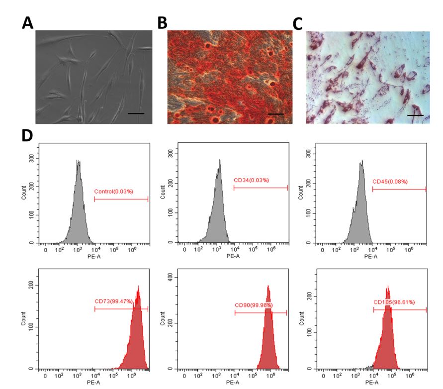
In order to evaluate the biocompatibility of the hydrogels, hPDLSCs were isolated and characterized, which are accorded with the identification of mesenchymal stem cells (Fig S1). HPDLSCs were co-incubated with the extracted liquids of the hydrogels. Cell proliferation was continuously detected over a period of 5 days (Fig. 4A). Under all conditions, the viability of the cells was maintained at a high level throughout the experiment, and the cells were capable of spreading and proliferating over time, indicating that the hydrogels did not have a marked detrimental effect on the long-term viability of hPDLSCs (Fig. 4A). Moreover, Calcein-AM and PI were used to monitor live and dead cells (Fig. 4B). These data verify that the different hydrogels proposed herein are conducive to cell growth and viability (Fig. 4B). These results further demonstrate the feasibility of using drug-loaded PDA NP-incorporated hydrogels with excellent biocompatibility for tissue engineering applications.
3.4 Antioxidant activity and oxidative stress resistance ability of the PEG-DA/PDA/PUE/FA hydrogel
Antioxidant activity of hydrogels is the key to inhibiting oxidative stress in tissue engineering. The antioxidant activity is measured by the DPPH•- and •OH-scavenging ability of hydrogels and quantified as a percentage of the suppression of free radical formation[36−37]. In the •OH-scavenging assay, the scavenging of •OH significantly improved after PEG-DA/PDA/PUE/FA hydrogel treatment (Fig. 5A). In the DPPH• test, the elimination rate of DPPH• also distinctly increased in the case of the PEG-DA/PDA/PUE/FA hydrogel group (Fig. 5B). More specifically, the PEG-DA/PDA/PUE/FA hydrogel demonstrated excellent antioxidant activity, with the maximum •OH- and DPPH•-scavenging rates of 79.27 ± 2.20 and 52.55 ± 2.98% (Fig. 5A and 5B), respectively.
To investigate the oxidative stress resistance ability of the PEG-DA/PDA/PUE/FA hydrogels in embedded cells, we used H2O2 (100 µM) to directly expose the cells to superoxide radicals. The results showed that the introduction of the drug effectively suppressed the generation of intracellular ROS, and the fluorescence intensity of 2,'7'-dichlorofluorescein in the PEG-DA/PDA/PUE/FA hydrogel was distinctly lower than that in the other hydrogels (Fig. 5C). Owing to the overproduction of ROS, cell biomolecules experience severe oxidative damage, causing disruption of the pro-oxidant-antioxidant balance[38]. SOD and GPx play a cytoprotective role under oxidative stress. Studies have shown that SOD is important for the oxidant and antioxidant balance in the body because it can eliminate superoxide anion free radicals as well as heal injured cells. GPx maintains the integrity of the cell membrane structure and is widely distributed in cells. Additionally, MDA produced by lipid oxidation can reflect oxidative stress injury caused by ROS. The PEG-DA/PDA/PUE/FA hydrogel effectively promoted the generation of SOD and GPx and inhibited the production of MDA (Fig. 5D–F). Therefore, the PEG-DA/PDA/PUE/FA hydrogel could protect the cells from oxidative stress damage.
PUE can react with free radicals because of its abundant surface electrons. It has been found to decrease Schwann cell apoptosis in a diabetic animal model owing to its antioxidant activity[39]. Furthermore, PUE significantly alleviates H2O2-induced oxidative stress injury and suppresses the apoptosis of neural cells[40]. FA has antioxidative, anti-inflammatory, and anti-hyperlipidemic properties[41]. The administration of FA reduces oxidative stress and DNA damage caused by lead acetate[42]. In our previous study, we showed that the hybrid hydrogel incorporated with PUE exerted excellent antioxidant effects, which promoted the regeneration and healing of damaged skin[43]. In the present study, we found that PUE and FA have a synergistic role in resisting oxidative stress damage in vitro and promoting wound healing in vivo. The PEG-DA/PDA/PUE/FA hydrogel also decreases cell death and enhances the survival capacity of hPDLSCs in an oxidative stress microenvironment, which is beneficial for wound healing.
3.5 In vivo wound healing effects of the PEG-DA/PDA/PUE/FA hydrogel
The wound healing properties of the hydrogels were further investigated by in vivo tests. The results demonstrated that the wounds treated with the PEG-DA/PDA/PUE/FA hydrogel healed faster than those treated with the other hydrogels (Fig. 6). On day 15, the wounds treated with the PEG-DA/PDA/PUE/FA hydrogel almost completely healed, whereas those treated with the control did not heal.
Wound healing involves several biological processes, including hemostasis, migration, proliferation, and remodeling. After treatment for 15 days, histopathological changes occurred in different skin samples. H&E staining revealed that the wound healed in the PEG-DA/PDA/PUE/FA hydrogel group is faster than other groups (Fig. 7A and B). Additionally, the PEG-DA/PDA/PUE/FA hydrogel-treated wounds showed distinct recovery, which possessed mature fibrous tissues, well-organized fibroblasts, and blood capillaries (Fig. 7A). Therefore, the lack of inflammation and pathological abnormalities confirmed the histocompatibility of the PEG-DA/PDA/PUE/FA hydrogel. Collagen fibers are produced by fibroblasts, and the remodeling of these fibers is necessary during wound healing[44–45]. Masson staining showed that the PEG-DA/PDA/PUE/FA hydrogels promoted the formation of collagen fibers (Fig. 7A and C). Moreover, the PEG-DA/PDA/PUE/FA hydrogels effectively upregulated the expression of CD34 protein, resulting in improved platelet-endothelial cell adhesion (Fig. 7A and D). VEGF had the highest expression in PEG-DA/PDA/PUE/FA hydrogel-treated wounds (Fig. 7A and E), which suggests that the vessel formation in PEG-DA/PDA/PUE/FA hydrogel is more than other groups.
The results of histological studies on wound healing showed that the PEG-DA/PDA/PUE/FA hydrogel dressings had antioxidant potential to promote wound healing. Moreover, the semipermeable nature and free radical-scavenging property of the PEG-DA/PDA/PUE/FA hydrogel wound dressings may be responsible for the early contraction of the wound and formation of fibrous tissue. These results demonstrate that the PEG-DA/PDA/PUE/FA hydrogel wound dressings can be used as candidate materials for wound applications such as repair and regeneration of damaged skin.