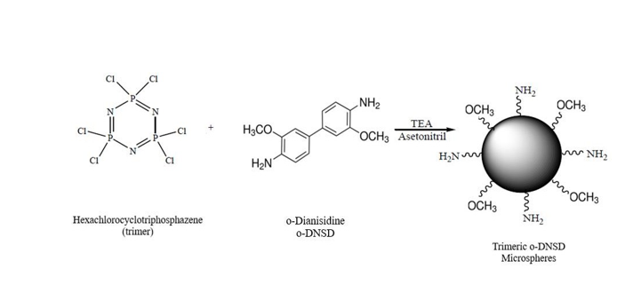
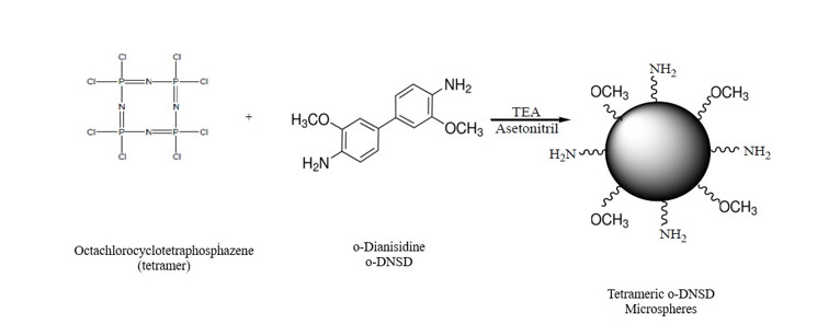
o-DNSD-microspheres were prepared via self- assembly one-pot polycondensation polymerization of hexachlorocylotriphosphazene (trimer, N3P3Cl6) trimer and tetramer octachlorocyclotetrahosphazene (tetramer, N4P4Cl8). In order to obtain the best morphological surface, the use of reagents in different mole ratios was tried. It was tested using chemicals in various mole ratios to get the optimal morphological surface. Utilizing SEM, FT- IR, and XRD, the polymeric microspheres were characterized. [15, 23, 24].
oDNSD-MS synthesis was accomplished via one-pot basic polymerization. Synthetic routes for the formation ODNSD-miscospheres were given in Scheme 1 and Scheme 2. The crosslinker molecules trimer and tetramer are flexible rings with six and eight chlorine atoms, respectively. So these features of both phosphazene rings give them high cross-linking ability against o-DNSD. Excessive amounts of TEA were used as an acid acceptor during polymerization operations, generating TEA.HCl. Stabilizing agent or surfactant was not needed for the polymerization process.
According to the previously described process, trimeric and tetrameric polyphosphazenes with cross-linked networks self-assemble. During the initial phase of polymerization, ODNSD, acting as the monomer, reacts with the crosslinker trimer/tetramer to create oligomers. Oligomers aggregate together to form primary nucleus particles at the next step. Subsequently, hydrogen bond interactions cause the primary nucleus particles to aggregate and become stable primary particles. Following that, oligomeric species are absorbed by the particles, causing them to grow larger than initial particles. The resulting microspheres are pore-free both inside and outside. SEM images of the cyclomatrix type o-DNSD-trimer microspheres are presented in Figure 2.
The crystalline or amorphous nature of the microspheres is ascertained using XRD spectral technique. The 1:1 o- DNSD-trimer XRD Spectrum is shown in Figure 3. The product's amorphous structure is displayed by the broad peak that emerged in accordance with the XRD diffraction pattern.
Figure 4 displays the FT-IR spectra of the trimer, o-DNSD, and 1:1 o-DNSD-trimer.
SEM images of the cyclomatrix type o-DNSD-tetramer microspheres are shown in Figure 5
The crystalline or amorphous nature of the microspheres is ascertained using XRD diffraction analysis. The 1:1 and 1:2 o-DNSD-tetramer XRD Spectrum is shown in Figure 6. The product's amorphous structure is displayed by the broad peak that emerged in accordance with the XRD diffraction pattern.
FT-IR spectra of 1:1, 1:2 1:3 and 1:4 o-DNSD- tetramer are given in Figure 7.
Four distinct types of crosslinked polyphosphazene were attempted to be synthesized in these studies using the precipitation polymerization process. Meanwhile, ratio experiments were carried out with o-DNSD as monomer and trimer and tetramer as crosslinkers. The aim of these ratio experiments is to obtain proper and homogeneous spheres. Some parameters were specifically tried to be kept constant like solvent amount, temperature and ultrasonic bath as reaction media. SEM morphologies were used by looking at the initial experiments to determine the reaction durations. These parameters were kept constant for each experiment.
For the o-DNSD-trimer and o-DNSD-tetramer tests, it was noted from the SEM images that the sphere surfaces were distorted in accordance with the increasing crosslinker concentration. Based on an assessment of the SEM images, it can be assumed that, for the most part, experiments are successful in generating microspheres. XRD analyzes of crosslinked polyphosphazene microspheres were performed. These analyses exhibit the crystalline or amorphous characteristics of the products. As a result, polymerization was seen, and the graph developed an amorphous structure with a large peak and a 2θ(o) value between 20 and 30. The peaks observed for some product types are an indication that small amounts of different non-polymeric products formed as a result of the reaction and/or effective work-up procedure is not performed during the purification of the products.
FT-IR spectra show the binding states of the monomer and the crosslinker. The spectra showed characteristic (P=N, N-H) peaks for polyphosphazene compounds. Besides, the P-Cl bond peak values in the trimer and tetramer either reduced their intensity or vanished entirely. The presence of the binding can be confirmed by looking for specific bands in the microsphere’s spectra which match those of monomers. Thus, in FT-IR spectra, the following stretching is attributed to microspheres with o-DNSD: the band at 3350-3410 cm-1 is thought to be related to N- H stretching; the band at 2932-2987 cm-1 to C-H aliphatic stretching; the band at 1610-1616 cm-1 to C=N stretching; the band at 1503 cm-1 to C-O stretching; the band at 1450-1460 cm-1 to C-C aromatic stretching; the band at 1390 cm-1 to C=C aromatic stretching; the band at 1240-1245 cm-1 to P=N stretching; the band at 810-930 cm-1 to C-H aromatic stretching; and the band at 490-508 cm-1 to P-Cl stretching. As a result, the desired polyphosphazene microspheres were synthesized and spectrally characterized.