Plants inoculation. The Iranian isolate of ToBRFV, Accession Nr. P557566.1, (kindly provided by Dr. K. Bananej) was used for mechanical inoculating four-week-old S. lycopersicum plants. An aliquot of 100 mg of frozen infected tissues was ground in a ball mill and homogenized in 20 volumes of pH 7.4 phosphate buffer [3.1 g NaH2PO4.H2O and 10.9 g Na2HPO4 (anhydrous) in ultra-pure water]. A cotton swab soaked in the virus extract was used to spread the drop of carborundum onto the adaxial side of a plant leaf. Photoperiods of 12 hours/day and 12 hours/night were maintained in a growth chamber at 25°C.
The preparation of samples. Systemic tissues (approximately 100 mg) collected from non-inoculated upper leaves at 10 days post-inoculation (DPI). These aliquots were purified using 1ml of ice cold RNX-Plus (SinaClon, Karaj, Iran) according to manufacturer protocol. Also, different dilutions of these original samples was prepared. Following the instructions of the manufacturer, cDNA was synthesized from 400 ng of extracted RNA using the SinaClon cDNA Synthesis Kit. Furthermore, cDNAs were stored in a refrigerator at -80°C until use. Presence of virus within the inoculated plants was confirmed by RT-PCR followed by gel electrophoresis.
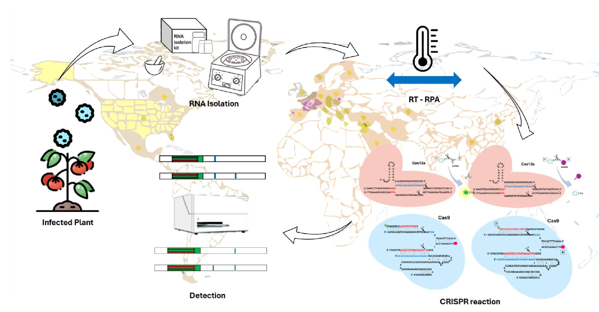
crRNAs and primers. For CRISPR-Cas12a-based detection, RT-RPA primers (Table 1) designed in the genomic region corresponding to the ToBRFV coat protein, ensuring that a PAM sequence (TTTV) is present in the amplification region at an appropriate place. Additionally, specific single-stranded DNA (ssDNA) probes were designed with fluorescein labeling at the 5′ end and a dark quencher at the 3′ end. Cas12a-dependent crRNAs were created with a spacer sequence complementary to the target sequence (Fig. 5) and a PAM position 47,55.
In the case of Cas9, the hybridizing ssDNA molecules that make up the gates were intended to establish a stable complex at 37°C and to have toeholds to initiate interactions with the displaced strands of the targeted DNA amplicon. The three strands incorporating a gate were heated to 95°C for two minutes and then cooled gradually to 25°C before being used in the CRISPR reactions. the sequences shown in Table 1. At a final volume of 20 µL, CRISPR procedures were carried out in 1× TAE buffer pH 8.5 (Invitrogen), 0.05% Tween 20 (Merck), and 12.5 mM MgCl2 (Merck). Combinatorial mixing was used to combine the input DNA amplicon of ToBRFV, 2 µL of each test samples came from infected samples. At 200 nM, the prehybridized gate was inserted. The reactions were incubated in an Eppendorf thermomixer at 37°C for one hour.
The crRNAs were synthesized through in vitro transcription using the TranscriptAid T7 High Yield Transcription kit (Thermo) with DNA templates obtained from IDT. Subsequently, the crRNAs were purified using RNA Clean and Concentrator spin columns (Zymo) and quantified using a NanoDrop spectrophotometer.
Table 1
cDNA sequences of crRNAs and primers used in this study.
| Description | Sequences (5’-3’) |
| ToBRFV forward primer for RT-RPA amplification | AGTAGATGACGCAACGGTGGCTATAAGGAG |
| ToBRFV reverse primer for RT-RPA amplification | TACGTGCCTACGGATGTGTATGAACCATAC |
| Cas12a crRNA to detect ToBRFV | GCAGGUGCAGAGGACCAUUGAUCUACACUUAGUAGAAAUUACCUAUAGUGAGUCGUAUUAGCGC |
| ToBRFV synthetic post strand biotin | /5Biosg/TTTGAAAGTGCATCCGGTTTACAATGGTCCTCTGCACCTGCATCTTGAGATAATCGAGATG |
| Cas9 gRNA to detect ToBRFV | AAAAGCACCGACTCGGTGCCACTTTTTCAAGTTGATAACGGACTAGCCTTATTTTAACTTGCTATT
TCTAGCTCTAAAACGTTTACAATGGTCCTCTGCACCTATAGTGAGTCGTATTAGCGC |
| ToBRFV beacon 1 (Hairpin) | CAGGTGCATATATGGTCCTCTGCACCTG/36-FAM/ |
| ToBRFV beacon 2 | TGGTCCTCTGCACCTGCATC/36-FAM/ |
Amplification of viral genomes using RT-RPA. The TwistAmp Basic kit (TwistDX) was used for RT-RPA amplification of desired viral genome as described by manufacturer. Briefly; forward and reverse primers (500 nM; sequences provided in Table 1), 500 U RevertAid (Thermo), and 50 U RNase inhibitor (Thermo) were combined with 29.5 µL rehydration buffer to reach a total volume of 43.4 µL (adjusted with RNase-free water). The TwistAmp Basic reaction pellet was resuspended in this mixture, and 21.7 µL, along with 2 µL of total RNA extracted from the plant (or 4 µL of crude plant extract without RNA purification), was added per reaction. The reaction was initiated by adding 280 mM magnesium acetate. Incubation took place at 40˚C for 5 min, followed by vortexing and spinning, and then re-incubation for an additional 25 min.
Reaction of CRISPR-Cas12a. The CRISPR-Cas12a reaction contained 50 nM Cas12a enzyme sourced from L. bacterium (NEB) and 62.5 nM crRNA which was allowed to incubate for 30 minutes in NEBuffer 2.1 (comprising 10 mM tris-HCl, 50 mM NaCl, 10 mM MgCl2, and 100 µg/mL BSA at pH 7.9; NEB) at room temperature. Subsequently, in a 96-well MicroAmp Fast plate from Applied Biosystems, 2 µL of amplified DNA was mixed with 17 µL of the CRISPR-Cas12a ribonucleoprotein complex and 500 nM of a fluorescein-quencher-labeled ssDNA probe (TTATT) to reach a total volume of 20 µL. The mixture was incubated at 37˚C for 1 hour, during which green fluorescence readings were taken every 5 minutes using a QuantStudio 3 real-time PCR machine from Thermo Fisher Scientific. Excitation and emission wavelengths were set at 470/15 nm and 520/15 nm, respectively. For multiplexed detection, each crRNA was loaded into a separate well, and distinct CRISPR reactions were concurrently performed on the same plate.
CRISPR-Cas12a Diagnostics process. Target identification based on Cas12a and isothermal amplification are two separate processes in the RT-RPA-CRISPR-Cas12a detection platform. Target detection requires transferring amplified products to the Cas12a cleavage mechanism. Due to the great fidelity of RT-RPA amplification, aerosols are easily produced and often lead to cross-contamination, or false positive reactions. Moreover, there is competition if RT-RPA reagents and CRISPR-Cas12a components are combined directly in the one-pot procedure, which might result in decreased sensitivity 56. Cas12a cleavage and RT-RPA amplification share the same DNA substrate. The reaction tube filled with Cas12a/crRNA components and RT-RPA amplification reagents to address these issues. To identify ToBRFV, the Cas12a/crRNA complex was manually shaken to the bottom of the tube during RT-RPA amplification and combined with the RT-RPA products. By eliminating the issue of lower sensitivity brought on by the rivalry of DNA substrates between Cas12a cleavage and RT-RPA amplification, the single-pot RT-RPA-Cas12a assay avoids aerosol contamination (Fig. 6).
Reaction of CRISPR-Cas9. CRISPR reactions were carried out in a final volume of 20 µL using 1× TAE buffer pH 8.5 (Invitrogen), 0.05% Tween 20 (Merck), and 12.5 mM MgCl2 (Merck). The CRISPR-Cas9 ribonucleoprotein was produced at a concentration of 100 nM after it had been formed for 30 minutes at room temperature. For test samples, each reaction contained 40 nM of amplified DNA (dsDNA amplified utilizing post-strand biotin labeled and the reverse one). For single or multiplexed detection, 2 µL of amplified product were utilized per reaction in the case of ToBRFV-infected samples. A volume of 2 µL of the refined RPA product was utilized for the limit of detection tests. The reactions were incubated in an Eppendorf thermomixer at 37°C for 25 minutes. After that, 100 nM of the molecular beacon (beacons) was (were) added, and it was incubated for 5 minutes at 37°C. To make the process simpler and get comparable outcomes, the beacons might alternatively be inserted at the start of the CRISPR response (Fig. 7).
CRISPR-Cas9-Based Detection. The lateral flow nucleic acid test was integrated with a CRISPR-Cas9 tool by Wang et al., 57 to eliminate the need for several nucleases, auxiliary proteins, and pricey fluorescent probes. This method combines the CRISPR-Cas9 system with a lateral flow device to detect target dsDNA. It is called the CRISPR-Cas9-mediated lateral flow nucleic acid assay (CASLFA). Prior to being dripped onto the sample pad, the target is first amplified using biotinylated primers in PCR or isothermal amplification techniques. It is then incubated with CRISPR-Cas9 RNPs. After that, CRISPR-Cas9-complexed biotinylated amplicons flow to the test line where the mixture is all caught by the streptavidin. At the test line, this colored signal is visible. Biotinylated probes hybridize with the precoated streptavidin at the control line to produce a colorful band. In conclusion, this method is a straightforward technique that finds target DNA with high specificity and sensitivity (LOD is ≈ multiple gene copies). It consists of a brief (3–5 min) lateral flow test followed by a PCR/RPA amplification step using biotinylated primers. To sum up, the authors propose a number of technological enhancements for the detection, including the use of distinct FAM-labeled beacons through the introduction of primers tagged with biotin, and the fusion of CALSFA and COLUMBO with some of the previous trends on a sophisticated diagnostic platform 39,57.
Molecular Beacons. In order to hybridize with the displaced DNA strands from the CRISPR reactions, various DNA oligonucleotides that folded into a stem-loop structure and were appropriately labeled were designed. These probes were made to have a seed region in the loop, high GC content, and a melting temperature higher than 50°C. The ability to correctly fold and hybridize (with the target DNA, but not with the sgRNA) was assessed using NUPACK.37 Molecular beacons that targeted the PAM-distal region were labeled in their 3′ end with a fluorophore (FAM); when targeting the PAM-proximal region, the beacon was labeled in its 5′ end with FAM. In order to guarantee appropriate folding, the molecular beacons were heated at 95°C for two minutes and then slowly cooled to 25°C before being used in the CRISPR reactions. Sequences provided in Table 1.
Lateral Flow Assay. This CRISPR-Cas12a reaction utilized the ssDNA probe TTATT labeled with fluorescent-biotin at its ends and at 200 nM and was incubated for 2 hours at 37°C. In an additional step, 80 µL of GenLine Dipstick buffer (Milenia) supplemented with 5% PEG 6000 were diluted 1:5 in water 55. Five minutes after dipping the lateral flow strip into the reaction tube (HybriDetect, Milenia), images were captured.
Following the acquisition of the CRISPR-Cas9 R-loop for the molecular beacon opening (COLUMBO), 20 µL of Cas9 reaction solution and 80 µL of running buffer (80 µL of GenLine Dipstick buffer (Milenia) supplemented with 5% PEG 6000 were diluted 1:5 in water) were added to the sample pad of the lateral flow device. Within two minutes, the bands in the test line and control line appeared. Following the test, a picture of the lateral flow device was taken.
Fluorescence Assay. 2 µL of RT-RPA-amplified dsDNA (of each concentration: 0, 10 pM, 100 pM, 1nM, and 10 nM) were combined with 17 µL of CRISPR-Cas12a ribonucleoprotein (already generated) and 1 µL of ssDNA probe (chemically performed by IDT, TTATT tagged with fluorescent-fam) in an Eppendorf tube. A 384-well Fluorescence Plate Reader (Clariostar Plus) was used to measure the fluorescence of each reaction concentration.