Cell culture and transfection
The GES-1 cells, GC cell lines, and embryonic renal cell line 293FT were purchased from the American Type Culture Collection (ATCC, Manassas, VA, USA). The HGC -27 cells was cultured in MEM (Minimum Essential Medium) supplement with penicillin and streptomycin(P/S) and fetal bovine serum (FBS); The other GC cell lines and GES-1 cells were cultured in RPMI-1640 (Roswell Park Memorial Institute-1640) mediums supplement with P/S and FBS. 293FT cell line was cultured as previously described [35]. The MEM, RPMI-1640 and DMEM meida, FBS, and antibiotics were obtained from Thermo Fisher Scientific, Inc. (Waltham, MA, USA).
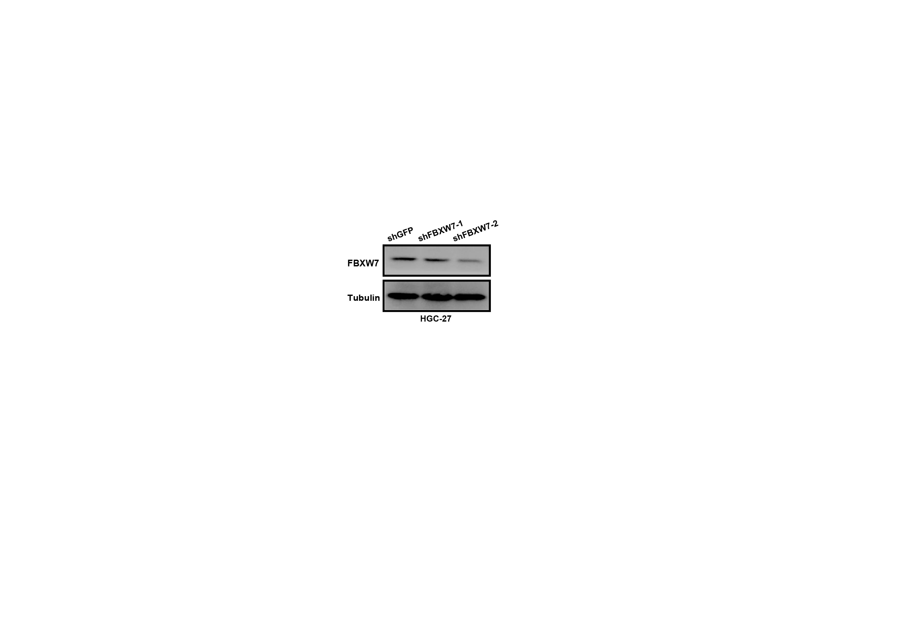
Sequences of the shZC3H15 and shFBXW7 were obtained from GenePharma Co., Ltd (Shanghai, China), and were listed as below:
shZC3H15#1: CAGATCCCAAGTCTGTAGTAT
shZC3H15#2: CCTAGAATCAACAGGATGTTT
shFBXW7#1: CCAGTCGTTAACAAGTGGAAT
shFBXW7#2: CCAGAGAAATTGCTTGCTTTA
Vector encoding of human ZC3H15 were constructed by PCR-based amplification, and the primers used were listed as below:
ZC3H15-F-(EcoRI): CCGGAATTCATGCCCCCCAAGAAAC
ZC3H15-R-(NotI): ATTTGCGGCCGCTCATTCTTCTAAATCAAGTGTATTT
Lentivirus was produced as previously described [35].
Reagents
Dimethyl sulfoxide (DMSO) was obtained from Sigma Aldrich (MO, USA). The ZC3H15 (cat. no. 26241), CyclinD1 (cat. no. 60186), c-Myc (cat. no. 10828) and Tubulin (cat. no. 11224) antibody were purchased from Proteinch (Wuhan, China); The FBXW7 (cat. no. ab74054) and Ki-67 (cat. no. ab92742) was purchased from Abcam (Shanghai, China); Flag (cat. no. 14793), CDK4 (cat. no. 12790), CDK6 (cat. no. 13331), MMP7 (cat. no. 3801) and N-cadherin (cat. no. 13116) antibody were purchased from Cell Signaling Technology (Beverly, MA, USA).
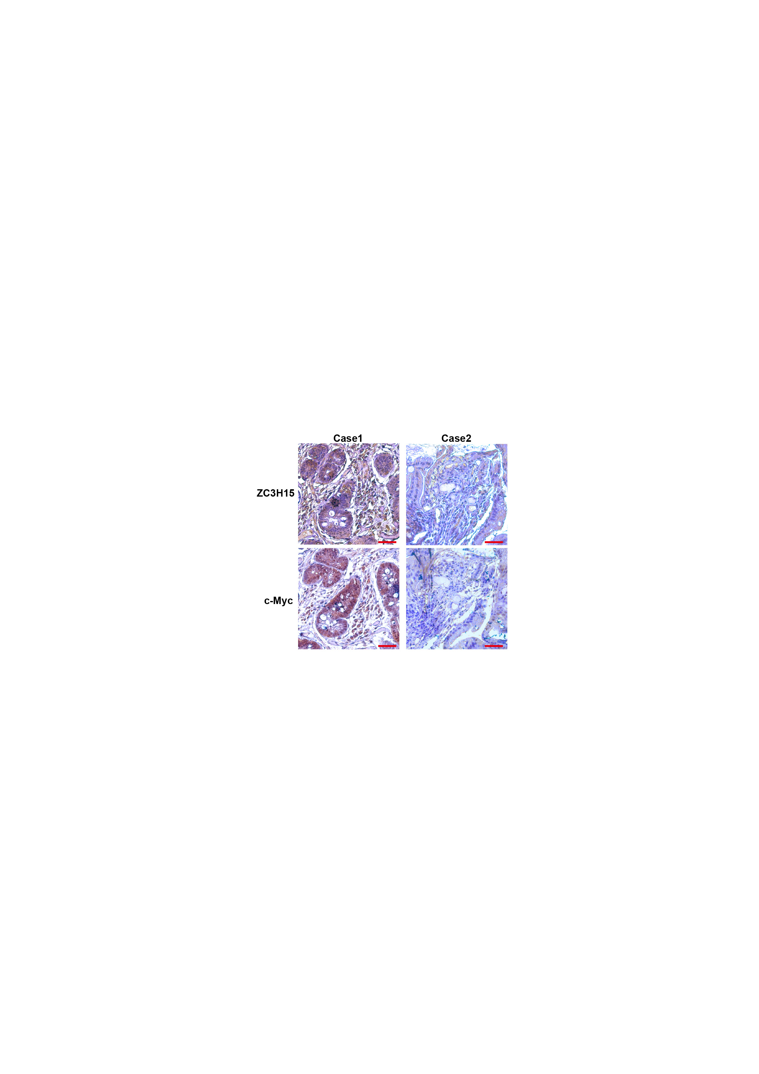
Immunohistochemistry staining
Tumor specimen was embedded in paraffin and sectioned into 5μm thick sections, and then deparaffinized and hydrated. The sections were performed by microwave heating for antigen retrieval, and then incubated with endogenous peroxidase and blocking with goat serum. After quenching for primary antibodies at 4℃ and secondary antibodies at room temperature, sections were covered with DAB (diaminobenzidine) for visualizing the staining.
Cell viability and proliferation assays
MTT assay was performed to examine the cell viability of indicated GC cell lines. Cells (1x10³ cells/ well) were cultured in the 96-well plates, and then were detected according to the manufacture’s protocol.
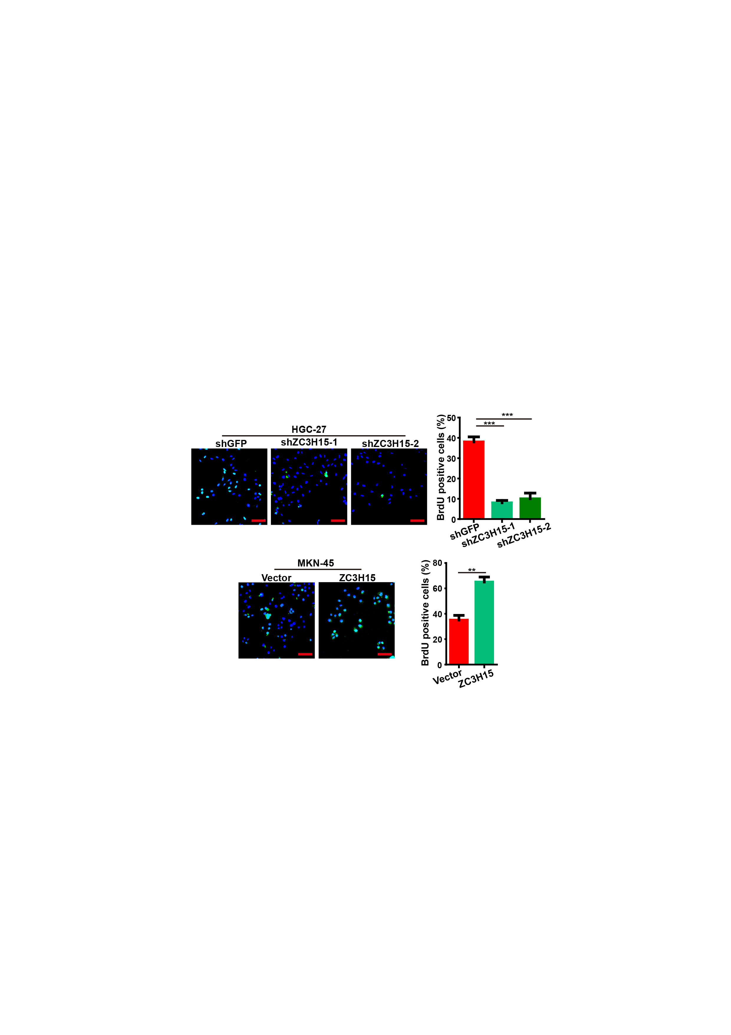
BrdU staining
For BrdU staining, indicated cells were seeded into 24-well plates. After incubated with BrdU (Sigma) and fixed in 4% PFA, cells were treated with 1 mol/l HCl and 5% goat serum. Then, cells were incubated sequentially with primary antibody against BrdU, and Alexa FluorR 594 secondary antibody. DAPI (4',6-diamidino-2-phenylindole) was used for nuclear staining.
Western blot analysis and Co-IP
For Co-IP assay, cells were lysed in IP lysis buffer (Sigma) and then incubated on a rocker with antibody as well as IgG at 4 ℃ overnight. After incubation with Protein A/G PLUS-Agarose, cell lysate were washed by PBS and resolved by sodium dodecyl sulfate-polyacrylamide gel electrophoresis analysis. Western blotting was performed as previously described [36].
Ubiquitination assay
For the ubiquitination assay, indicated plasmids were transfected into the 293FT cells. MG132 (50 μg/ml, Selleck, Houston, TX, USA) was added into the cells for 6h before harvesting. Cells were lysed and then performed following the same protocol used in Co-IP.
Turnover assay
The cells were transfected with indicated plasmids, and then a final concentration (100μg/ml) of CHX was added into the media. After harvesting at the indicated time points, cell were lysed and analyzed by Western blotting.
Quantitative and RT-PCR
Total RNA was harvested from the indicated cells and then reversely transcribed into cDNA by iScript cDNA Synthesis Kit (BioRad, #170-8891). The expression of mRNA was measured by using a Roche LightCycler Real-Time PCR System. Primers for RT-PCR assays were listed as Table II.
Luciferase reporter assay
Cells were transfected with shZC3H15, ZC3H15 or miR-124-3p mimics together with the indicated reporter (FBXW7, ZC3H15-WT, or ZC3H15-Mut) or control plasmid. Dual luciferase assay was performed by using the Dual-Luciferase® Reporter Assay System (Promega, #E1910). The promoter fragments of FBXW7, ZC3H15-WT, and ZC3H15-Mut were purchased from Wuhan GeneCreate Biological Engineering Co., Ltd.
Chromatin immunoprecipitation
Chromatin was isolated from 2x10⁷ 293FT/Vector and 293FT/Flag-ZC3H15. ChIP assays were performed using the EZ-ChIPTM kit (Millipore, CA, USA), and then detected according to the manufacture’s protocol. The primers used in ChIP assays are listed as Table II.
Soft agar assay
For the soft agar assay, 0.4 × 10³ cells were mixed with 0.6% agarose (Sigma-Aldrich, USA) in RPMI-1640 medium and then plated into 12-well plates containing a solidified bottom layer (0.3% agarose in medium).
Animal experimental procedures, tumour xenograft experiment, and lung metastasis assay
All animal studies were approved by the Institutional Animal Care and Use Committee of Southwest University. Four-week-old female nude mice were purchased from Beijing Animal Research Center and were housed in the SPF room. For the tumor xenograft experiment, mice were randomly divided into three groups. HGC-27 cells (1×10⁶) stably transfected with shGFP, shZC3H15-1 and shZC3H15-2 were subcutaneously injected into the mice in 18 November, 2019. Isoflurane anaesthesia system, which could help animals enter an anaesthetised state faster and recover quickly, was used reduce the pain of the mice. Isoflurane anaesthesia, is an inhalation general anesthesia, and the anesthesia-induction is stable, rapid, comfortable, fast recovery, good muscle relaxation, no sympathetic nervous system excitatory effect. In addition, isoflurane has a low metabolic rate in the liver, so it has little toxicity to the liver, and repeated use has no effect obvious side effects. Isoflurane was purchased from Reyward Life Technology Co., Ltd. (Shenzhen, China), and the concentration was MAC 1.6%. After subcutaneous injection, the mice were sterilised with 75% medical alcohol. The mice were observed and weighed every 3 days, and the feeding conditions were strictly standardized. The volume of tumors was calculated as follows: V = (length × width2)/2. Before the tumors were collected, the isoflurane anaesthesia system was also used to reduce mice’s pain, and then the mice were killed by cervical dislocation and the tumors were harvested. The bodies of mice were frozen at -20 °C and then transferred to Laibite Biotech Inc. (Chongqing, China) for incineration.
For the lung metastasis model, mice were randomly divided into three groups. HGC-27 cells (5×10⁵cells/ml) stably transfected with shGFP, shZC3H15-1 and shZC3H15-2 were injected subcutaneously into the tail vein of the mice in 18 November, 2019. Isoflurane anaesthesia system was used to reduce the mice’s pain during this experiment. The mice were observed and weighed every 3 days. Before the lungs were collected, the isoflurane anaesthesia system was also used to reduce mice’s pain, and then the mice were killed by cervical dislocation and the lungs were harvested. The bodies of mice were frozen at -20 °C and then transferred to Laibite Biotech Inc. (Chongqing, China) for incineration. The lungs were fixed with paraformaldehyde for H&E staining.
Transwell assay
For the transwell assay, cells in serum-free MEM or RPMI-1640 Mediem were seeded into the 24-well Boyden chambers (8μm pore size, Corning). MEM or RPMI-1640 Mediem with 10% FBS was added to the lower chamber. Cells were fixed in 4% paraformaldehyde (PFA) and then stained with crystal violet. Then, Cells were imaged and calculated.
Patient data analysis and patient tumor tissues
Bioinformatics analyses were performed using these specific programs: TCGA (https://cancergenome.nih.gov), UCSC Xena (https:// xena.ucsc.edu/public/), starBase (https://www.starbase.sysu.edu.cn/), and Kaplan Meier-plotter (http://kmplot.com/analysis/). Clinical samples were obtained from Chaoying Biotechnology Co., Ltd. (Henan, China). All the studies were approved by the Medical Ethics Committee of Tongxu County People’s Hospital of Henan Province. All of the patients were informed consent.
Gene set enrichment analysis (GESA)
To gain insight into ZC3H15 expression associated with the biological processes in GC, GSEA was performed using the Broad Institute GSEA version 4.0.3 software. The TCGA database was downloaded from UCSC Xena (https://xena.ucsc.edu/public/). The gene sets used for the enrichment analysis were downloaded from the Molecular Signatures Database (MsigDB, http://software.broadinstitute.org/gsea/index.jsp).
Statistical analysis
All experiments were performed at least three independent experiments, and the quantitative data were expressed as mean ± SD. Two-tailed Student’s t- test was performed to calculate significance, and a value of P < 0.05 was considered statistically significant, *P<0.05, **P<0.01, ***P<0.001.