Fungal strains
Aspergillus ochraceus, Aspergillus parasiticus, Aspergillus niger, Fusarium acuminatum, Fusarium graminearum, Fusarium equiseti, Fusarium oxysporum, Paecilomyces lilac, Penicillium crustosum, and Penicillium chrysogenum strains were used in this study. The fungal strains were stored at +4 ℃ in Hacettepe University Culture Collection Laboratory, Beytepe, Ankara, Turkey.
Preparation of inoculum for petroleum degradation
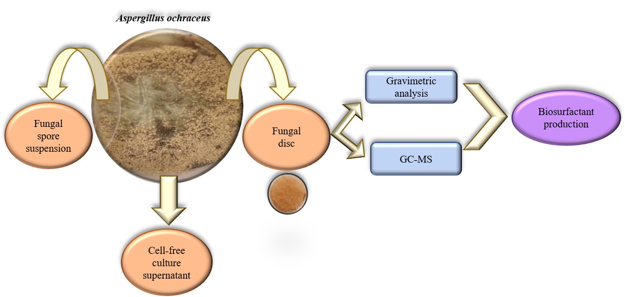
Fungal spore suspension
The fungal strains were inoculated on PDA (potato dextrose agar) (Merck) and incubation was performed at 30 ℃ in static condition for a week. Following the fungal cultures were suspended in 0.9% NaCl (pH 7.0) solution, and the spores were counted on a Thoma slide (Asemoloye et al. 2020).
Fungal disc
The fungal strains were inoculated on PDA and incubation was performed at 30 ℃ in static condition for a week. Following the incubation, 7 mm diameter of fungal discs were cut from the surface of an actively growing fungi on PDA (Benguenab and Chibani 2020).
Fungal pellet
The fungal strains were inoculated into PDB (Potato dextrose broth) (Merck) and incubation was performed at 30 ℃ in a rotatory incubator for a week (Mipro MCI, Turkey). Then, cultures were filtered using Whatman No:1 filter paper under sterile conditions. Obtained pellets were dried at 30 °C under steril conditions (Tugrul Yucel 2018).
Cell-free culture supernatant
The fungal strain was inoculated into Bushnell Haas (BH) (Sigma-Aldrich) medium containing 1% (v/v) of petroleum and incubation was performed at 30 °C and 150 rpm for a week (Mipro MCI, Turkey) Following this, the culture was filtered using Whatman No:1 filter paper under sterile conditions.
The petroleum biodegradation assay
The BH medium (g/L: 0.2 MgSO4, 0.02 CaCl2, 1 KH2PO4, 1 K2HPO4, 1 NH4NO3, 0.05 FeCl3) containing 1% (v/v) of Triton X:100, 0.1% (w/v) of glucose, 0.1% (w/v) of yeast extract and 0.1% (v/v) of trace element in 50 mL was sterilized at 121 ℃ for 15 minutes. Following the cooling of BH medium to 45 °C, 1% (v/v) of petroleum sterilized with 0.22 μm cellulose acetate syringe filter (Millipore, Sartorius) was added. 5% (v/v) of fungal spore suspension (1.5 × 10⁷ CFU mL-1), 1 g/100 mL of live biomass and 5% (v/v) of sterile culture supernatant were inoculated into BH medium and incubation was performed at 30 °C and 150 rpm under dark condition for 7 days (Mipro MCI, Turkey). All experiments were performed in triplicate (Maddela et al. 2015; Bilen Ozyurek and Avcioglu 2020a).
Colorimetric method
The redox solution was prepared by dissolving 1 g (w/v) of 2,6-dichlorophenol indophenol (DCPIP) in 1 L of distilled water. Following the inoculation of the fungal spore suspensions into BH medium containing 1% (v/v) of petroleum and 1% of redox indicator, the cultures were incubated at 30 °C and 150 rpm under dark condition for 7 days (Benguenab and Chibani 2020). Accordingly, the potent fungal strain in petroleum degradation will be determined with this method. The change of the DCPIP redox indicator from blue (oxidized form) to colorless (reduced form) indicates that the fungi are effective in hydrocarbon degradation (Lima Souza et al. 2016).
Gravimetric method
Following the incubation period, extraction was carried out with dichloromethane (DCM) (CH2Cl2) (1:2) (Sigma-Aldrich). The flasks containing petroleum + DCM were left in the water bath (Memmert, Schwabach, Germany) at 90 °C for 1 hour and DCM was removed. The degradation of petroleum was also calculated as:
D (%) = (p0 – p1 –p2)/p0 × 100
where p0 and p1 show the initial and remaining concentrations of petroleum at different incubation periods, p2 indicates the abiotic loss (Barnes et al. 2018; Benguenab and Chibani 2020).
Petroleum degradation kinetics
The degradation data applied to first order kinetic model according to Maletić et al (2009):
lnct = lnco - kt,
where ct is the residual petroleum concentration at time; co is the initial petroleum concentration; k is the first-order kinetic degradation constant (day-1), and t is time (day). The half-life period of petroleum was calculated as follows:
t1/2 = ln2/k
Investigation of optimal physiological conditions on petroleum degradation with fungal spore suspension
To optimize physicological conditions, initial pH (3.0 - 8.0), petroleum concentration (0.5% - 4%), inoculum concentration (2.5% - 10%) (v/v) and incubation period (7, 14, 21 and 28 days) parameters were investigated. The degradation of petroleum was obtained by gravimetric analysis.
Investigation of optimal physiological conditions on petroleum degradation with fungal live biomass
By comparing the efficiencies of fungal pellets and fungal discs in petroleum biodegradation, the most effective fungal live biomass was determined and used in further optimization studies. Accordingly, initial pH (3.0 - 8.0), petroleum concentration (0.5% - 5%), inoculum amount (0.5 - 2.5%) (g/100mL) and incubation period (7, 14, 21 and 28 days) parameters were investigated. The degradation of petroleum was obtained by gravimetric analysis.
GC/MS analysis
The petroleum was extracted with DCM from the culture with the highest degradation ratio under optimized conditions. The analysis was carried out by the Petroleum Research Center at Middle East Technical University (Turkey) using TRB-1 GCMS-QP-2020 (Shimadzu, Tokyo, Japan) to obtain the removal of n-alkane fractions in petroleum with fungal disc of potent strain. The procedure was performed according to Bilen Ozyurek and Seyis Bilkay (2020b).
Screening assay for biosurfactant production
The assay was aerobically carried out with 50 mL of sterile BH medium supplemented with 1% (v/v) of petroleum and 1% (w/v) yeast extract (Merck). 1 g (w/100mL) of live biomass was inoculated into BH medium and incubation was performed at 30 °C and 150 rpm for a week. Following the incubation period, the whole broth was centrifuged at 4650 ×g for 10 min (Eppendorf 5810R) and at 10752 ×g for 10 min (Eppendorf 5417C, Sigma-Aldrich, USA). Then, the supernatant was also filtered with 0.45µm pore size filter paper (Millipore, Sigma-Aldrich). The cell-free culture supernatants were transferred to a clean test tube and used for biosurfactant screening assays (Parthipan et al. 2017).
Drop-collapse method
The drop-collapse method was carried out as described by Bodour et al. (1998). 5µL of petroleum was added into 96-microwell plate and left in room temperature for 2 h. 5 μL of cell-free culture supernatant was added on petroleum in 96-microwell plate. The change in the drop size was observed after 1 min. Deionized water and Triton X:100 (a chemical surfactant) was used as negative and positive controls, respectively (Parthipan et al. 2017).
Oil-spreading method
The oil spreading method was carried out as described previously Hassanshahian (2014). 50 mL of distilled water was added into petri plate followed by addition 20 µL of petroleum. Then, 10 µL of culture supernatant was added on petroleum-coated water surface. The diameter of the clear zone on petroleum surface was measured after 30 s. Deionized water and Triton X:100 was used as negative and positive controls, respectively.