1.1 Materials
The female gametophyte BAC library of S. japonica was constructed and preserved in our laboratory. It contains 31872 BAC monoclones with an average length of 115 kb, covering 6.57 times of the whole genome of S. japonica gametophyte.
1.2 Methods
1.2.1 BAC Clone screening and sequencing
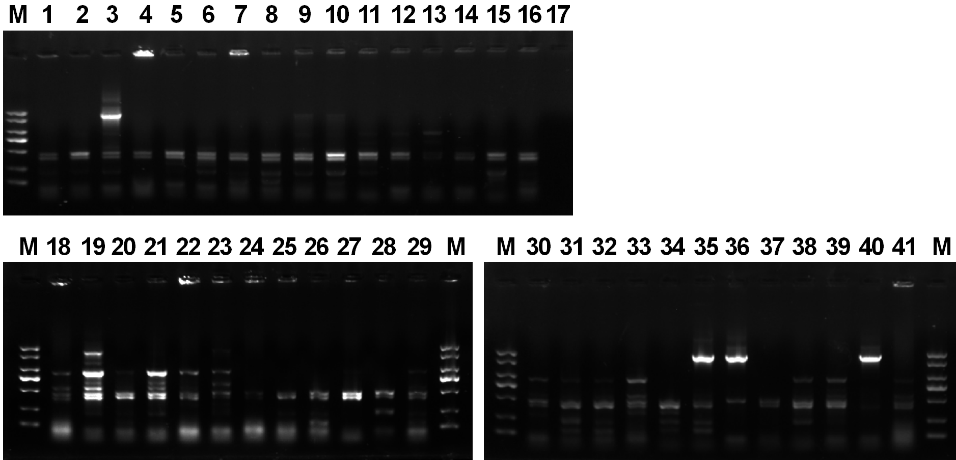
For screening BAC clones containing 45 S rDNA repeat units, three-dimensional PCR was conducted with primers designed with the 18S rRNA gene of S. japonica (GenBank accession number:EU293553) as template. The primers were 18F: 5'- tcggacggtttgtggtga-3' and 18R: 5'- ccttccttggatggtggtagcc-3'. 25µL PCR reaction system included 12.5µL 2 × dream Taq PCR mix [0.1 U/µL Taq polymerase, 500 µmol/µL dNTP, 20 mmol/L Tris-HCl (pH 8.3), 100 mmol / L KCl, 3 mmol/L MgCl2] (Thermo Fisher Scientific Inc., USA), 0.5 µL upstream and 0.5 µL downstream primers of 10 µmol/L, 1 µL bacterial solution as template DNA. The amplification procedure included 94℃ pre denaturation for 3 min, followed by 30 cycles of 94℃ denaturation for 45 s, 57℃ annealing for 45 s, 72℃ extension for 1.5 min, and 72℃ extension for 10 min. The products of PCR were detected by 1% agarose gel electrophoresis.
The high molecular weight BAC plasmid DNA of the screened clones was extracted witha DNA Extraction Kit (Qiagen, Germany).Pulsed field gel electrophoresis was performed for measure the size of the inserted DNA fragment after NotI digestion of the extracted plasmid. The inserted fragments were sequenced using the third generation of PacBio sequencing technology (Wuhan Eight Star Bio-tech Co., Ltd., Wuhan, China).
1.2.2 DNA Sequence analysis of BAC clone
The obtained sequence was pre-processed to remove cloning vector sequences from the reads using Vec Screen(https://www.ncbi.nlm.nih.gov/tools/vecscreen/) of NCBI. Sequence similarity analysis was conducted by using Blastn༈https://blast.ncbi.nlm.nih.gov/Blast.cgi༉. The Plant-CARE database (http://bioinformatics.psb.ugent.be/webtools/plantcare/html/) was employed to predict the conserved cis-element motifs presented in IGS. CpG islands in the IGS were predicted by using CpGPlot [17]. The physical map of the inserted fragment of the positive BAC clone (625-E18) was constructed according to the genome sequenceof S. japonicaby using IBS1.0 [18] and MapChart2.32 [19].
1.2.3 FISH on BAC clone molecule
The positive BAC clones were cultured in LB liquid medium containing 12.5 µg/mL chloramphenicol. The BAC plasmid DNA was extracted with plasmid DNA Extraction Kit (Qiagen, Germany). 5 µL BAC-DNA (about 50 ng) was diluted in 5 µL sterilized deionized water and then pipette onto a poly-L-lysine glass slide. A 18 mm⋅18 mm coverslip was used to spread the molecular to prevent molecular broken [20]. Slide was air-dried for 30 min at room temperature, and then the coverslip was washed off in water. After air dry, slide was fixed in the Carnot's fixative (Ethanol / glacial acetic, 3:1, v/v) [21] for 2 min, and dried at 60℃ for 30 min.
The 18S rDNA probes were labeled by nick-translation with Alex flow green-5-dUTP (PerkinElmer, Boston, USA). FISH was performed according to the procedure described by Liu et al. [22].Slide with BAC-DNA was cross-linked for 2–3 times in a ultraviolet cross linker, and then 8 µL of hybridization solution, including 6 µL of mixture (2×SSC + 1×TE buffer) and 2 µL of 18S rDNA probe, was pipette on the slide. After covering with a coverslip, the slide was water bath at 100℃ for 5 min. Then put it in an oven at 55℃ for hybridization overnight. After hybridization, the slide was washed with 2 × SSC at 42 ℃ for 10 min and then dried at 55 ℃ for 15 min. At last, 5 µL DAPI staining solution was added. After incubation for 10 min, the results were observed under a fluorescence microscope. LeicaDM4000 fluorescence microscope (Germany) and Orca ER camera were used to take pictures, and Adobe PhotoShop CS6.0 was used to process the pictures.