Cell culture and construction of SAH model
Rat PC12 cells were used in this study and cultured with RPMI-1640 medium supplemented with 10% heat-inactivated horse serum and 5% fetal bovine serum. Cells were treated with 10 mM oxygen hemoglobin (OxyHb) to construct SAH cell model. BAY11-7082 (5µmol/L; S2913, Selleck), the inhibitor of NLRP3, was dissolved in PBS and added into culture medium.
Quantitative real-time PCR
Total RNA was extracted using TRIzol Reagent (Invitrogen, USA). The cDNA synthesis kit (Thermo, USA) was used for cDNA synthesis. The cDNA was subjected to the following procedures: 95°C for 10 minutes followed by 40 cycles of 95°C for 15 seconds, 60°C for 45 seconds. GAPDH was used as the reference gene. Gene expression was calculated by the 2−ΔΔCt method. All data represented three replicates. All primers were listed as follows: APC, F: CACCCCTTTCAGTTCAAGTAGC, R: AAGACCCAGAATGGCGTTTAG; NLRP3, F: ACCTCAACAGACGCTACACCC, R: GCTGCTCCCTGGAACACC; GAPDH, F: CGGTCAGGTCATCACTATC, R: CAGGGCAGTAATCTCCTTC.
Western blot
Total protein of cells or brain tissues was obtained using RIPA lysis buffer with protease inhibitor Cocktail (MCE, China). The protein was separated on 10% SDS-PAGE and transferred to a nitrocellulose membrane. After the application of primary antibodies and secondary antibodies, the protein content was detected by ECL. The primary antibodies used in this study were provided as follows: APC (ab40778, abcam), NLRP3 (ab263899, abcam), active Caspase-1 (ab74279, abcam), GSDMD-N (#39754, CST), and β-actin (#3700, CST).
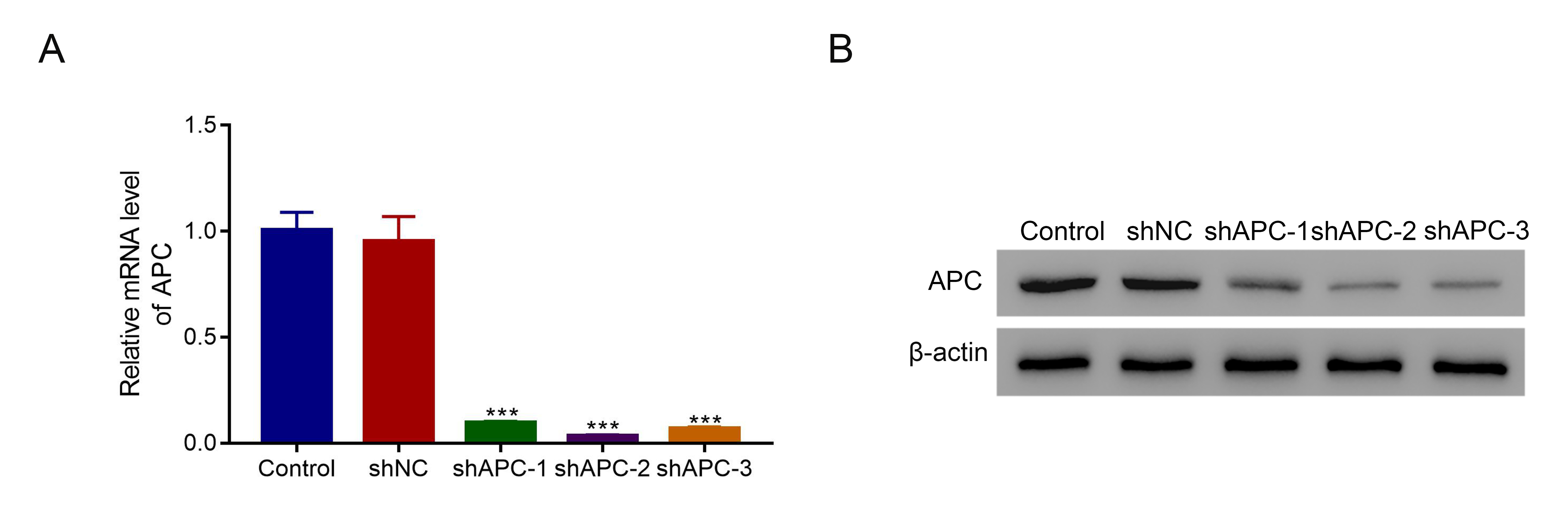
Knockdown of APC
Three short hairpin RNAs (shRNA) were designed targeting human APC (shAPC-1: GCAUGAAACUGCCUCCCAUTT; shAPC-2: GCAGGAAGCCCAUGAACAATT; shAPC-3: GCCACUGACAUAUCUUCAUTT. The negative control shRNA (shNC) was taken as the reference with the following sequence: UUCUCCGAACGUGUCACGUTT. In 48 hours prior to experiments, cells were transfected with shRNAs using Lipofectamine 2000 (Invitrogen).
ELISA
The levels of IL-1β and IL-18 were estimated using corresponding ELISA kits according to the protocol of the manufacturer. Briefly, IL-1β and IL-18 antibodies were applied at 37°C for 2 h. After the rinse of scrubbing solution, the secondary antibody was applied. Then, the stop solution was added and the optical density at 450 nm was estimated.
Estimation of cell pyroptosis
The activated caspase-1 was estimated to reflect the level of cell pyroptosis using FAM-FLICA Caspase-1 Assay Kit (ImmunoChemistry Technologies, USA). In brief, cells were incubated with caspase-1 detection probe for 1 h in the dark. After the rinse to remove unconjugated FLICA reagent, cells were stained with propidium iodide (PI) for 20 min. Flow cytometry was used to define pyroptosis cells as double positive for cleaved caspase-1 and PI.
Construction of SAH rat model
The SAH rat model was constructed by internal carotid artery puncture. After anesthesia, the external carotid artery was ligated and cut off. A nylon thread was put through the external carotid artery into the intracranial part of the internal carotid artery. When the branch met the resistance, advance the nylon thread to puncture the blood vessel of the internal carotid artery, which could lead to SAH in rats. Then, 2 mg/kg 3K3A-APC was intraperitoneally injected every 12 h. After the intervention for 24 h and 72 h, brain tissues were collected for pathological analysis. There were six rats at each time point in different groups.
Neurological function score
The scoring of neurological functions was estimated using modified Garcia scoring and balance beam test. Modified Garcia scoring was composed of spontaneous activity, the extension of four limbs, climbing on the metal mesh wall, and touch response and whisker response of both sides of the trunk. Each of the first three items was 0–3 points, and that of the latter three items was 1–3 points, and total scores were 3–18 points. Balance beam test was defined as rats walking on a wooden rod (20 cm wide), with 0–4 points.
SAH hemorrhage score
The scoring of SAH hemorrhage was estimated by modified Sugawara's scoring. The head of rats was decapitated under anesthesia, the basal cistern and Willis’ circle were photographed. The basal cistern was divided into 6 parts, and the scores for each part are as follows: 0 stood for no SAH; 1 was defined as a small amount of SAH; 2 was classified as medium SAH with several distinguishable blood vessels; 3 was defined as a large amount of SAH without distinct blood vessels. The final score was the sum of the score of six parts, among which 0–7, 8–12, and 13–18 points were defined as mild, moderate, and severe hemorrhage.
Statistical analysis
GraphPad Prism version 7.0 (CA, USA) was used for data analyses and visualization. Quantitative data were represented as mean ± standard deviation for more than three samples. The difference between groups was estimated by Student’s t-test or one-way ANOVA. The P-value < 0.05 indicated statistical significance.