The mRNA and Protein Expression Levels of FAM166B in Different Types of Normal and Tumor Tissues
To determine differences of FAM166B expression in tumor and normal tissues, the FAM166B mRNA or protein levels were analyzed by using multiple online databases. Firstly, to evaluate FAM166B expression in human tissues, we examined FAM166B expression via the TIMER website (Fig. 1A). We analyzed the FAM166B expression profile across diverse tumors in TCGA database and found that FAM166B had a significantly lower expression relative to the corresponding normal samples (Fig. 1B). Analysis of both TCGA and GTEx datasets indicated significantly lower levels of FAM166B in 9 tumor types compared to the normal specimens (Fig. 1C). The analysis revealed that the FAM166B expression was at a significantly lower level in BRCA (Breast Cancer), KICH (Kidney Chromophobe), HNSC (Head and Neck squamous cell carcinoma), KIRP(Kidney renal papillary cell carcinoma), LIHC(Liver hepatocellular carcinoma), LUAD (Lung Adenocarcinoma), LUSC(Lung squamous cell carcinoma), PRAD(Prostate adenocarcinoma) and in KIRC ( Kidney Renal Clear Cell Carcinoma), compared to adjacent normal tissues, which implied that FAM166B could play a role in cancer suppressing. To further evaluate FAM166B mRNA levels differential expression in human cancers, we analyzed it by using the Oncomine database. This analysis revealed that the FAM166B expression was higher in Brain and CNS Cancer, Colorectal Cancer, Kidney Cancer, and Prostate cancer. However, lower expression was observed in bladder, breast, head and neck cancer, lung cancer, lymphoma, and melanoma (Fig. 1D). The fact that FAM166B shows a significantly lower expression in 2 breast cancer databases caught our attention (Fig. 1E), thus in the following studies we focused on the correlation between FAM166B and breast cancer.
Prognostic Potential of FAM166B in Breast Cancer
To further dig out the impact of FAM166B expression on BRCA, we stratified all BRCA patients in TCGA database into the FAM166Bhigh and FAM166Blow groups and found that increased FAM166B expression was correlated to longer OS in BRCA patients (Fig. 2E). By using the Kaplan-Meier plotter database, we then divided breast cancer patients into different subgroups, trying to get a more specific prognostic potential of FAM166B, shown in Fig. 2A-D, which indicated that a high FAM166B expression leads to a significantly better prognosis in luminal A breast cancer patients. Next, we performed univariate and multivariate Cox regression analyses on the TCGA dataset to discover the prognostic value of FAM166B expression and other clinical variables in BRCA. Univariate Cox regression analysis indicated that FAM166B expression, age, stage, tumor size, lymph nodes, metastasis were significantly correlated with the OS of BRCA patients, whereas gender did not reveal any correlation (Fig. 3A). By multivariate analysis, only two parameters were further identified as independent prognostic factors including FAM166B expression [P < 0.001, hazard ratio (HR) = 0.48] and age [P < 0.001, hazard ratio (HR) = 1.04] (Fig. 3B). We also investigated how FAM166B was involved with the prognosis of various stages of breast cancer and with the lymph nodes status, the results turned out to be insignificant (data not shown). Based on the results above, we drew the conclusion that FAM166B could be an independent factor to predict Breast Cancer patients’ prognosis and an elevated expression of FAM166B indicates a better prognosis.
GSEA identifies a FAM166B-related signaling pathway
In order to better understand how FAM166B is involved in the BRCA pathogenesis, we conducted Gene Set Enrichment Analysis (GSEA) with KEGG analysis (based on absolute value of Spearman Score) to identify the signaling pathways activated between high and low FAM166B expression data sets. As we considered that the absolute value of NES > 1.5 and P value < 0.05 were with statistical significance, four significantly enriched biological pathways caught our attention which were fructose and mannose metabolism pathway, sugar and nucleotide sugar metabolism pathway, pentose phosphate pathway and glycolysis gluconeogenesis pathway (Table 1; Fig. 4). The above biological pathways are closely related with glucose converting and cell metabolisms. Therefore, it is a reasonable deduction that FAM166B could play a role in reducing tumor progression by restraining cell metabolism.
FAM166B Expression Is Correlated with Immune Infiltration Level in Breast Cancers
According to previous studies, tumor-infiltrating lymphocytes could be an independent predictive factor of sentinel lymph node status and survival in cancers [15]. Hence, we explored whether FAM166B expression was correlated with immune infiltration levels in multiple cancers. Tumour mutation burden (TMB) and microsatellite instability (MSI) are sound biomarkers for immune therapy response in various kinds of tumor[16]. We analyzed the correlation between FAM166B and TMB. The results revealed that a significant association between targeted gene and TMB in BRCA (p = 8.6e-05), shown in Fig. 5A, while FAM166B has limited influence on MSI with the P value of 0.4 (Fig. 5B). Furthermore, we investigated if FAM166B expression was correlated with immune infiltration levels in different breast cancer subtypes from TIMER. Tumor purity is an essential factor that affects the analysis of immune infiltration in tumor samples, hence we selected the subtypes of breast cancer in which FAM166B expression levels have an evidential correlation with tumor purity in TIMER(Figure 5C). We found that FAM166B expression level correlates with favorable prognosis and high immune infiltration in luminal and in basal breast cancers. FAM166B expression level is significantly correlated with infiltrating levels of B cells (P = 0.0166), CD4 + T cells (P = 0.0274), macrophages (P = 0.0007) in BRCA-Basal sub-group. Similarly, there were significant correlations with tumor purity (P = 0.0004) and with infiltrating levels of CD4 + T cells (P = 0.0185) in BRCA-Luminal. These findings suggest that FAM166B plays a role in immune infiltration in luminal and basal breast cancers.
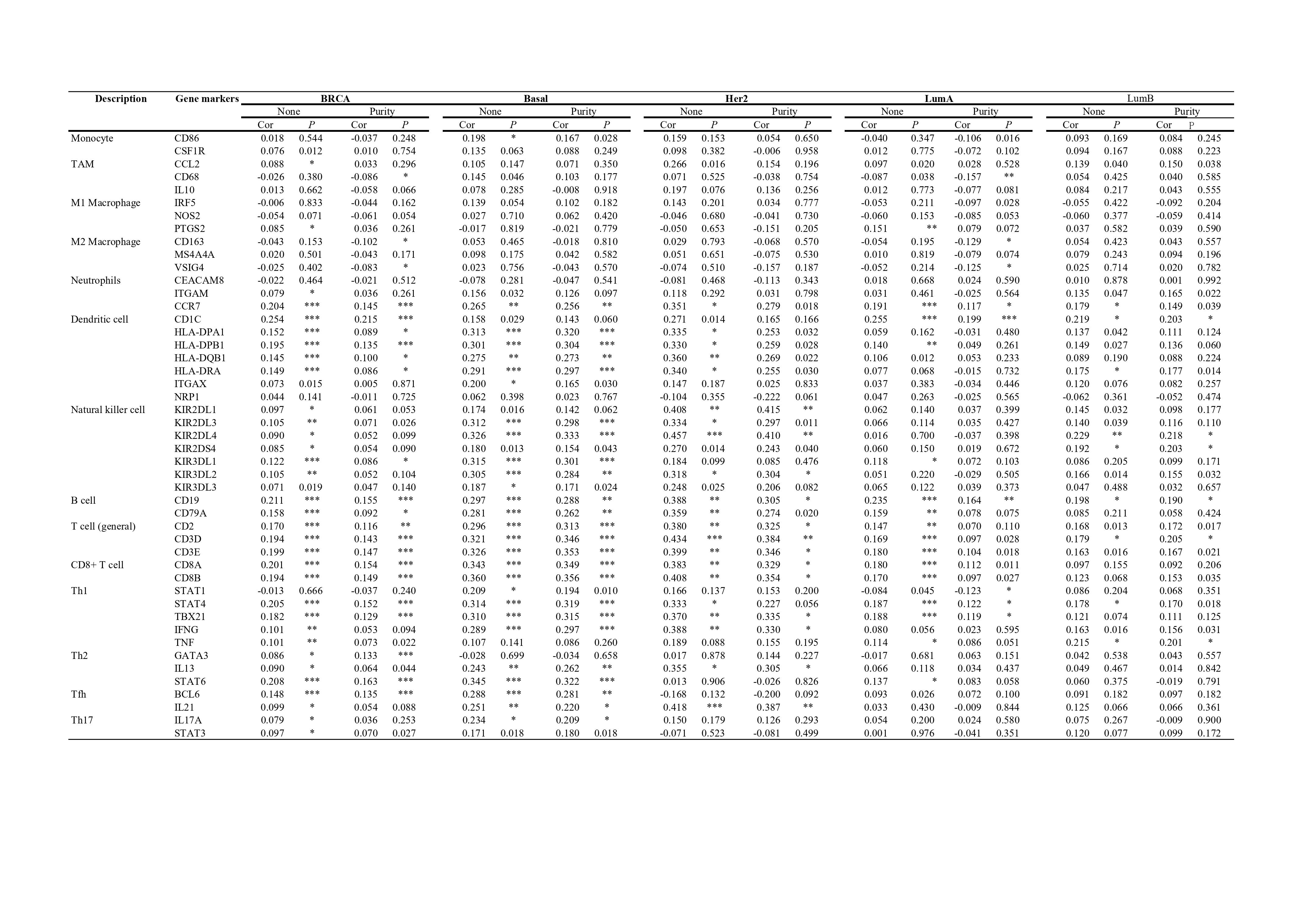
To further investigate how FAM166B is related to the diverse immune infiltrating cells, we used TIMER to research the correlations between FAM166B and immune marker sets of different immune cells in breast cancer. The results were shown in Table 2. We found that the expression levels of most marker set of CD8 + T cells, B cells, T cells, and dendritic cells have positive correlations with FAM166B expression while M2 macrophages have a negative correlation with FAM166B expression in BRCA. Specifically, we found out that CCR7 of neutrophils, STAT4, TBX21 of Th1 phenotype, STAT6 of Th2 phenotype, BCL6 of Tfh are significantly correlated with FAM166B expression in BRCA (P < 0.0001). We further analyzed the correlation between FAM166B expression and the above markers between different subtypes of breast cancer, including luminal, basal, and Her-2 subtypes. Correlation results between FAM166B and markers of B cells, T cells, CD8 + T cells, and Th1 cells are accordant to those in BRCA. These findings suggest that FAM166B may regulate macrophage polarization and T cells in breast cancer. Based on TIMER, the results showed that high FAM166B expression relates to the high infiltration level of DCs in all types of breast cancer, DC markers such as HLA-DPB1, HLA-DQB1, HLA-DRA, HLA-DPA1 also show significant correlations with FAM166B expression. Moreover, FAM166B expression has a strong correlation with the high infiltration level of natural killer cells, NK markers like KIR2DL1, KIR2DL3, KIR2DL4, KIR3DL1, KIR3DL2, KIR3DL3, KIR2DS4 are positively related to FAM166B expression.