The TREM2 protein has been partly crystallised from a mammalian cell system (10, 11), this structure contains the protein’s extracellular ligand binding domain (ECD) which is amino acids 19-134. This structure was used as the basis for molecular dynamic modelling experiments in this study. The TREM2 ECD domain is a V-type Ig domain which contains nine ß-strands and two short α-helices, all of which are characteristic of an Ig protein domain. Both variants, R47H and R62H, can be found on the TREM2 protein surface and the surface of this Ig domain, on one of the ß-strands.
Predicted mutational effects;
The Have your Protein Explained server (HoPE) was run to predict the possible mutational effects of both variants on the protein (16). Results from the server suggest that the wildtype amino acid (arginine) at position 47 forms a hydrogen bond with amino acids at positions 66 (threonine) and 67 (histidine) which would not occur with the histidine variant in this position. These bonds may be important for protein structural integrity. The wildtype residue is conserved at position 47, though a histidine is observed here in some species brought up in the blast search. Residue 62 on the other hand is less well conserved, but histidine is not observed here in any species searched. There is an obvious loss of charge and size with both variant amino acids, shown schematically in figure 1. The SIFT online tool was used to predict the tolerance of the two variants in the protein, this does not predict the effect of binding, or function, but whether the variants will be tolerated in the protein structure. Results from this show R47H would be tolerated with a score of 0.06 and R62H to be tolerated with a score of 0.10, this was based on 13 sequences. A score of <0.05 would be classed as a damaging prediction (17). The I-mutant server results showed a decrease in stability for both the R47H and R62H variants (18).
Molecular Dynamic Simulations;
Structures containing the variants were constructed as detailed in the methods section and along with the original structure, each was run in triplicate, for 300n.s. The stability of the simulations was checked, and volume, pressure and root mean square deviation (RMSD) remained stable throughout.
Local structural changes;
Local structural changes were investigated in the three models. The region surrounding both variants, amino acids 43-65, were viewed using VMD, figure 2 shows the starting structure of the wild type (WT) simulation with CDR loops and variant amino acids highlighted. The R62H variant alters this local structure with a shift in the beta sheet and a large movement of the random coil (figure 3c). However, the R47H variant does not appear to alter the structure surrounding the amino acid in any way. As well as altering the local structure the flexibility of the individual residue, i.e. the amount of movement it has, was also altered for the R62H variant. This flexibility was measured using the gromacs rmsd module. Results show the WT and R47H to have an average flexibility of 0.23 +/- 0.02 and 0.01 respectively at amino acid 62, the R62H variant on the other hand has a reduced flexibility of 0.17 +/- 0.01 (figure 3a). There is also, to a lesser extent, a reduction of flexibility across neighbouring amino acids which surround the R62H variants. A further change to the local structure seen in the MD simulations is a change in positioning of the wildtype to variant amino acid, the wildtype pertruding from the molecule in both cases and the mutated amino acid being visually far more buried within the structure, figure 3 (d-g).
Wider structural changes;
Solvent accessible surface area (SASA) for the whole model, and the individual mutated residues were measured. Overall SASA was reduced from 71 to 70, this small change is not significant and would not have any effect on the protein function. Amino acid specific SASA was measured for the WT and mutated proteins, at the 47 and 62 sites. Here a SASA change can be seen at the mutated site in each protein, with a reduction of SASA, figure 3b. This correlates with the observed positional change of the amino acid from one protruding from the structure to a more buried position.
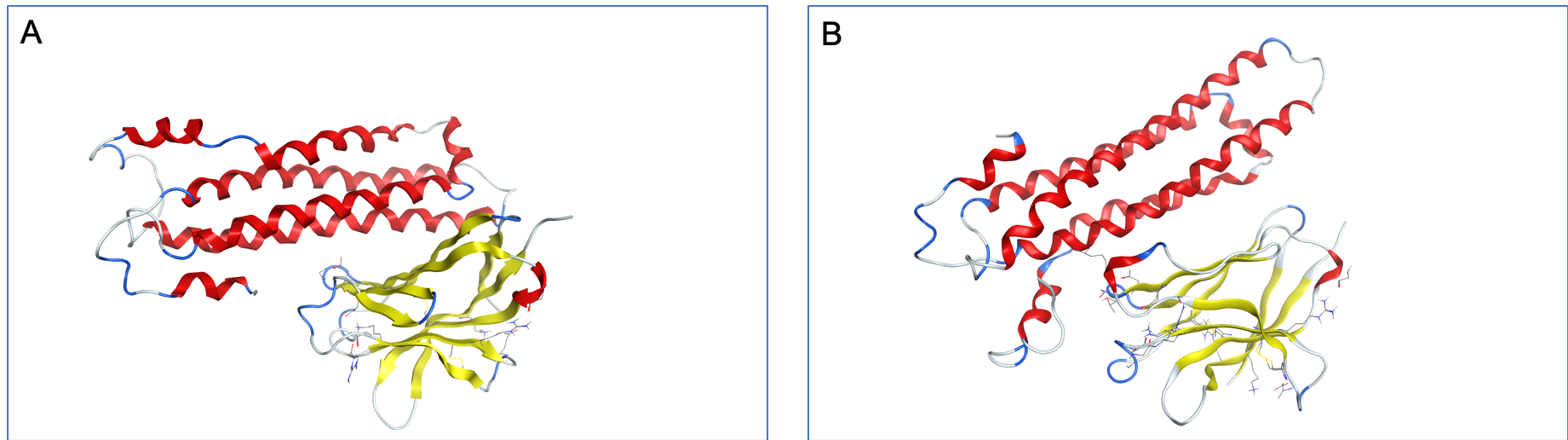
Structural alteration can be seen in the CDR2 loop, figure 4 shows the R47H variant to cause a loss of beta sheet and a changing of alpha helix position in this binding loop. The effect of the R62H variant is subtler, where there is a change in loop structure to the left and right of the alpha helix. There is a further effect on the flexibility of the loop, figure 4d, here the variants differ with the R47H becoming more flexible and the R62H variant less flexible, when compared to the wild type simulation. All three images are taken from the representative structures created from the entire simulations and show the loop in the same position and therefore should all be identical if no structural change was observed.
Resulting structures from the MD simulations were taking forward for an in-silico docking study. APOE crystal structure (PDB ID: 2KC3) was docked with TREM2 WT and variant containing protein structures which were generated from each of the MD simulations. First a protein-protein docking simulation was run using the CDR2 loop as the binding region. The top hit from this was taken forward into a general dock to assess the overall score of the binding relationship and the combined complex’s energy. The docked complex can be found in supplementary figure 1. The WT protein, APOE complex had an ‘S-score’ of 18.77 with the R47H variant and APOE complex exhibiting a very high ‘S-score’ of 6288.41.