Periodontal bacteria and polymicrobial inoculum
In this study, the following periodontal pathogens were tested, P. gingivalis FDC 381, T. denticola ATCC 35405, T. forsythia ATCC 43037, and F. nucleatum ATCC 10953. They were cultured anaerobically (85% N2, 10% H2, 5% CO2) at 37oC in an anaerobic chamber as described previously[12,83]. P. gingivalis and F. nucleatum were grown for 3 days on Tryptic Soy Broth (Becton Dickinson, Franklin Lakes, NJ) containing 5 mg/ml yeast extract, 0.5mg/ml L-cysteine hydrochloride, 5 μg/ml hemin, 1μg/ml menadione and 5% FBS (Gibco Thermo Fisher Scientific, Waltham, MA). T. denticola was cultured in Oral Treponeme Enrichment Broth (OTEB) media (Anaerobe systems, Morgan Hill, CA) for 5 days. T. forsythia was grown in Tryptic Soy Broth containing 5 mg/ml yeast extract, 0.5mg/ml L-cysteine hydrochloride, 5 μg/ml hemin, 1μg/ml menadione, 10 μg/ml N-acetylmuramic acid (Sigma-Aldrich, St. Louis, MO), and 5% FBS (Gibco) for 7 days. Bacterial concentration was determined quantitatively and each organism was resuspended in phosphate-buffered saline (PBS) at 1 × 10¹⁰ bacteria per ml for experimentation.
For the oral polymicrobial infection, P. gingivalis was mixed with an equal volume of T. denticola for 5 min. Subsequently, T. forsythia was added to the culture tubes containing P. gingivalis and T. denticola, and the bacteria were mixed gently for 1 min and allowed to interact for an additional 5 min. Lastly, F. nucleatum was added and mixed well with P. gingivalis, T. denticola, and T. forsythia. After 5 min, the four bacterial consortium was mixed thoroughly with an equal volume of sterile 4% (w/v) carboxymethyl cellulose (CMC; Sigma-Aldrich) in PBS, and this mixture was used for the oral gavage[12,83].
Lactococcus lactis growth conditions
Two L. lactis strains were used in this study; nisin-producing L. lactis (ATCC 11454) was obtained from ATCC and non-nisin producing L. lactis (NZ9800) was kindly provided by Dr. Paul Cotter, Head of the Food Biosciences Department in the Teagasc Food Research Center, Cork Institute of Technology, Ireland. L. lactis strains were grown in Brain Heart Infusion (BHI, Sigma-Aldrich) media overnight in a 37oC shaking incubator. The L. lactis strains were then pelleted by centrifugation, resuspended in PBS to a concentration of 1 ×10¹⁰ CFU/ ml, and mixed with an equal volume of sterile 4% CMC. This mixture was used for oral inoculation .
Nisin preparation
An ultra-pure (>95%) food grade form of nisin Z ( NisinZ® P) also referred to as nisin ZP was purchased from Handary (S.A., Brussels, Belgium), a primary manufacturer of nisin in the food industry. From here forward, nisin ZP will be referred to as nisin. The stock solution was prepared at a concentration of 600 or 200 μg/ml in sterile water, filter sterilized, and stored at 4◦C for a maximum of 5 days for use in experiments. For oral treatment of mice, the nisin solution was mixed with an equal volume of sterile 4% CMC to reach the final concentration (300 or 100μg/ml).
Infection and treatment of mice
A total of 60 eight-week old BALB/cByJ female mice (The Jackson Laboratories, Bar Harbor, ME) were housed in microisolator plastic cages and randomly distributed into 10 groups (6 mice per group). The description of the experimental groups and infection and treatment protocols are shown in (Figure 1A and B). The experimental protocols were approved by the Institutional Animal Care and Use Committee of the University of California, San Francisco (IACUC APPROVAL NUMBER: AN171564-01B). All the mice were given trimethoprim (0.17 mg per ml) and sulfamethoxazole (0.87 mg per mL) daily for 7 days in the drinking water and their oral cavity was rinsed with 0.12% chlorhexidine gluconate (Peridex) mouth rinse to inhibit the native oral microbiota[12,14]. The polymicrobial inoculum (5×10⁹ combined bacteria per ml; 1×10⁹ cells in 0.2 ml per mouse; 2.5×10⁸ P. gingivalis, 2.5×10⁸ T. denticola, 2.5×10⁸ T. forsythia and 2.5×10⁸ F. nucleatum) was administered topically in the morning for 4 consecutive days every week for a total of 8 weeks. Nisin (100 or 300 μg/ml, 0.2 ml per mouse) and L. lactis (5×10⁹ bacteria per ml; 1×10⁹ cells in 0.2 ml per mouse) were administered every day in the evening every week for a total of 8 weeks. A sterile 2% CMC solution was administered as the control treatment.
Following 8 weeks of polymicrobial infection, oral swab samples were collected to evaluate the microbial status and to examine the effect of nisin on periodontal pathogens. The samples were collected from the oral cavity of the mice using a sterile micro sized cotton swab. The teeth and surrounding gingival tissue were swabbed and the cotton tip was immersed in 10:1 Tris-EDTA buffer immediately and stored at -80°C until further processing for DNA isolation. Then mice were euthanized and the blood was collected for analysis of antibody response to the periodontal pathogens. The maxillae and mandibles were resected from each mouse for morphometric, histologic, immunologic, and sequencing analysis.
DNA isolation from oral swabs, ethanol precipitation, and real time PCR to confirm bacterial infection
DNA isolated from oral swabs was used to evaluate and confirm infection in the mice using methods described in our previous study[12]. In brief, DNA was isolated from the swabs and purified using the QIAamp® DNA Mini kit (Qiagen, Germantown, MD, USA). Ethanol precipitation of DNA was then performed to prepare the samples for subsequent real time polymerase chain reaction (PCR). Lastly, standard real-time PCR was used to quantify the periodontal pathogens in the oral swab samples.
Morphometric analysis of periodontal alveolar bone loss
After autoclaving and de-fleshing to remove all the soft tissues, the left maxillae and mandible from each mouse were immersed in 3% hydrogen peroxide overnight and then stained with 1% methylene blue. Digital images of both buccal and lingual/palatal root surfaces of all molar teeth were captured under a stereo dissecting microscope (SMZ1000, Nikon) at the magnification shown in the images, then the line tool of Image J software (NIH Image) was used to measure the alveolar bone loss from the cementoenamel junction (CEJ) to alveolar bone crest (ABC). For bone loss measurements, the distance between CEJ and ABC were measured from a total of 28 sites on the buccal and lingual/palatal surfaces of the molars (3 sites on the first molar, 2 sites on the second molar, and another 2 sites on the third molar)[12,84,85]. Two blinded examiners (experienced periodontists) performed all measurements twice at separate times. Both horizontal bone loss and intrabony defects were detected under the stereo dissecting microscope. The intrabony defects were marked as present or absent[12,14].
Histopathological evaluation of periodontal inflammation and cellular content
The right maxilla was resected from each mouse and immediately fixed in 4% paraformaldehyde for 24h, then decalcified with diethyl pyrocarbonate-treated 0.5M ethylenediaminetetraacetic acid (pH 8) for 28 days at room temperature. The decalcified specimens were then dehydrated and embedded in paraffin using a fully-enclosed tissue processor (ASP300S, Leica Biosystems, Buffalo Grove, IL, USA). The tissue blocks were cut into serial sections (4 μm) parallel to the mesiodistal plane using a microtome, then sections were stained with Mayer’s hematoxylin (Sigma-Aldrich, St. Louis, MO, USA) and eosin Y solution (Sigma-Aldrich) for assessment of inflammation. The sections were examined with a stereomicroscope.
The number of inflammatory cells (round-shaped nuclei) and gingival fibroblast (spindle-shaped nuclei) within a square field (100 × 100 μm) in connective tissue adjacent to the gingival epithelium between first and second molars were morphologically evaluated and counted in three tissue sections per mouse specimen (n = 3 per group). Similarly, the number of periodontal ligament (PDL) cells (spindle-shaped nuclei in the PDL space) and alveolar bone lining cells (cell nuclei on bone surface) were counted. All cell counts were averaged for each group, and data were expressed as the mean number of cells per 1.0 mm2 of connective tissue in the maxillary specimens.
PCR evaluation of immune cytokine profiles from gingival tissues
The gingival tissue was treated overnight at 4 ºC with RNA stabilization solution (RNAlater, Invitrogen) after tissue harvesting. Samples were powdered with a mortar and pestle under continuous liquid nitrogen, and total RNA was then isolated from each sample using the RNeasy mini Kit (QIAGEN). The purity and quantity of the RNA were evaluated using the NanoVue Plus spectrophotometer (Biochrom Ltd.). Subsequently, total RNA was synthesized into cDNA using the SuperScript VILO Master Mix (11755050; Invitrogen).
To assess the immune cytokine profiles in gingival tissues, relative gene expression was evaluated by real-time PCR as in our previous study[86] using the following TaqMan primers and probes (TaqMan Gene Expression Assays; Applied Biosystems): interleukin-1β (IL-1β; Mm00434228_m1), IL-6 (Mm00446190_m1), tumor necrosis factor-α (TNF-α; Mm00443258_m1), interferon gamma (IFN-γ; Mm01168134_m1), C-C Motif Chemokine Ligand 2 (CCL2; Mm00441242_m1), C-X-C Motif Chemokine Ligand 2 (CXCL2; Mm00436450_m1), and transforming growth factor beta 1 (TGF-β1; Mm01178820_m1). Glyceraldehyde 3-phosphate dehydrogenase (GAPDH; Mm99999915_g1) was used as a housekeeping gene to normalize the amount of mRNA present in each reaction. PCR was performed in 20 μl reaction mixtures containing the TaqMan Fast Advanced Master Mix, cDNA template (20 ng/well), primers, and probes using a QuantStudio 3 Real Time PCR system (Thermo Fisher Scientific). The optimized thermal cycling conditions were as follows: 20 min at 95°C, followed by 40 cycles per 1 min at 95°C, and 20 min at 60°C. To compare the expression levels among different samples, the relative expression level of the genes was calculated by the comparative CT (ΔΔCT) method using QuantStudioTM Design & Analysis Software.
Serum antibody analysis
Serum from all 60 mice was collected on the day of euthanasia and used to determine the host response in the form of immunoglobulins (IgG) against P. gingivalis, T. denticola, T. forsythia, and F. nucleatum by an enzyme-linked immunosorbent assay (ELISA)[12,14]. For positive controls for the ELISA, each of these pathogens was grown to a cell density of 7×10⁸ cells/mL and harvested by centrifugation (7000 x g, 30 min, 4 °C). After washing, the cells were treated overnight with 0.5% formalin in buffered saline, then diluted to 0.3 (OD600nm) in 0.05 M carbonate‐bicarbonate buffer and used as the coating antigen. The amounts of specific IgG antibodies present were determined by using a mouse IgG ELISA quantification kit (Bethyl Laboratories, USA). A pilot test was first performed with diluted (1:100) mouse serum to confirm reactivity with the bacterial antigens after 1 h at room temperature. Then, after coating with the formalin-fixed bacterial cells, wells were incubated with diluted mouse serum (1:100) for 1 h at room temperature. Wells were then washed with PBS containing 0.05% Tween-20 (PBST), and alkaline phosphatase-conjugated goat anti-mouse IgG and the 3,3′,5,5′-tetramethylbenzidine chromogenic substrate reagent were added for detection and measurement of antibody response. Samples were assayed in duplicate and the purified mouse IgG was used to establish a standard curve on each plate for specific IgG quantification.
DNA isolation from gingival tissue for next generation shotgun sequencing
DNA was extracted from the mandibular gingival tissue of all mice (6 mice/group) using the QIAamp® DNA Mini kit (Qiagen, Germantown, MD, USA) as follow. The gingival tissue was ground in liquid nitrogen with a mortar and pestle and 180 μl of Buffer ATL was mixed with 25 mg of tissue powder by vortexing. Then, 20 μl of QIAGEN proteinase K was applied to each sample and samples were incubated at 56°C for 3 hours in a shaking water bath. Subsequently, 20 μl of the RNase reagent (20 mg/ml) was added to the samples followed by incubation for 2 min at room temperature. After adding 200 μl of Buffer AL, the samples were incubated at 70°C for 10 min. In addition, 200 μl of pure ethanol was mixed with each sample. This entire mixture was then applied into the QIAamp Mini spin column and centrifuged at 6000 x g for 1 min. Next, 500 μl of Buffer AW1 were added to the spin column and samples centrifuged at 6000 x g for 1 min. Then, 500 μl of Buffer AW2 was added and samples were centrifuged at full speed (20,000 x g) for 3 min, followed by centrifugation (20,000 x g) for 1 min again to eliminate the chance of possible Buffer AW2 carryover. Lastly, samples were incubated with 200 μl of Buffer AE in the spin column, which was placed in a clean 1.5 ml microcentrifuge tube at room temperature for 5 min, then DNA were eluted by centrifugation at 6000 x g for 1 min.
The purity and quantity of the DNA were evaluated using the NanoDrop™ OneC Microvolume UV-Vis Spectrophotometer (Thermo Scientific), which met quality control measures for subsequent shotgun sequencing analysis.
Metagenome shotgun sequencing and microbiome data production and analyses
Shotgun metagenomic sequencing library preparation was performed by Novogen, Inc. The libraries were prepared according to a standard protocol from Illumina, and at least 1 Gb of 150 bp pair-end reads per sample were sequenced on the Illumina Hiseq4000 machines. FASTQ files were generated from the sequencing machines and used for the analyses of the bacteriome/microbiome and virome as described below.
Data Processing
The following criteria were used for processing and cleaning up the raw data. Low quality bases (Q-value ≤ 38), which exceeded a certain threshold (40 bp by default) were trimmed. Reads which contained N nucleotides over a certain threshold (10 bp by default) were trimmed. Reads which overlapped with adapter over a certain threshold (15 bp by default) were trimmed.
Metagenome assembly
We utilized de novo assembly for each sample as follows. Samples passing quality control were assembled initially using SOAPdenovo (http://soap.genomics.org.cn/soapdenovo.html). The Scaffolds were cut off at “N” to get fragments without “N”, called Scaftigs. Clean data for all samples were then mapped to assembled Scaftigs using SoapAligner (http://soap.genomics.org.cn/soapaligner.html) and unutilized paired-end reads were collected. Mixed assembly was conducted on the unutilized reads with the same assembly parameter. The scaftigs of each sample and mixed assembly, which were less than 500 bp, were trimmed.
Taxonomy annotation
The following taxonomy annotation scheme was used. We aligned unigenes to the NCBI nonredundant database with DIAMOND to taxonomically annotate each metagenomic homolog (MEGAN). According to the abundance table of each taxonomic level, various analyses were performed using custom scripts by R and Python.
Statistical analysis
SPSS 21.0 statistical software (IBM, Chicago, IL, USA) was used for statistical analysis of the non-sequencing data. Student’s t-test was used to compare two independent groups. For comparison of intrabony defects, data were expressed as frequency and percentage, and a chi-square test was used for analysis. Further, analyses of the PCR data from the oral swabs and quantification of inflammatory cells were performed using an ANOVA followed by a Tukey's test. Data were presented as means ± standard deviations (SD). Values of p < 0.05 were considered significant.
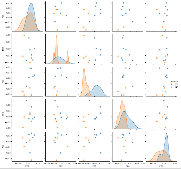
For the microbiome/virome analyses, we normalized the data to have 1 million reads per sample (reads per million, RPM). We filtered out taxa with average read counts less than 1 RPM. We removed 5 samples, namely Infection 1, Infection 6, Non-nisin L. lactis + Infection 4, Non-nisin L. lactis + Infection 6 and Nisin + Infection 3, that have low sequencing coverage. We used the Shannon diversity index to quantify bacterial and viral diversity across different groups. In order to compare the difference of bacterial contents, viral contents, and Shannon diversities between different groups, we computed the p-values using a two-sample t-test assuming equal variance of samples from the two groups. For the Principal Coordinates Analysis (PCoA), we further restricted to species with RPM<500 to avoid the result being dominated by commonly present species. Three species, namely Mouse Intracisternal A-particle, Chlamydia abortus, Chlamydia trachomatis, were filtered out under this criterion. We used the Bray Curtis dissimilarity to quantify the difference between microbiome composition of different samples. The 95% confidence ellipses were computed assuming that the data in each group followed a two-dimensional normal distribution. For the differential abundance analysis, we performed a log transformation (log10 (RPM + 0.1)) for the bacterial and viral read counts and used a two-sample t-test to compute the p-values, assuming equal variance in the two groups. We further used the Benjamini–Hochberg procedure[87] to correct for multiple comparisons. We reported the corresponding false discovery rate (FDR) for conducting pair-wise comparisons (e.g., Infection versus Control), and the multiplicity is the total number of taxa. For correlating microbial species with the immune markers, we considered the data in log space for both read counts and immune marker measurements (log10(x+0.1)). We considered only microbial species that are significant in at least one differential abundance comparison (comparison v.s. control or v.s. infection). We computed Pearson’s correlation with a p-value based on t-test. We performed the Benjamini–Hochberg procedure88 for multiple testing for each immune marker (across all microbial species) separately.