Cell culture
Murine OCY454 osteocyte cells were purchased from the Centre for Skeletal Research Bone Cell Core, an NIH-funded program (P30AR075042), which is supported by NIAMS. OCY454 cells were expanded on type I collagen (0.15mg/ml in 0.02M acetic acid) coated T-175 flasks in α-MEM supplemented with 1% L-glutamine, 2% antibiotics, and 10% FBS. OCY454 cells were routinely cultured at the permissive temperature of 33°C, cells were then trypsinized and placed in non-collagen coated T-175 flasks. After 3 days, cells were differentiated by being transferred to the semi-permissive temperature of 37°C for 15 days [51] before being treated with 17β-estradiol, cells were maintained in a humidified environment at 5% CO2.
Osteoclast differentiate from cells of the monocyte/macrophage lineage [19], based on this bone marrow macrophages (BMM) were isolated from 8 month old female C57BL/6 mice based on [75]. All animal work was carried out under license from the Animal Care and Research Ethics Committee (ACREC) of the National University of Ireland Galway and the Health Products Regulatory Authority (HPRA), the national authority for scientific animal protection in Ireland. Briefly, hind limbs were removed of muscle and tissue, each end of the bone was sectioned, bone marrow aspirate was centrifuged and plated at 5x10⁶ cells in 10cm bacteriological petri dishes. The bone marrow macrophages were differentiated in the presence of α-MEM supplemented with 1% L-glutamine, 2% antibiotics, 10% FBS and 20% L929 fibroblast cell conditioned media (source of M-CSF). Once macrophages were confluent (approximately 1 week after isolation), cells were trypsinized and scraped, and frozen down or used straight away. BMM cells were used at passage 3 or below. Additionally, RAW264.7 cells were purchased from the American Type Culture Collection (ATCC, Manassas, VA, USA), to assess whether the commonly used male derived cell line responded similarly to primary BMM cells which were isolated from female mice. RAW264.7 cells were expanded in Dulbecco’s modified Eagle’s medium with 1% antibiotics, 1% L-Glutamine, and 10% heat-inactivated FBS (HyClone). BMM and RAW264.7 cells were cultured in a humidified atmosphere at 37°C in 5% CO2.
Estrogen treatment regimes
The effect of supplementing culture media with pre-menopausal levels of estrogen (E) on OCY454 osteocyte behaviour under oscillatory flow conditions in vitro was investigated. Following 15 days of expansion in α-MEM culture media at 37°C, OCY454 cells were treated with estrogen (E: 10 nM 17β-estradiol) for 6 days. To simulate postmenopausal conditions (ED), we implemented a model that first accustomed osteocytes to estrogen before a subsequent period where estrogen supplementation was discontinued, known as estrogen withdrawal, based on our previous established postmenopausal model using MC3T3-E1 and MLO-Y4 cells [27, 60]. OCY454 cells were treated with 17β-estradiol (10 nM) for 3 days before it was withdrawn from the media for a further 3 days. In total OCY454 cells were culture at differentiated 37°C for 21 days before being exposed to oscillatory fluid flow.
Sclerostin inhibition
Sclerostin antibody (Scl-Ab VI) was kindly provided by UCB pharma (UCB pharma, UK/Amgen Inc. USA) and was stored at 5mg/ml aliquots at -80°C. In order to obtain sufficient neutralisation of sclerostin, we treated with ≥30x higher concentration than the amount of sclerostin produced by OCY454 cells. A previous study reported that OCY454 cells cultured at 37°C for 14 days produce approximately 75pg/ml of sclerostin [51], based on this, a non-cytotoxic concentration of 300ng/ml was chosen to ensure sufficient sclerostin neutralisation. Untreated groups received PBS as a vehicle, similar to in vivo Scl-Ab studies [9-11, 72, 76].
Mechanical stimulation
Osteocytes are the main mechanosensors in bone and are subjected to various forms of mechanical loading [15, 77, 78]. Therefore to capture the in vivo environment more accurately, all osteocytes were subjected to oscillatory fluid flow. Prior to mechanical stimulation OCY454 cells from each of the treatment groups (E and ED) were seeded at 200,000 cells per collagen coated glass slide (76 mm x 26 mm) (collagen coating was used to prevent cell detachment during fluid flow) and were cultured for a further day either under (a) continued estrogen (E) or (b) estrogen deficient conditions (ED). A subset of these groups, were also treated with 300ng/ml Scl-Ab, 24 hours before stimulation. Laminar oscillatory fluid flow was applied to OCY454 cells using a parallel plate system, which comprised of a syringe pump (NE-1600, New Era Pump Systems, Farmingdale, NY, USA), parallel plate chambers and individual media reservoirs connected through gas-permeable silicone tubing (Cole-Parmer, Vernon Hills, IL, USA) [52]. The OFF loading regime subjected the OCY454 cells to a shear stress of 1 Pa at 0.5 Hz for 1 h, which is within the range of shear stresses experiences by osteocytes in vivo [77, 79-81]. Before and after oscillatory fluid flow, slides were rinsed with PBS (x3) to remove residual media, to ensure that conditioned media collected did not contain 17β-estradiol. After flow, fresh media (without estrogen supplementation) was applied and cells were then cultured for 24 hours before conditioned media was collected for further experiments (described below). Cells treated with Scl-Ab 24 hours prior to flow continued to be treated with 300ng/ml Scl-Ab (i.e. when CM was collected OCY454 cells had been exposed to 300ng/ml Scl-Ab for 48 hours).
Conditioned media experiments
Conditioned media from all cell treatment groups (E, E+ Scl-Ab, ED and ED-Scl-Ab) was centrifuged at 1500 rpm and then frozen at -80°C in 1 mL aliquots. RAW264.7 cells and BMM were seeded at 5000 and 12,000 cells respectively per well in a 96 well plate and then treated with 50% conditioned media and 50% expansion media (DMEM). Cells were then cultured in the presence of 15ng/mL of RANKL for 5 days.
Bone resorption assessment
6mm bone discs with a thickness of between 0.4-0.6mm were created from bovine metatarsals based on [82]. Bone discs were soaked in 70% ethanol and placed in ultrasonic bath (VWR, Dublin, Ireland) at room temperature for 15 minutes. Prior to culture, discs were sterilised under UV light, BMM cells were seeded on discs at 14,000 cells per discs in a 96 well plate. Cells were cultured with 50% CM and 15ng/ml RANKL for 10 days, on day 7 concentrated hydrochloric acid (HCL) was added to the culture media to achieve pH 6.9 to induce a more acidic environment necessary for osteoclast resorption [82]. On day 10 the experiment was terminated and cells were removed by sonication in 0.25M ammonium hydroxide for 5 minutes. Resorption pits were stained using 1% Toluidine blue in 1% sodium borate solution. Images were acquired using a light microscope (Olympus BX43, Olympus, Tokyo, Japan) and quantified using ImageJ software, in which images were colour-thresholded and percentage area resorbed was quantified.
Tartrate-resistant acid phosphatase (TRAP) staining
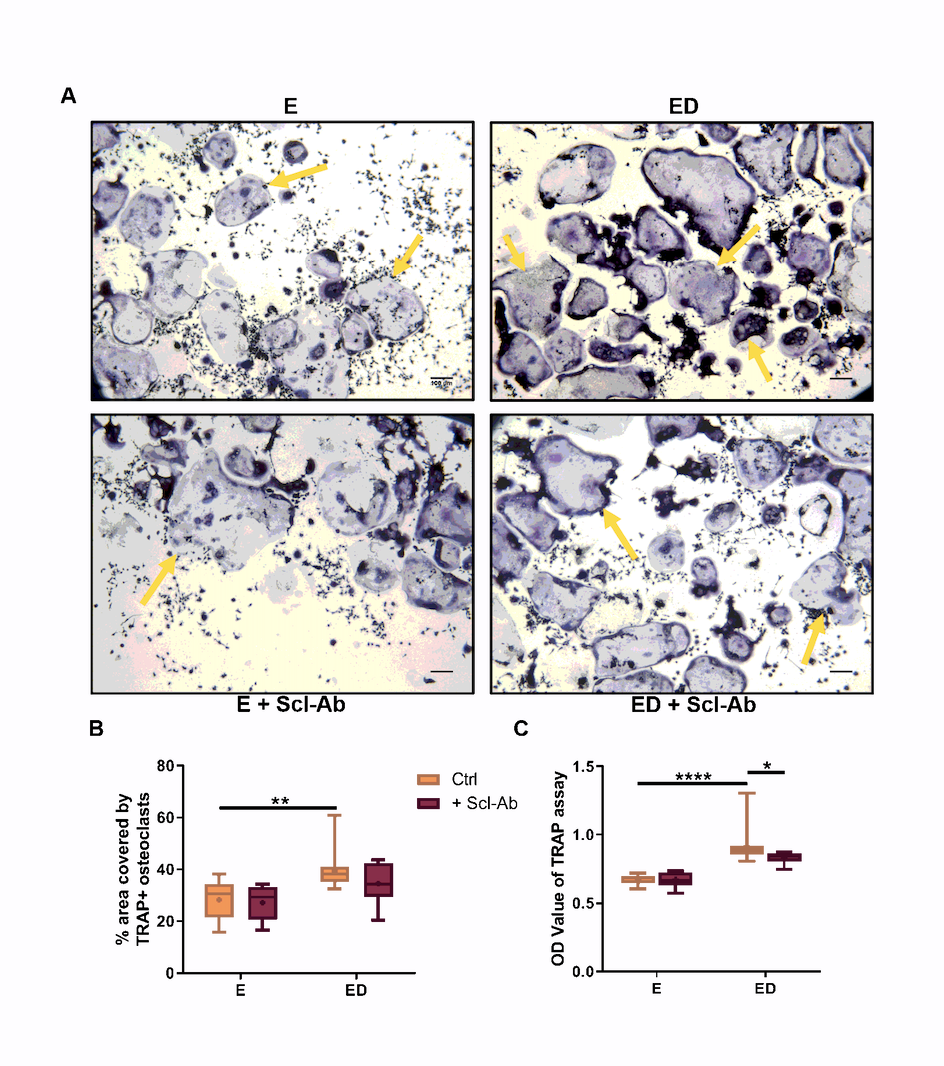
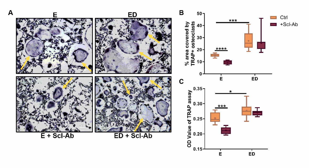
At the end of the co-culture and conditioned media experiments RAW 264.7 cells and BMMs were rinsed with PBS and fixed with 4% Paraformaldehyde. They were then rinsed with PBS and stained for tartrate-resistant acid phosphatase (TRAP) activity with a commercial kit and counterstained with Gill No. 3 hematoxylin for 3 minutes. Staining was also performed on RAW264.7 and BMM cells, which received expansion media only (negative control) and cells that received expansion media with only supplementation of 15ng/mL RANKL (positive control). Images were acquired using a light microscope (Leica DMi1, Leica Biosystems, Wetzlar, Germany) and quantified using ImageJ software, in which images were colour thresholded and the percentage area covered by osteoclasts was quantified. TRAP-positive cells with 3 or more nuclei were considered to be osteoclasts.
TRAP activity assay
At the end of the conditioned media and co-culture experiments, culture media was collected from wells and secreted TRAP activity was detected. The TRAP activity assay involved measuring enzyme activity by the conversion of p-nitrophenylphosphate (20 nM) to p-nitrophenol in the presence of 80 mM sodium tartrate and was expressed as optical density at 405nm using a microplate reader (Synergy HT, Biotek Instruments Inc., Winooski, VT, USA).
Co-culture experiments
Mechanically stimulated OCY454 cells from each group (E, E+ Scl-Ab, ED and ED+Scl-Ab) were seeded at 25,000 cells per well in a 48 well plate. After 24 hours RAW264.7 cells were seeded on top of OCY454 cells at 10,000 per well based on a previous study [60] and BMM cells were seeded at 34,000 cells per well. Cells were co-cultured in the presence of 15ng/mL of RANKL for a further 6 days. To mimic systemic administration in humans, Scl-Ab treatment groups continued to receive 300 ng/ml Scl-Ab throughout the 6 days of co-culture (i.e. both osteocytes and osteoclast precursors were cultured in the presence of Scl-Ab)
qRT-PCR
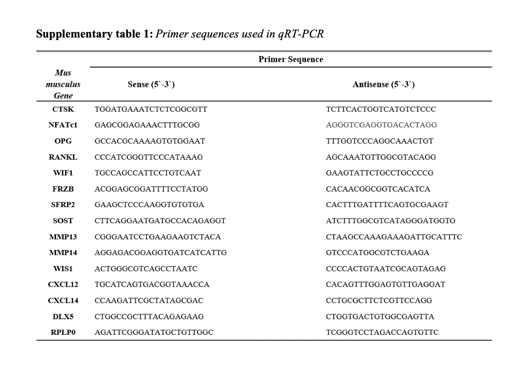
BMM cells were exposed to 50% CM, as described above, and cultured for 5 days. At day 5 mRNA was isolated using High pure RNA isolation kit (Roche Applied Science, Mannheim, Germany). RNA concentration and quality was assessed with a Spectrophotometer/Fluorometer (DS-11 FX, DeNovix, Wilmington, DE, USA)). cDNA was generated using the QuantiNova Reverse Transcription Kit. qRT-PCR was performed on resultant cDNA template on a StepOne plus real-time PCR machine (Applied Biosystems, Foster City, CA, USA) using a QuantiNova SYBR Green PCR Kit and custom designed primers (IDT, Coralville, IA, USA) for Nuclear factor of activated T-Cells cytoplasmic 1 (NFATc1), and Cathepsin K (CTSK) (Primer sequences are shown in Supplementary table 1).
mRNA from OCY454 cells was isolated 24hrs after exposure to oscillatory fluid flow and transcribed to cDNA using the same protocol as described above. Expression of the following genes were assessed; Receptor activator of nuclear factor kappa-Β ligand (RANKL), Osteoprotegerin (OPG), Wnt inhibitory factor 1 (WIF1), Frizzled-related protein (FRZB), Secreted frizzled related protein 2 (SFRP2), Sclerostin (SOST), Wnt1-inducible signalling pathway protein-1 (WISP1), C-X-C motif chemokine ligand 12 (CXCL12), C-X-C motif chemokine ligand 14 (CXCL14) and Distal-less homeobox 5 (DLX5) (Primer sequences are shown in Supplementary table 1). The normalised relative quantities (NRQ) of each sample (BMM and OCY454 samples) were calculated with reference to ribosomal protein large subunit P0 (RPLP0) (Reference gene was identified through qbase+ software). Data was analysed using the Pfaffl method [83].
Statistical analysis:
Data are representative of 3 independent experiments performed in triplicate and are presented as mean ± SD. Statistical analysis was performed by unpaired two-tailed Student’s t-test to examine the effect of estrogen treated groups vs. estrogen deficient groups or Scl-Ab treated group vs. untreated groups. In addition, the interaction between the effects of estrogen and Scl-Ab was also tested though two-way ANOVA analysis. A value of p< 0.05 was regarded as statistically significant. Technical replicates are represented as N, while biological replicates are represented as n.
Availability of data and materials:
The datasets generated during and/or analysed during the current study are available from the corresponding author on reasonable request.