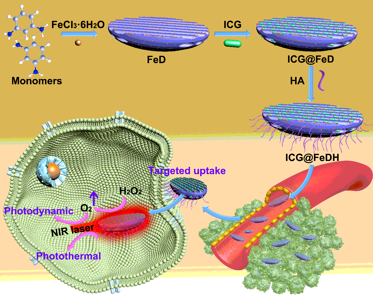
Synthesis and characterization of FeDH
As depicted in Scheme 1, the FeD was first prepared by iron ions-initiated polymerization of DAP monomer [37]. TEM images show that as-prepared FeD nanoparticles exhibit fusiform-like structure with a length of 71.4 ± 3.8 nm and width of 16.9 ± 2.1 nm (Figure 1A). Unlike the spherical nanoparticles, fusiform-like morphology probably endows the nanocarriers with some specific advantages on biological effect. For instance, the mesoporous silica nanorods have been reported with enhanced cell internalization, higher drug loading capacity and improved tumor accumulation in comparison with mesoporous silica nanospheres [38-40]. However, the systematical investigation of the shape effect of covalent organic polymers has rarely been reported, which is mainly due to that the controlled synthesis of covalent organic polymers in different morphology and size still remains a huge challenge to be overcame. The porosity of FeD nanoparticles can be clearly seen in high-magnification TEM image. Moreover, SEM image demonstrate the uniform morphology of FeD in large scale (Figure 1C). Due to the abundant amino groups on FeD, the targeted molecule HA can be modified onto it by amide reaction. The obtained FeDH maintains the original structure, and the pores are still visible in TEM image (Figure 1D-F). Further, the element mapping of FeDH is recorded on scanning TEM. Notably, strong signals from iron element can be observed, the energy dispersive X-ray spectrum also confirms the existence of iron ions in FeDH (Figure 1G-H). It has been reported the iron ions (Fe3+) can react with hydrogen peroxide by Fention-like reaction [41, 42], modulating the tumor microenvironment and producing sufficient oxygen to enhance the therapeutic efficacy. Thus, ICG is selected as photosensitizers to be loaded in FeDH. Accordingly, the typical absorption peak of ICG at NIR region for ICG@FeDH verifies the efficient loading process (Figure 1I and Figure S1). The free ICG exhibits maximum absorption peaks around 780 nm. It is noted that the characteristic peak of ICG shows a red shift to around 810 nm for ICG@FeDH, which is probably resulted from the interactions between loaded ICG molecules and metal-doped FeDH as well as the self-aggregation of ICG during the loading process [43]. The encapsulation efficiency and loading content is calculated to be 52% and 11.5% on the basis of UV-vis spectrophotometer, respectively. Additionally, the zeta potential of prepared samples is also measured. The bare FeD displays strong positive surface due to the amino groups of DAP (Figure 1J). Zeta potential of FeDH turns to -20.3 mV after modification of HA and the ICG@FeDH also exhibits negative surface, which is expected to benefit the colloidal stability of nanoparticles [44]. Then the colloidal stability of FeDH is inspected by DLS. As expected, the size distribution of FeDH in different media, including water, PBS and cell culture medium, shows no abnormality but slight increase in PBS and cell medium (Figure 1K and S2). The larger size of FeDH in cell medium can be attributed to the absorption of albumen in cell medium onto its surface as demonstrated by previous report [45]. Moreover, the average size of FeDH exhibits no obvious change in one week, and no agglomeration appears after one week storage (Figure 1L), suggesting the excellent colloidal stability of FeDH.
Photothermal effect of ICG@FeDH
Since the ICG@FeDH shows strong NIR absorption, its photothermal property is evaluated next. First, the ICG@FeDH solutions with different concentrations were irradiated with 808 nm laser for 5 min. The temperature of the solutions exhibit an obvious concentration-dependent increase (Figure 2A), which gradually elevates to 42.3 °C at a low concentration of 100 μg/mL and power density of 1.0 W/cm−2. However, the temperature of free FeDH solutions changes 1.2 °C after 5 min of irradiation, and the pure water only 0.3 °C under the same conditions (Figure 2B and C). The strong contrast of pseudo-color in thermal imaging photos also demonstrates the superior photothermal effect of ICG@FeDH than that of free FeDH (Figure 2D). Furthermore, the photostability of ICG@FeDH and ICG@FeD is assessed. The dispersions are subjected to three rounds of repeated laser irradiation. Remarkably, the elevation of temperature maintains well without any decrease for both ICG@FeDH and ICG@FeD (Figure 2E and S3). The excellent photostability can be attributed to the loading of ICG in FeDH considering the susceptible photobleaching of free ICG [46]. In addition, the photothermal conversion efficiency of ICG@FeDH and ICG@FeD is also calculated to be 19.7% and 19.5%, respectively (Figure 2F and S3). The values are apparently lower than previously reported semiconducting polymeric nanoparticles (SPNs) [47, 48], which is due to that the polymerization of small molecules into macromolecules can improve their optical properties [49, 50]. Anyway, the results confirm that the prepared ICG@FeDH can be used as an excellent and stable photothermal agent.
In vitro photodynamic effect of ICG@FeDH
In addition to the enhanced photothermal effect, ICG can also generate singlet oxygen under NIR laser irradiation [51]. Thus, the photodynamic effect of ICG@FeDH is investigated. On the other hand, the doped iron ions of FeDH can catalyze the decomposition of hydrogen peroxide. The oxygen level of FeDH solution rapidly increases with the addition of hydrogen peroxide, while the pure water or hydrogen peroxide alone cannot produce oxygen under the same conditions (Figure 3A). The catalase-like activity of FeDH is further confirmed in living cells by a commercial O2 sensing probe RDPP [52]. No surprisingly, the green fluorescence of RDPP in cells treated with FeDH was dramatically weakened as compared to the cells without treatments (Figure 3B). The result evidences that iron ions doped in FeDH can efficiently catalyze the decomposition of hydrogen peroxide to generate intracellular oxygen, thereby relieving the hypoxia condition in tumor microenvironment. Based on this, the photodynamic effect of ICG@FeDH is also expected to be improved due to the elevated oxygen level. To illustrate this issue, another probe DPBF is used to evaluate the singlet oxygen generation ability of ICG@FeDH under excitation of NIR laser. As expected, the absorption peak of DPBF decreases with the irradiation time of NIR laser (Figure 3C), implying the generation of singlet oxygen. Moreover, the decrease of the absorption value becomes faster when hydrogen peroxide is added into the ICG@FeDH solution (Figure 3D). Specifically, after 5 min irradiation, 64.8% of DPBF is oxidized in the presence of hydrogen peroxide (Figure 3E), which is much high than that of ICG@FeDH alone (57%). Further, the singlet oxygen generation ability of ICG@FeD and ICG@FeDH is further evaluated by SOSG. The fluorescent intensity enhancement (F/F0) is calculated according to the previously reported method [53, 54]. The value of F/F0 for ICG@FeDH and ICG@FeD is determined to be 3.82 and 3.79 (Figure S4), respectively, suggesting that the modification of HA would not affect the singlet oxygen generation ability of ICG. Meanwhile, the value increases to 4.57 with the participation of H2O2 for ICG@FeDH, which is consistent with the results of DPBF. Combined with the decomposition of H2O2 catalyzed by FeDH, the result can demonstrate that the oxygen-evolving capacity of FeDH can improve the ROS generation of ICG@FeDH under NIR laser irradiation.
Targeted ability of ICG@FeDH
With specific affinity towards CD44-receptor [55, 56], the attachment of HA on FeDH can render it with targeted ability towards tumor cells. To demonstrate this, the intracellular uptake of ICG@FeDH is investigated by CLSM using PC-3 cells. After the cells were co-incubated with ICG loaded nanoparticles for 4 h, the red fluorescence of ICG is very weak for non-targeted ICG@FeD. In sharp contrast, strong red fluorescence can be observed around the nucleus for ICG@FeDH group (Figure 4), which can be attributed to the specific binding of HA with CD44 receptors on PC-3 cells. Once the receptors are inhibited by treating PC-3 cells with free HA, the intracellular fluorescent intensity for ICG@FeDH obviously decreases, further confirming the receptor-mediated endocytosis of ICG@FeDH. Taken together, the modification of HA endows FeDH with excellent targeted ability for tumor therapy.
In vitro evaluation of ICG@FeDH
Next, the tumor cell killing effect of ICG@FeDH is assessed using PC-3 cells. With the aforementioned photothermal and photodynamic effect, ICG@FeDH is supposed to exert favorable killing effect on tumor cells under NIR laser irradiation. DCFH-DA, a probe molecule that can be oxidized to emit fluorescence at 488 nm [57], is applied to assess the cellular amount of reactive oxygen species (ROS), the major killing mechanism of photodynamic therapy. As shown in Figure 5A, the cells treated with laser irradiation or ICG@FeDH alone display very weak fluorescence, while the bright green fluorescence can be observed upon laser irradiation for both ICG@FeD and ICG@FeDH treated cells. In particular, the fluorescent intensity of ICG@FeDH with laser irradiation is determined to be 108.3 (Figure 5B), which is much stronger than that of ICG@FeH with laser irradiation (~75.4), demonstrating the higher ROS level for ICG@FeDH. The result can be ascribed to the enhanced cellular uptake of targeted ICG@FeDH as mentioned above.
To further evaluate the cell killing efficacy, the cytotoxicity of free FeDH is investigated first. The result of CCK-8 demonstrates that the cells treated with various concentrations of FeDH all show negligible toxicity even at a ultra-high concentration of 1600 μg/mL, suggesting the outstanding biocompatibility of FeDH. Thus, this polymer-based nanocarrier is highly suitable for biological applications in comparison to those non-degradable inorganic nanomaterials [58]. Afterwards, the killing effect is assessed using PC-3 cells. The time-dependent mortality can be observed upon the tumor cells subjected to ICG@FeDH plus NIR laser irradiation (Figure 5C), demonstrating the outstanding phototherapeutic efficacy of ICG@FeDH. Moreover, cells treated with ICG@FeDH or laser irradiation alone exhibit high cell viability over 95% (Figure 5D), suggesting the minimal damage of NIR laser alone. The superior cell killing efficacy of ICG@FeDH is probably due to its excellent photothermal and photodynamic effect under laser irradiation. Combining with the modification of HA, the ICG@FeDH can serve as a targeted phototherapeutic agent for killing tumor cells.
In vivo antitumor effect of ICG@FeDH
Next, the in vivo therapeutic efficacy of ICG@FeDH was investigated on PC-3 tumor-bearing mice. To implement the treatments, the mice are intravenously injected with ICG@FeDH and NIR laser irradiation is conducted. Figure 6A shows the variation of relative tumor volume in the period of treatment. The tumors treated by ICG@FeDH with 808 nm laser irradiation were remarkably inhibited and displayed a relative tumor volume of 0.38 on day 6 without any recurrence, implying the superior phototherapeutic efficacy of ICG@FeDH under NIR laser irradiation. As for control groups of PBS and NIR laser alone, the size of tumors are rapidly increased within two weeks (Figure 6B), showing negligible therapeutic effect. Notably, the ICG@FeD with laser irradiation shows an inferior tumor inhibition rate in comparison to the counterpart of ICG@FeDH, which confirms that the modification of HA can improve its in vivo antitumor effect. Besides, the body weight of experimental mice shows no obvious change in the period of treatments (Figure 6C), indicating that the well-tolerance and excellent biocompatibility of the applied samples. Furthermore, the tumors are harvested at the end of treatments and stained with H&E for histological analysis. The images show that no apparent necrosis appears in control group (Figure 6D). Moreover, the group of ICG@FeDH plus laser irradiation exhibits much more necrosis and karyolysis in slice than that in the group of non-targeted ICG@FeD plus laser irradiation. It is deduced that the better therapeutic efficacy of ICG@FeDH is mainly resulted from targeted ability of HA as well as the improved photodynamic effect because of the oxygen-evolved capacity of FeDH. To better understand this, the immunofluorescent staining of hypoxia-inducible factor 1α (HIF1-α) is conducted to detect the oxygen level in tumor. The green fluorescence indicative of hypoxia remarkably reduces in the group of ICG@FeDH (Figure 6E and Figure S5). It should be noticed that the fluorescence in the group of ICG@FeD is also weakened as compared with control group and NIR laser group, but still stronger than ICG@FeDH, which is probably due to that the efficient targeted ability of ICG@FeDH render it with higher accumulation at tumor site. Taken together, ICG@FeDH effectively integrates several advantages, including excellent biocompatibility and targeted ability as well as oxygen-evolved capacity for enhanced phototherapeutic efficacy, showing great potential for biomedical applications.