Since the discovery of Aquaporin (AQP), the role of AQP protein in water transport and tissue edema formation in different tissue cells has received significant attention. AQP proteins are a family of membrane proteins responsible for the rotation of water inside and outside the cell and some other small molecular substances. It can increase the water permeability of cell membranes, thereby driving the transport of water under the action of osmotic pressure. A total of 13 AQP proteins have been found in mammals (0–12). It is widely distributed in organs such as red blood cells, kidneys, lungs, brain and eyes(1)
The kidney is an organ that maintains water balance in the body. Its main function is to regulate the concentration and dilution of urine, and to regulate water reabsorption through different AQPs. Of the 13 aqp (AQP0-AQP12) currently found, nine aqp (AQP1-8 and AQP11) were detected in human kidneys. Dysfunction of AQPs, especially AQP1-4, can cause disorders with water balance. Among them, AQP2 is the most researched aquaporin, which is mainly expressed in the main cells of the renal junction tubules and collecting tubules. It is one of the most important channel proteins involved in regulating urine concentration. Rojek A et al.(2) after genetic research, compared with wild mice, AQP2-deficient mice lost weight and increased their urine output ten-fold in adulthood. Moreover, the urine output decreased significantly after dehydration, and urine osmotic pressure did not change significantly. Human water balance is critical and cannot be compensated by other mechanisms. In fact, the expression of AQP2 in rats increased under water-restricted conditions, while the expression of AQP2 decreased in conditions of water overload(3). The water reabsorption function of AQP2 is mainly regulated by arginine vasopressin (AVP)(7). There is no circadian rhythm in the secretion of AQP2(4).AVP is a hormone released from the posterior pituitary, which can stimulate the expression of AQP2 in the apical plasma membrane(5, 6). When AVP stimulation was removed, AQP2 returned to the cytoplasm and restored the cell impermeability(7). In normal animals, the opposite effect could be observed by using AVP antagonists(8).
In addition to AVP, there are also some factors including osmotic pressure, inflammation, insulin, aldosterone / prostaglandin and other factors that also affect AQP2 transcription, transport and post-translational modification(9). A study in the 20th century showed that higher levels of insulin can reduce urinary excretion(10). The water permeability of the isolated medullary collecting duct was increased after insulin infusion(11) Using mpkCCD (CL4) cells(12), it can be proved that insulin can induce a slight increase in AQP2 abundance in whole cells(13).
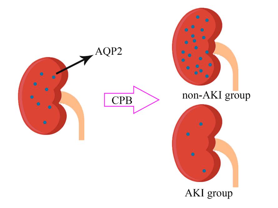
Acute kidney injury (AKI) is one of the most important postoperative complications of cardiac surgery, and despite complete recovery of renal function after surgery, AKI is independently associated with high mortality within 10 years after surgery(14). In AKI caused by ischemia-reperfusion, AQP2 decreases after acute kidney injury(15). A recent study showed that aquaporin-2 contained in urine extracellular vesicles in patients after renal transplantation Significantly reduced on the first postoperative day, accompanied by high urine volume and low osmotic pressure. Since then, AQP2 levels have gradually increased to control levels on the 6th day(16).However, some studies have pointed out that the content of aquaporins in urine is significantly increased after extracorporeal circulation(17). Therefore, the purpose of this study is to further determine the relationship between AQP2 and the occurrence of AKI after cardiopulmonary bypass, and to provide us with directions for further prevention and treatment of AKI.
Although it has been found that some indicators such as cystatin C(18), neutrophils(19), urinary uromodulin(20), α 1-antitrypsin(21) and urinary IL-18(22) can predict the occurrence of AKI, creatinine is still the gold standard. As a result, we have no recognized indicators to judge AKI in the early stage. And we have not found an effective way to intervene the occurrence of AKI. In this context, we chose aquaporins for further study.
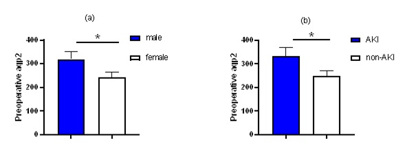
According to relevant literature reports,aquaporin-2 was detectable in the urine in both soluble and membrane-bound forms(23). Membrane bound AQP2 is mainly located in a structure called exosomes(24). However, due to the complexity of exosomes separation, it is not easy to apply in clinical practice. Therefore, this paper attempts to directly detect the content of AQP2 in urine after ordinary centrifugation.