5.1 Materials preparation and characterization
The preparation method of Ti-NW-Zn surfaces and the identification of surface element was described previously[6]. After being washed and dried, the obtained samples were randomly assigned to 6-well plates with 2 ml ddH2O.
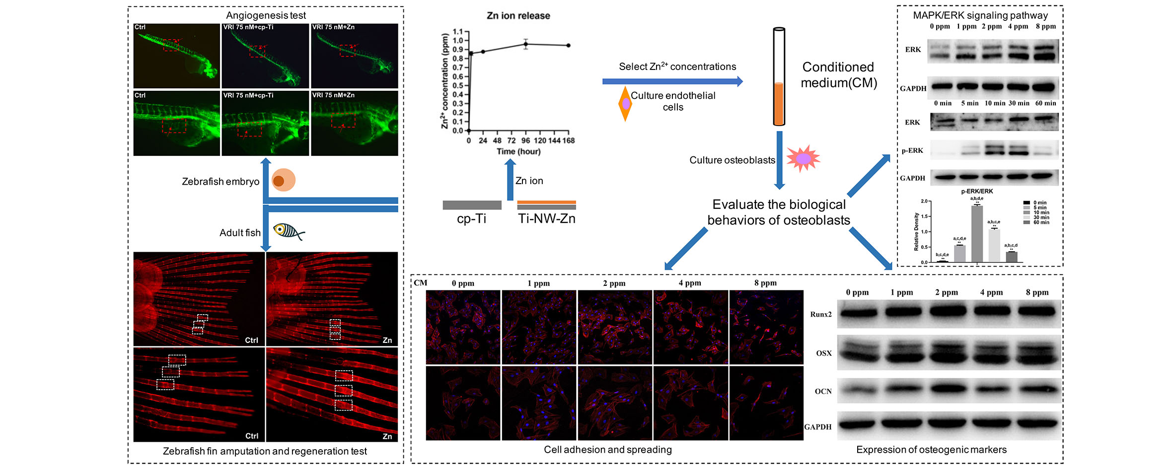
5.2 Zinc ion release assay
Ti-NW-Zn samples were immersed in 6-well plates (2 ml PBS/well) at 37°C for 1 h, 1 day and 10 days. The concentrations of the released zinc ions in PBS solution were quantified using a Zinc assay kit (E011-1-1; Nanjing Jiancheng Bioengineering Institute, Nanjing, China). The absorbances were measured using a microplate reader (SpectraMax 190, MD, USA) at 630 nm wave-length. Three samples of each group were used in this assay.
5.3 Zebrafish embryo collection
Wild-type Zebrafish (Danio rerio) embryos were obtained from Wild-type AB strain adult zebrafish (Danio rerio), while Tg (Fli-1:EGFP)y1 zebrafish embryos were obtained from outcrosses of Tg (Fli‐1:EGFP)y1 parents[18]. The spawning adults were offspring of parents obtained from Model Animal Research Center of Nanjing University and maintained in an aquatic animal breeding and reproduction system (HAISHENG, Shanghai, China), under standard conditions. All zebrafish studies were approved by the Institutional Animal Care and Use Committee at Nanjing Medical University. Groups of 1 male and 2 females were mated in translucent plastic tanks and embryos were obtained within 30 min after the onset of light in the morning. The eggs were collected immediately after fertilization and washed several times for 1 min using a 0.5 % bleach solution for disinfection. After this, clean eggs were incubated in 6-well plates at 28.5°C.
5.4 Zebrafish Rearing
Wild-type AB strain adult zebrafish (Danio rerio) were maintained in a recirculating aquatic system at 28.5°C with a 10/14-h dark/light cycle according to standards[18]. Circulating water in the aquarium was filtered by reverse osmosis (pH 7.0–7.5). All zebrafish was fasted for 1 day before starting the experiment. Male and female zebrafish were randomly used in experiments.
5.5 Angiogenesis observation of transgenic Zebrafish embryo
24 hpf (hours-post-fertilization) Tg (Fli-1:EGFP)y1 zebrafish embryos were pretreated with 75 nM vascular endothelial growth factor receptor-2 tyrosine kinase inhibitor to inhibit the normal angiogenesis for 8 hours, then co-cultured on Ti-NW-Zn surfaces randomly pre-assigned to 6-well plates (10 embryos per well with 2 ml medium) at 28.5°C for 3 days to detect the defective vessels regeneration. In the 48 hpf, zebrafish were anesthetized (4% Tricaine) and the intersegmental blood vessels (ISVs) were observed under the inverted fluorescent microscope; In the 72 hpf, the subintestinal vessels (SIVs) of the yolk sac region gross morphological changes were observed under the inverted fluorescent microscope. Three parameters indicative of angiogenesis or vasodilation were measured: variation in the number of vessels, vessel thickness and the subintestinal venous plexus (SIVP) branching (angiogenic phenotype).
5.6 Survival test in embryos and adult fish
According to the concentration of zinc ion release from Ti-NW-Zn surfaces, all embryos were cultured in 6-well plates (10 embryos per well with 2 ml medium) at 28.5°C with different concentrations of zinc ions, including a nominal concentration of 0 (control group), as well as concentrations of 2, 4, 8, 16, 24 and 32 ppm zinc ions, which last for 120 hpf (n = 10 for each testing concentration), respectively. LC50 tests were then carried out according to these series groups. Adult fish were exposed to ddH2O with different concentrations of zinc ions, including a nominal concentration of 0 (control group), as well as concentrations of 1 and 2 ppm zinc ions for 7 days for chronic exposures (n = 5 for each testing concentration), respectively. LC50 tests were also carried out according to these series groups. The exposure water was changed daily. The zebrafish were fasted during the entire experiment’s duration.
5.7 Zebrafish fin amputation and regeneration test
7–10 months-old adult zebrafish with body weights of 0.3–0.5 g were initially anesthetized the with tricaine (160 mg/L) for 5 minutes, then caudal fins were partially amputated using a #11 blade. All fish were then allowed to recover in open tank for 2 hours and randomly assigned to 500 ml culture vessels with different concentrations of zinc ions, where one was placed in each vessel and the solution was changed every 3 days and fed twice a day. Then, Alizarin Red Stain was used to detect the skeletal calcification of Zebrafish fin after executed. The experiment was carried out for 9 days and repeated three times.
5.8 Cell culture
Commercially available osteoblast-like cell line MC3T3-E1 (Cell Bank of Chinese Academy of Science, Shanghai, China) and human umbilical vein endothelial cells (HUVECs, ATCC, USA) were used in this study. MC3T3-E1 cells were cultured in α-Minimum Essential Medium (α-MEM; Gibco, USA) supplemented with 10% fetal bovine serum (FBS; Gibco, USA) and 1% penicillin/streptomycin (Gibco, USA). HUVECs were cultured in Dulbecco's Modified Eagle's Medium (DMEM; Gibco, USA), which containing 10% fetal bovine serum (Gibco, USA) and 1% penicillin/streptomycin (Gibco, USA). Both MC3T3-E1 and HUVECs were maintained in an incubator containing 5% CO2 and 95% air at 37°C. The fresh complete medium changed every two days. When reaching 80% cell confluence, the cells were passaged every three or four days.
5.9 Collection and preparation of conditioned medium (CM)
HUVECs were seeded in the 6-well plates and incubated with Zn ions at a range of doses (0, 1, 2, 4, 8 ppm). When the cell confluence was up to 80%, the culture medium of each group was collected and centrifugated (1000 rpm, 15 min) under sterile conditions. After collecting the supernatant, it was mixed with α-MEM containing 10% FBS and 1% penicillin/streptomycin at a ratio of 1:1 to get conditioned medium (CM). CM was placed in a -20°C refrigerator for later use.
5.10 Cell proliferation assay
CCK-8 kit was used to assess the cell proliferation. MC3T3-E1 cells (3×10³ cells/well) and HUVECs (3×10³ cells/well) were seeded in the 96-well plates and treated with Zn ions at different concentrations (0, 1, 2, 4, 8 ppm). MC3T3-E1 cells were cultured for 1, 3 and 6 days, while HUVECs were cultured for 1, 2 and 3 days. Afterwards, 100 µl fresh medium which containing 10 µl of CCK-8 solution (Beyotime, Shanghai, China) was added in each well, incubating for another 2 hours at 37˚C. The absorbance of each well was measured by a microplate spectrophotometer (Spectramax 190, CA, USA) at the wavelength of 450 nm.
5.11 Cell adhesion and spreading assay
Commercially pure titanium (99.5 wt% purity, Alfa Aesar, USA) disks were polished with 600, 800, 1200 and 1500-grit silicon carbide abrasive papers. MC3T3-E1 cells (5×10³ cells/well) were seeding on the surface of polished titanium disks in the 96-well plates, treating with or without CM as described above for 8 hours. Afterwards, each sample was washed with PBS and fixed with 4% paraformaldehyde at room temperature for 10 minutes. To observe cell morphology on titanium, cells were stained with 100 nM Rhodamine phalloidin (Cytoskeleton, USA) for 30 minutes and 4’, 6’ -diamidino-2-phenylindole (DAPI; Beyotime, Shanghai, China) for 2 minutes in the dark. The cell morphology was observed under a laser scanning confocal fluorescence microscopy (LSM710NLO; Zeiss, Jena, Germany) at 100 x and 200 x magnification.
5.12 Western blotting
MC3T3-E1 cells (2×10⁵ cells/well) were seeded in 6-well plates and cultured with CM described above, after which cells were washed with pre-cooled PBS and lysed with RIPA buffer containing 1% PMSF. Protein samples were separated after electrophoresis, transferred to PVDF membranes (Millipore, Billerica, MA, USA), blocked in protein free rapid blocking buffer (EpiZyme, Shanghai, China) for 10 minutes, and incubated with primary antibodies specific for Runx2 (12556, CST, USA), OSX (ab22552; Abcam, USA), OCN (ab93876, Abcam, USA), ERK (4695, CST, USA), p-ERK (4370, CST, USA) and GAPDH (BM0627, Boster, China) at 4°C overnight. Afterwards, PVDF membranes were incubated with secondary antibodies (ZB-2301; Goat anti-Rabbit IgG, ZSGB-BIO, China) for 2 h at room temperature and exposed to ECL substrate solution (NCM Biotech, Suzhou, China). GAPDH served as a loading control. The cell experiments were performed in triplicate.
5.13 Statistics
Statistical analysis was performed on SPSS 22.0 software (SPSS, Inc., Chicago, IL, USA) using one-way analysis of variance (ANOVA) with Student-Newman-Keuls (SNK) method for multiple comparisons. The significant changes were set as *P < 0.05, **P < 0.01.