Insectary
The study was carried out at the CIRDES insectary in Bobo-Dioulasso, Burkina Faso. This is a mass rearing insectary for G. p. gambiensis set up in 1975 to support national and regional eradication of tsetse flies [16]. More than 250 000 females are reared and produce over 30 000 male pupae per week that are currently shipped to the eradication program in the Niayes of Senegal as chilled irradiated pupae [14]. Tsetse flies are maintained at 24-25°C, 75-80% relative humidity, and a photoperiod of 5:19 h (L:D).
Biological material
Teneral flies of the G. p. gambiensis colony were used in this study. This tsetse colony was established in 1972 at Maison- Alfort (France) with pupae collected from Guinguette (Bobo-Dioulasso, Burkina Faso). In 1975 the colony was transferred to the Centre de Recherche sur la Trypanosomiase Animale (CRTA) (renamed later CIRDES) from 5333 pupae [16]. In 1981 the colony was replenished with wild material from the “Marre aux hippopotames” [17]. Flies are maintained on fresh irradiated bovine blood collected from the local slaughterhouse using in vitro silicon membrane system [18]. The feeding frequency is six times per week, from Monday to Saturday.
Blood collection and conditioning
Bovine blood from different animals (cattle and pigs) were collected at the Bobo-Dioulasso slaughterhouse using 10 liters sterile containers and were defibrinated using a custom-made stain-less steel electric paddle stirrer. Upon arrival to the insectary, glucose was added to blood as a phagostimulant at the dose of 1g/L and was previously diluted in 10ml of distilled water [19]. Blood was irradiated with 500 Gy in a 137Cs source for 1 hour and 40 minutes and stored in 2 liters containers at 4°C. After irradiation, blood was tested for bacterial contamination. One milliliter of blood samples was poured with a syringe on agar petri dishes and incubated for 72 hours. After incubation, if the irradiated blood contained no more than 10 bacterial colonies, the blood is conserved for feeding, and discarded if more than 10 colonies are found. All blood manipulations were performed under a laminar flow hood to avoid bacterial contamination.
Experimental procedure
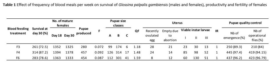
The effect of blood feeding frequency was evaluated through the measurement of several biological parameters routinely check in tsetse colonies for performance evaluation. Female’s survival (percentage alive on day 30), productivity (number of pupae produced at day 30), fecundity, first larviposition date, pupal size, pupal emergence rate and flight ability of newly emerged flies were assessed for each blood feeding treatment. Among these, female survival, fecundity and pupal size were used in a formula to calculate a Quality Factor (QF) of the blood feeding frequency as a comprehensive indicator for colony production [20,21]. The detailed formula is described in De Beer et al. [22]. A QF factor above 1 indicates that blood is suitable for colony maintenance.
To evaluate the effect of blood feeding frequency, three experimental treatments were performed. Flies were fed three times per week (F3) on Monday, Wednesday and Friday, four times per week (F4) on Monday, Tuesdays, Thursday and Friday, and finally six times per week (F6) from Monday to Saturday. Newly emerged flies were blood fed using an in vitro silicon membrane feeding system with the appropriate blood treatment during 30 days. Bioassays were conducted using 10 males and 30 females per cages of six and three days old respectively. Females were monitored daily for survival and productivity (pupal production and abortion). Pupae produced were sorted in five class sizes (A to E) calibrated for G. p. gambiensis according to their weight (mg): A (<22), B (22 <28), C (28<32), D (32<36), E (>36) [21]. After 30 days, all surviving females were dissected to determine their reproductive status (presence/absence of egg/larvae in the uterus and insemination status). For each feeding frequency, pupae produced were put in petri dishes under ~1cm of sand and covered by a flight cylinder (see Seck et al. 2015 for details). The inner wall of the cylinder was coated with unscented talcum powder to prevent the flies from crawling out. This method was used to assess the number of flies able to fly out and thus “available for the SIT”. After emergence, the number of pupae that did not emerge was counted. This study was performed between January and June 2018, and four cohorts of flies were studied for all treatments. For each cohort, all bioassays were replicated three times, leading to an overall of twelve replicates per treatment.
Statistical analyses
The survival of flies fed with different feeding frequencies was analyzed using Kaplan-Meier survival curves. Survival curves were compared using the “coxme” function where the blood treatment was used as explanatory variable, the survival as the response variable and cohorts and replicates were used as random effects.
Female’s productivity, first larviposition date and pupal classes were analyzed using a generalized linear mixed effects model with a “poisson” family. Treatments were used as fixed effect and cohorts were used as random effect.
Quality factors were tested using linear mixed effects models. The treatments were used as fixed effect and cohorts as random effect.
The adult emergence rate and percentage of flies able to fly were analyzed using binomial generalized mixed effects models. The treatments were used as fixed effect and cohorts were used as random effect. For each model, the best model was selected on the basis of the lowest corrected Akaike information criterion, and the significance of fixed effect was tested using the likelihood test ratio [23,24]. The R software (version 3.5.0) was used for data analysis [25].