Fabrication of elastic organic crystals. Synthesis of β-COPVs was performed by Knoevenagel condensation of aryl dicarboxaldehyde with aryl acetonitrile in the presence of a strong base.24 As a result of screening, an elastic organic crystal based on the molecule with a methoxyphenylene core and bromophenyl end groups (Fig. 1a) was obtained from 4-bromobenzyl cyanide and 2,5-dimethoxybenzene-1,4-dicarboxaldehyde.25 Reddish orange-colored needle-like crystals were grown from 1,2-dichloroethane (Fig. 1b). The optical properties, i.e., UV-vis absorption (λabs) and emission (λem) wavelengths, full-width half-maximum (FWHM), absolute fluorescence quantum yield (ΦF), fluorescence lifetime (τ), and radiative (kr) and nonradiative (knr) decay rate constants, were measured from solution and crystals. The β-COPV with methoxy groups on the phenylene core exhibited charge transfer (CT) interactions. UV-vis absorption spectrum showed two peaks corresponding to π-π* and CT transitions (Fig. 1c). The emission spectrum of the crystal was red-shifted compared with that in CH2Cl2 (ca. +100 nm) owing to formation of an intermolecular donor-acceptor system and a more planar π-system (Fig. 1c). The compound had a higher quantum yield (ΦF) in its crystal form (ΦF = 0.95) than in CH2Cl2 solution (ΦF = 0.41) owing to suppression of nonradiative processes from the excited state. Moreover, the emission spectrum of the crystal was very narrow, with a FWHM of 65 nm, which was much smaller than that in solution (74 nm). When compared with the solution results (e.g., CH2Cl2: kr = 1.86 × 10⁸ s− 1, knr = 2.68 × 10⁸ s− 1), the crystal had a comparable kr of 2.38 × 10⁸ s− 1 but a much smaller knr of 0.12 × 10⁸ s− 1. The suppression of knr likely contributed to the enhanced ΦF of the crystal. These results suggest that the desired intermolecular interactions in crystal produced efficient and low-energy emission.
The crystal structure of the molecule is shown in Fig. 2 (triclinic, space group = P-1). The torsion angle (θ) between the core Ar1 and terminal Ar2 units is 19.37° (Figs. 2a and 2b). The packing of the molecules has a slip-stacked assembly along the a-axis that results in close π-π-stacking interactions (lp = 0.3651 nm) (Fig. 2c). The center-to-center distance of the molecular planes (ls) is 0.2798 nm. The fibril lamella morphology originates from the slip-stacked molecular packing wires through the self-assembly of planar molecules (Fig. 2c).
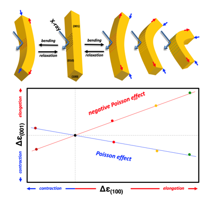
Elastic bending for macroscopic deformation. Individual crystals were mechanically tested to assess their elastic properties (Fig. 3a and Supplementary Video S1). A single crystal (thickness: 168 µm, width: 320 µm, length: 12 mm) was fixed to a metal pin with adhesive. Figure 3b and Video 1 show the mechanical bending–relaxation performance of the crystal. Bending stress was applied by pushing the crystal with a glass plate. The straight crystal bent under an applied stress in the c direction and then recovered its original shape upon releasing the stress. Notably, the reversible bending–relaxation of the crystal could be repeated many times (Figs. 3b-l). This mechanical behavior clearly indicates that the crystal is an elastic (bendable) organic single crystal. The crystal bending angle exceeded 70° (Fig. 3k). The elastic organic crystals did not exhibit slipping between the planes and on visual inspection there was no detectable change in the angle between the ends of the crystals. Thus, the crystal underwent elongation at the outside and contraction at the inside of the bend (Supplementary Fig. S1). The elastic strain (εn) of the crystal in the c-direction was estimated from the curvature of the bent crystal and the equation εn = d/2r, where d corresponds to the width of the (001) plane (d = 168 µm) and r is the radius. The values of εn were 1.02%, 1.19%, 1.40%, 1.70%, and 2.80% (Figs. 3c, 3 g, 3i, and 3 k). In contrast, the crystal did not bend under an applied stress in the b-direction, i.e., the elasticity was limited to one direction (directional-specific elasticity). Figure 3m schematically illustrates the elastic bending test using a pair of tweezers and a needle. The crystal bent without breaking when the (001) plane was face-up and the bent crystal quickly recovered its original straight shape without any breaking or crack formation upon withdrawal of the force (Figs. 3n and 3o). In contrast, under elastic bending, when the (010) plane was face-up the crystal was brittle (Figs. 3p and 3q). In this case, the inflexible crystal fractured. This direction-depended elasticity is related to anisotropy of the molecular arrangement in the crystal (Fig. 2c).
X-ray analysis of nanoscopic structural changes. To link the directivity of the crystal to its crystal structure, one-dimensional (1D) X-ray diffraction (XRD) analysis of the crystal was performed with an original set-up (Supplementary Fig. S2). Patterns derived from the lamellar structure for the c-axis were detected when the crystal was positioned parallel to the substrate (Fig. 4a). The Bragg equation was used to calculate the length of the original crystal (010) face up to be 12.367 Å (7.12°), which corresponds to one lamellar layer in the c-direction (Fig. 4b). Thus, the face parallel to the substrate is the (001) face. To determine the changes in the structure induced by bending and relaxation, 1D XRD analysis of the front and back of the straight (original and relaxed) and bent crystal was conducted by performing measurements with the crystal set at different curvatures (Fig. 4a). The elastic strain (εn) of the crystal in the c-direction was also estimated from the width of the (001) plane (d = 168 µm) and the radius (r = 3, 5, and 7 mm) of the curved jigs. Upon bending (εn = 1.2%), the patterns at the front of the crystal shifted to a lower angle (7.04°), which corresponded to a distance of 12.556 Å (Fig. 4b). The patterns returned to their original positions upon releasing the stress. Upon re-bending (εn = 1.68%), the patterns shifted to an even lower angle (6.98°, 12.657 Å). Recovery of the pattern upon relaxation was also observed. In the case of a greater bending (εn = 2.8%), a further shift to a low angle was observed at the front of the crystal (6.94°, 12.723 Å). Furthermore, the patterns from the back of the bent crystal (εn = 0.84%), shifted to a higher angle (7.18°, 12.303 Å). These analyses point to a change of the lamellar distance in the c-direction, d0 → dx upon bending (Fig. 4c).
Plots of strain (%) from curvature [Δε(100)] against the degree of change, Δε(001), from XRD results, and εn showed a correlation between strain and the change of lamellar distance (Fig. 4d). Hence, the elongation or contraction in the a-direction induced elongation or contraction in the c-direction, respectively (Fig. 4e). The calculated Poisson’s ratio, v, is defined as the ratio of the change in the width per unit width of materials (plastics, metals), to the change in its length per unit length, as a result of strain. Typically, v values are in the range of 0.2–0.5 associated with a contraction of the width of a material when it is stretched (Fig. 4f). Here, the v value of the cell unit (001) can be estimated from the a-axis elongation, as calculated from the bending strain and the variation ratio of c-axis: v(001) = − Δε(001) / Δε(100).16 Notably, the v(001) value of this crystal was approximately − 1.0 (Fig. 4d). A negative value of Poisson’s ratio (negative Poisson effect) represents an unusual deformation mode in material science,18 but characteristic examples are based on molecularly dense and well-organized organic crystals.
To investigate the applicability of this method to other crystals, similar measurements were performed for an elastic 9,10-dibromoanthracence crystal. A reversible peak shift occurred as the shape changed from straight to bent (Supplementary Fig. S3a). Importantly, the degree of change in the peak shifts of [Δε(001)] and [Δε(100)] changed with the curvature. In this crystal, contraction of the c-axis occurred rather than elongation of the a-axis. However, because elongation of the c-axis occurred with respect to contraction for a-axis, the behavior was different from that of the elastic β-COPV crystal (Supplementary Figs. S3b and S3c). Hence, this crystal showed a typical Poisson effect with a v(001) value of 0.23–0.25.