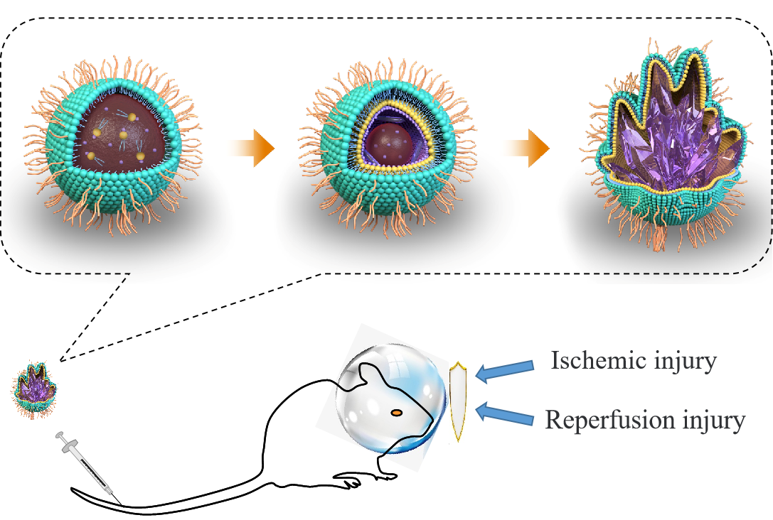
Preparation and characterization of OSDP LNPs
A key feature of the research is the precursor mediated, controlled drug crystallization inside the liposomes. In order to obtain nanosized crystals in the liposomes, the traditional nanoprecipitation technique were optimized. The hydrogen bond were formed between OEA and lecithin (SPC) via our previous method [18, 20], which was confirmed by H1NMR and XRD. In the H1NMR spectra (Fig. 1A), the peak at 7.76 ppm (ascribed to the –NH– of OEA), the peak at 4.63 ppm (ascribed to the –OH of OEA), and the peak at 3.47 ppm (ascribed to phosphatidylcholine of SPC) appeared to obviously weakened in the OEA–SPC complex, illustrating the formation of hydrogen bonds. The disappear of the sharp peaks of bulk OEA in the XRD pattern of OEA-SPC also confirm that the hydrogen bonds might have formed between OEA and SPC molecules (Fig. 1B).
Afterwards, extra OEA and OEA–SPC were dissolved in dichloromethane, which was then dispersed in DI water containing DSPE-PEG under ultrasonic conditions. After stirring for one hour, the system turned into a stable, white O/W suspension with the dispersion of the organic droplets in the continuous phase. In this stage, there was an arrangement of DSPE-PEG molecules with the hydrophobic groups of DSPE around the organic droplets and the PEG groups in the continuous phase. The droplets could be seen as precursors, which might proceed via non-classical crystallization routes. Lastly, with dichloromethane rapidly removed by rotary evaporation, the multistage crystallization process proceed. Due to the evaporation of the organic phase, water molecules entered the droplets, resulting in the precipitation and arrangement of SPC-OEA complex. Hence, phospholipid bilayers were prepared according with the arranged DSPE-PEG molecules. The bond of OEA to SPC molecules would serve many nucleation sites, leading to a number of OEA crystal nucleus. The rapid evaporation of the organic phase would induce a high supersaturation, under which the OEA crystal nucleus grew into OEA nanocrystals and rapidly reached the thermodynamically stable crystalline form. Since the phospholipid bilayers were flexible, their shapes might be changed by OEA nanocrystals. Followed by extrusion through polycarbonate membranes (0.22 µm pore diameter), irregularly non-spherical shaped liposomes, with drug crystal inside and phospholipid bilayers outside, were successfully prepared. The representative structure illustration of the OSDP LNPs is depicted in Fig. 1D.
As illustrated in Fig. 1C and Figure S1, the OSDP LNPs were around 100 nm with an irregularly non-spherical shape. It could be noticed that the black fields inside of the liposomes appear darker than the fields forming the contour of the liposomes, which appear to be a paler shade of grey (Fig. 1C-b and c). The inside dark field might owe to the existence of OEA crystals, whose molecules arranged well-ordered and closely. Since the electrons could not easily pass through the closely packed crystals, the inside field of the liposomes showed a deep color. On the contrary, the phospholipid bilayers were loosely arranged, leading to a higher electron transmittance and hence a light shade. The two different colored sections also illustrated the delicate hierarchical architectures of the OSDP LNPs.
X-ray diffraction was also employed to detect the form of OSDP LNPs. As shown in Fig. 1B, the sharp peaks of bulk OEA would disappear with the formation of hydrogen bonds in the OEA-SPC complex. Nevertheless, a majority of sharp peaks of OSDP LNPs belongs to OEA, suggesting its high crystallinity. Although, the OEA within OSDP LNPs shows the same polymorphic form as their bulk counterparts, the width of the peaks have reduced a lot, which might attribute to the nanoscaled crystal size of OEA within OSDP LNPs. Meanwhile, the coverage of lipid bilayer on the OEA nanocrystals led to the weakness of OEA.
Stability test and in vitro drug release study
The biggest advantage of the OSDP LNPs lies in the sustained drug release property with a high drug loading. The drug loading of OSDP LNPs could reach up to 15.9 ± 1.2 wt%, while that of the OEA-SPC LNPs collected from the traditional method ranged from about 0.5 wt% to 3.0 wt%. With the formation of hydrogen bonds, the drug loading increased to about 8.0 wt%. When the nanoscaled OEA crystal appeared within the liposomes, the drug loading was redoubled to 15.9 ± 1.2 wt%. Although the drug loading of many liposomes might come up to more than 50 wt%, the retention of highly hydrophobic drugs is still a critical bottleneck.20 The drug loading of many liposomes was less than 10 wt%, or even less. In this research, the formation of hydrogen bonds, plus the crystallization within the liposomes, greatly enhanced the retention of OEA with highly hydrophobic property. A well-established study showed that drug release could be controlled by the drug-loading content of the particles. High drug loading means severe burst release, while poor drug loading brings well controlled drug release [21]. As to OSDP LNPs (Figure S2), higher drug loading also led to a little burst release within the first 8 h, which might come from the exposed OEA crystals and the OEA on the surface of the liposomes. Compared to the OEA crystals without the coverage of lipid bilayer, this slight burst release could be neglected. Just like the OEA-SPC NPs with a drug loading of 8.2 wt%, the OSDP LNPs exhibited a remarkably prolonged and controlled drug release in the next 40 h, which could almost be seen as zero-order release. That is to say, the enhanced drug loading did not reduce the controlled release property of the liposomes. This was owing to the unique construction of the liposomes. Unlike other drug crystals, the OEA crystals was located in the center of the OSDP LNPs, covered with lipid bilayers and bonding with the SPC molecules, which would largely limit the diffusion of the drug. Hence, the OSDP LNPs possessed a steady sustained release pattern throughout the release period. Moreover, the lipid bilayers, the PEG chains, the monodisperse, the high Zeta potential (-16.1 mV, Figure S3) and the small particle size contributed to the stability of the OSDP LNPs. With minor size changes, the OSDP LNPs could preserve stability in an aqueous environment for 48 hours (Figure S4). On the contrary, the OEA-SPC NPs (d = 213 nm) without coating of PEG chains would aggregate within 6 hours. And the lyophilized OSDP LNPs redissolved easier than OEA-SPC NPs. The redissolved dispersion of lyophilized OSDP LNPs possessed a size of about 180 nm and was still applicable for intravenous injection within 1 month, while the OEA-SPC NPs couldn’t (Figure S5). The reason was the strong hydrophilia of PEG, which could stabilize the liposomes and make them more soluble in water.
Assessment of the neuroprotective effect of the OSDP LNPs in vivo.
To assess the in vivo neuroprotective effects of the OSDP LNPs on ischemic cerebral injury, systematic experiments were performed on rats. According to our previous study [30, 35], OEA could obviously reduce the cerebral infarct volume of the brains from the MCAO rats. Hence, it could be speculated that a lot of ischemic tissues were preserved to be ischemic penumbra with the administration of OEA, which could be salvaged by timely reperfusion. To visualize the ischemic penumbra and its recovery, positron emission tomography (PET) of [18F]fluoro-2-deoxy-D-glucose ([18F]FDG) was employed to assess the cerebral glucose metabolism [36, 37]. PET data were acquired for 20 minutes at 0 h, 1 h, and 2 h after reperfusion. Since the inflammation-related high-[18F]FDG uptake might appear around 3 d post-reperfusion [38], PET data were collected within 2 h post-reperfusion to eliminate the effect of inflammation. As shown in Fig. 2a, the right cerebral hemisphere of all the operated rats exhibited significantly reduced glucose metabolism compared with the left cerebral hemisphere at 0 h. The reason was that the blood supply of the right cerebral hemisphere was cut off and [18F]FDG could not arrive. Then, with 1 h of reperfusion, the hypoperfused area began to recover in all the operated rats, and their uptake of [18F]FDG began to increase. Interestingly, about 41.7% of the right cerebral hemisphere from the rats administrated with OSDP LNPs exhibited significantly elevated signals compared with the normal tissues (the blue dashed outline in Fig. 2a). And the enhancement extended to 83.6% of the right cerebral hemisphere at 2 h post-reperfusion. The PET intensity of these regions was about 63.24 ± 2.43 kbp/mL, which was much higher than that of the left cerebral hemisphere (51.76 ± 2.58 kbp/mL). In these regions, the cutting-off blood flow and thereby glucose supply is compensated by an increase of glucose uptake and phosphorylation rate to maintain cellular energy consumption [39], which might be used for repairing the cell damage caused by hypoperfusion. The enhanced glucose metabolism also stated the good cell viability in the region, and the region was called “the ischemic penumbra” in clinic. The ischemic penumbra would recover and functioned well afterwards, which could be verified via the TTC-stained brain slices (Fig. 2b). It could be observed that almost all the hypoperfused brain tissues, whose blood supply had been cut off for 90 min, were not infarcted via the administration of the OSDP LNPs. The results indicated that the hypoperfused brain tissues with elevated PET signals after reperfusion were mostly likely to recover. On the contrary, the other MCAO rats did not exhibited enhanced glucose metabolism in the ischemic brain tissues. Although the brains were recovering with reperfusion, the low-level glucose metabolism stated their terrible cell viability at 1 h post-reperfusion. What’s worse, an obvious infarction core appeared at 2 h post-reperfusion (The red dashed outline in Fig. 2a). While the other hypoperfused brain tissues possessed similar glucose metabolism to that of the left cerebral hemisphere, they had suffered from irreparable damage owing the acute ischemia and might ultimately die. Many hypoperfused brain tissues with certain PET signals at 2 h post-reperfusion were infarcted as shown in the TTC-stained brain slices of 24 h post-reperfusion (Fig. 2b). Then the cerebral infarct volume and the cerebral edema degree of all the rats were calculated (Fig. 2c-d). With a cerebral infarct volume of about 372.2 ± 26.9 mm3 and a cerebral edema degree of 13.6 ± 1.0%, the operated rats without treatment had a severe brain damage, which would lead to a serious neurological deficit. With the treatment of free OEA, the cerebral infarct volume of the rats was reduced to 332.2 ± 35.3 mm3 and the cerebral edema degree to 11.3 ± 1.1%. However, there was no significant difference between the OEA and MCAO groups (P > 0.05), which was in according with our previous studies [30]. Nevertheless, the cerebral infarct volume was decreased to 78.2 ± 18.4 mm3, and the cerebral edema degree was also decreased to a slight level. The data stated that the rats treated with OSDP LNPs suffered from much less brain damage than the rats of the other groups. What’s more, the photos of the TTC-stained brain slices, plus the PET data, forcefully demonstrated that the acute ischemic brain tissues could be preserved as penumbral tissues to a great extent via the administration of OSDP LNPs. And the preserved penumbral tissue would bounce back with reperfusion and possess an increased glucose metabolism within hours to compensate the reduced supply, leading to a better and more rapid recovery.
As an acute disease, ischemic stroke possesses a high mortality, and reducing death rates must be the primary task to a formulation for stroke. As shown in Fig. 2e, only 45.8% of the MCAO rats could survive for 14 d, and the administration of free OEA could not obviously change this data. In comparison, OSDP LNPs could effectively protect from MCAO and increase the survival rate to 83.3%. Furthermore, we speculated that the rats administrated with OSDP LNPs were more likely died from the complications of MCAO, not from the cerebral injury. The small cerebral infarct volume illustrated that they suffered from a little cerebral injury (Fig. S6). The result indicated that the administration of OSDP LNPs might extend the time window for beneficial reperfusion.
Since the behavior of the stroke patients was one of the most important evaluation indicators in clinic, the behavior ability of the rats was evaluated via Garcia method. The Garcia scores of the rats were evaluated at 1, 3, 5, 7, and 14 d after operation. As shown in Fig. 2f, the sham-operated rats got full marks, while the operated rats only got about 10 points in the 1st assessment, illustrating the neurological function deficit of the models and the success of our operation. The rats administrated with OSDP LNPs performed much better than those of the other two operated groups throughout the assessment (p < 0.01). In addition to getting higher scores, the rats administrated with OSDP LNPs maintained enhanced recovery rate within the first week after operation, which might come from the preserved penumbral tissue. Under the protection of OSDP LNPs, more penumbral tissues were preserved and revived with reperfusion, leading to a better and more rapid recovery. On the contrary, OEA seemed to have no effect on improving neurological function deficit, owing to its extremely low bioavailability.
Morris water maze task.
Learning and memory are one of the most important computational strategies of the brain, which might be impaired by ischemia stroke. To examine the effects of OSDP LNPs on spatial learning and memory, rats were exposed to the water maze task. Between day 15 and 19 after operation, the rats were trained with five trials per day. Spatial learning ability was assessed by escape latency (the time required to find the platform in training). Compared with the sham group, the operated rats without treatment exerted much longer escape latency, illustrating their impaired spatial learning ability (Fig. 3A). Owing to their severe brain damage, two rats even could not find the platform within 120 seconds in the third training, while all the sham rats could reach the platform within 60 seconds in the first training. Compared with those of the MCAO group, the rats treated with OSDP LNPs needed less time to find the platform, stating their improved spatial learning ability. Moreover, the training seemed to be more effective to the rats of the nanodrug group, leading to a result that the escape latency was decreased from 75 s in the first training to 23 s in the fourth training (Fig. 3A). The result also illustrated their strong learning ability. At last, the data had no significant difference with that of the sham group, forcefully illustrating that the OSDP LNPs treatment effectively protected the spatial learning ability of the operated rats. Interestingly, the sham-operated rats exhibited significantly smarter than the operated rats after the first training. When entering water in the second training, many of them looked around to confirm the position of the platform, and straightly swam to it. Two rats treated with OSDP LNPs began to have the same performance in the fourth training, while the rats of the other groups just swam around to find the platform throughout the training.
When the platform was removed, the rats of the sham group and the nanodrug group exhibited markedly increased target crossing times (Fig. 3B), indicating their stronger potential memory for the removed platform. Moreover, it could be seen from the traces that the rats of the sham group and the nanodrug group always swam around the position of the removed platform (Fig. 3C). This strongly indicated that they had significant purpose and strong memory for the removed platform. On the contrary, the rats of the other groups possessed much less target crossing times and almost random traces, indicating their little memory for the platform. The results stated that the OSDP LNPs greatly ameliorated ischemia-induced spatial memory impairment.
Immunofluorescence staining and cell counting.
The intractable sequelae of stroke mostly resulted from the injury of neurons, which were really hard to recover. Here, TUNEL staining was employed to evaluate the apoptosis of the neurons in hippocampal CA1, which mainly involved in memory and cognition. As shown in Fig. 3D, none apoptotic cell was detected in the hippocampal CA1 of the sham-operated rats, while a significant increase in apoptotic cells could be found in those of the MCAO group and the OEA group. The results stated that the MCAO was likely to induce the apoptosis of the neurons, and this could not be improved by the administration of free OEA. In contrast, only one apoptotic cell was observed in the field of the nanodrug group. This significant decrease was likely related to the improved learning and memory ability of the rats of the nanodrug group. The result also indicated the nice neuroprotective effect of OSDP LNPs.
According to our previous studies [35, 40, 41], OEA could inhibit the inflammation of reperfusion, which was one of the most primary causes of the brain injury. Microglia and astrocyte play paramount roles in the brain inflammation, which could be specifically marked by Iba-1 and GFAP, respectively. Hence, the quantity of expressed Iba-1 and GFAP was used to evaluate the inflammation of reperfusion. As depicted in Fig. 4A and Fig. 4B, both markers suggested that the inflammation was slight in the sham-operated rats, while a significant increase could be observed in the cortex around the ischemic focus of the MCAO group and OEA group. With the administration of OSDP LNPs, the inflammation was decreased to a low level. The result indicated that the OSDP LNPs could greatly alleviate the inflammation induced by ischemic reperfusion, and therefore provide significant neuroprotective effects.

