3.2 Single-cell transcriptomics of mADSC-treated and untreated pulmonary resident cells
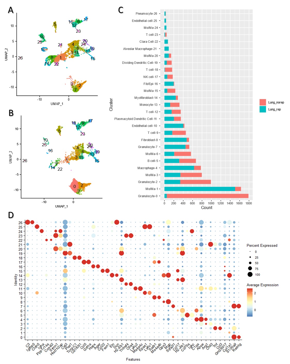
In order to understand the effects of mADSCs in the inflammatory lung microenvironment, we compared the single-cell transcriptomic profiles of pulmonary resident cells from the BLM + mADSCs group with those from the BLM + PBS group. We injected the GFP-labeled mADSCs intratracheally into BLM-treated mice on day 3, and the mADSCs and lung parenchyma cells were sorted from mouse lungs on day 7 by flow cytometry (Fig. 2a).
Firstly, we compared single-cell profiles of mouse lung parenchyma cells from the BLM + PBS and BLM + mADSCs groups (Fig. 2b), with libraries comprising 8181 and 6405 cells, respectively. A total of 27 cell clusters were identified in each group of lung parenchyma cells (supplementary Fig. 1a, b), and we identified 12 discrete cell types for each group based on cell markers with no cell cycle effects on data architecture (Fig. 2b). The identified lung cell types included pneumocytes, bronchiolar exocrine cells, natural killer cells, fibroblasts/epithelial cells, myofibroblasts, dendritic cells, endothelial cells, T cells, B cells, monocytes/macrophages, and granulocytes (Fig. 2b, c). Notably, the immune cells mainly included monocytes/macrophages in the BLM + mADSCs group (48.78%), and granulocytes in the LM + PBS group (44.87%) (Fig. 2d). Fibroblasts, endothelial cells and myofibroblasts were the main non-immune cells in the BLM + mADSCs group, accounting for 7.45, 6.83, and 3.80%, respectively.
Monocytes/macrophages were the dominant cell type in both the BLM + mADSCs group (48.78%) and the BLM + PBS group (27.28%) (Fig. 3a), and were clustered into 9 subclasses based on their gene expression profiles (Fig. 3b). The Mo/Ma-1 subclass, characterized by the expression of the marker genes MPG, COL1a1, and SPARC, was the most abundant subpopulation (48.71%). The macrophage-4 subclass, expressing GPNMB, PF4, and APOE, was the second most abundant subpopulation in the BLM + mADSCs group (20.42%). By contrast, the alveolar macrophage-21 and the Mo/Ma-24 subclasses were practically not detectable in the BLM + PBS group (Fig. 3b), 0 and 0.07%, respectively.
We next examined the function of genes exhibiting significant changes of expression levels in the Mo/Ma-1 and macrophage-4 subclasses based on gene ontology (GO) and KEGG pathways analysis (Fig. 3c, d). The results indicated that the Mo/Ma-1 subclass was related to the regulation of the extracellular matrix (ECM) and proteoglycans, non-integrin membrane-ECM interactions, collagen chain trimerization, collagen degradation, and crosslinking of collagen fibrils, with high expression of COL1a1, COL1a2, and SPARC genes.
3.3 scRNA-seq of mADSCs retrieved from mouse lungs
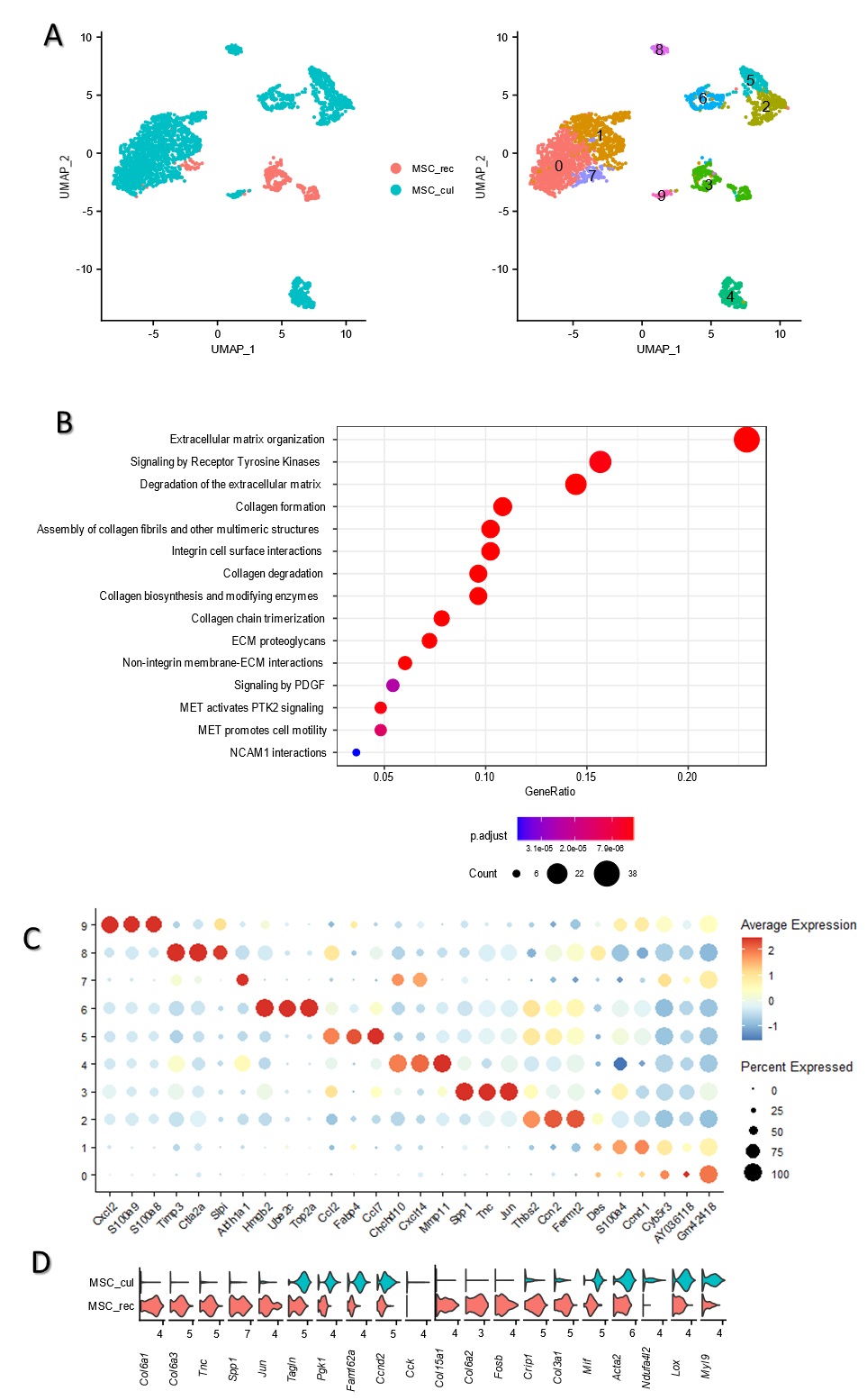
To investigate the potential therapeutic mechanism of intratracheally injected mADSCs in the BLM-injured inflammatory lung microenvironment, we analyzed the single-cell transcriptomic profiles of retrieved mADSCs from mouse lungs on day 7, i.e., 4 days post-injection of mADSCs. We intratracheally injected the GFP-labeled mADSCs (5×10⁵ cells in 50µL of PBS per mouse) into BLM-treated mice on day 3 and the mADSCs were retrieved from mouse lungs on day 7 by fluorescence-activated cell sorting (FACS). On day 7, the moue lung was extracted and a single-cell suspension was prepared by enzymatic digestion to retrieve the GFP-expressing mADSCs by FACS. The retrieved mADSCs were subjected to for 10× scRNAseq analysis, with in-vitro cultured mADSCs as control.
We retrieved a total of 417 GFP + cells from three mice lungs and pooled them together. At the same time, 2801 culture cells were sorted, and a single mRNA sequencing library was constructed for each group. When comparing the retrieved mADSCs with in-vitro cultured cells using UMAP cluster analysis, we found a great heterogeneity among the retrieved mADSCs (Suppl. Figure 2a). The gene expression pattern of retrieved and in-vitro cultured mADSCs is highlighted in Suppl. Figure 2d. Notably, the retrieved mADSCs exhibited high expression level of COL6a1/2/3, TNC, AAP1, FOSB, CRIP1, and COL3a1, which were practically not expressed by in-vitro cultured mADSCs.
We further analyzed the functions of genes with significantly changed expression levels in the retrieved mADSCs using gene ontology (GO) analysis. The upregulated genes of the retrieved mADSCs were found to be related to ECM remodeling in the BLM-injured lung microenvironment (Suppl. Figure 2b), with GO terms such as Extracellular matrix organization, Degradation of the extracellular matrix, Collagen formation, Assembly of collagen fibrils and other multimeric structures, Collagen degradation, Collagen biosynthesis and modifying enzymes, Collagen chain trimerization, as well as Non-integrin membrane-ECM interactions pathways reflected in the significant upregulation of APP, BMP1, CD44, COL2a1, and COL5a1 (Fig. 2d).
3.4 mADSCs alleviate lung fibrosis by promoting the polarization of macrophages to an anti-inflammatory phenotype and downregulating pro-inflammatory cytokines in the early stage
Although the retrieved mADSCs did not show significant expression of anti-inflammatory cytokines, we were able to confirm that the abundance of TREM2+ macrophages (cluster 4 identified by scRNA-seq) was increased at day 7 in the BLM + mADSCs group, as indicated by the stronger fluorescence signals of TREM2 and SPP1(OPN) (Fig. 4a). However, APOE, which was highly expressed by most of the monocyte/macrophage subgroups, was not found to be upregulated, suggesting that it may not participate in the anti-inflammatory activity of these macrophages.
Similarly, qPCR showed that the pro-inflammatory cytokines tumor necrosis factor-alpha (TNF-α) and interleukin-1 beta (IL-1β) were significantly downregulated in the mouse lungs at day 7, while anti-inflammatory or functional cytokines IL-10 and IL-6 exhibited the opposite trend (Fig. 4b).
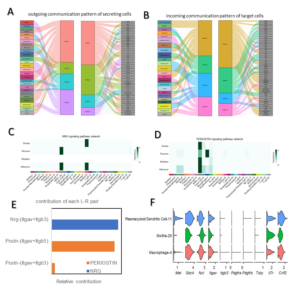
To investigate the interplay between the injected mADSCs and the lung cells in the BLM-injured lung microenvironment, we first analyzed the lung cells and retrieved mADSCs together, and visualized their outgoing and ingoing communication patterns. We identified the global communication patterns and the key signals in different cell subgroups by employing the pattern-recognition algorithm CellChat. The four outgoing communication patterns were analyzed and four incoming communication signals were identified (Suppl. Figure 3a). Pattern #1 accounted for the largest portion of outgoing signaling, which represents TGF-β, GDF, VEGF, XCR, and IL2, produced by immune cells such as Mo/Ma-20, macrophage-4, NK cell-17 and T-cell-23. The outgoing signaling of recovered mADSCs was assigned to pattern #3, which included the ncWNT, EGF, PDGF, PTN, and PERIOSTIN signaling pathways (Suppl. Figure 3a). On the other hand, the incoming communication pattern of target cells showed that the injected mADSCs received BMP, WNT, EGF, FGF, PDGF, and OSM signals in the BLM-injured lung microenvironment, which are assigned to incoming communication pattern #2 (Suppl. Figure 3b).
We detected 51 significant ligand-receptor pairs among the 28 cell groups/clusters including the retrieved mADSCs, which were categorized into NRG, PERIOSTIN, TGF-β, HGF, PDGF, TNF, WNT, non-canonical WNT (ncWNT), SPP1, PTN, CXCL, and CCL pathways. We next found that the retrieved mADSCs received Mo/Ma-20 subgroup signals via the NRG signaling network (Suppl. Figure 3c) Using network centrality analysis (NCA), and ITGAV/ITGB3 were identified as the contribution receptors on mADSCs in the BLM-injured lung microenvironment (Suppl. Figure 3d). Similarly, the PERIOSTIN signaling pathway was identified as the main communication channel from mADSCs to the Macrophage-4 subgroup (Suppl. Figure 3d). Postn-ITGAV/ITGB5 was the dominant contributing ligand-receptor pair (Suppl. Figure 3e). In addition to these pathways, the mADSCs were also sending HGF to plasmacytoid dendritic cell-11 and pneumocyte-26 subgroups through the HGF signaling pathway, and HGF-MET was the main contributing ligand-receptor pair (data not shown).