Model preparation and grouping of beagle dogs with rapid atrial pacing
For the purpose of establishing a sustained AF model, nine beagles purchased from Hunan Silaikejingda Experimental Animal Co.,Ltd(China)were fasted and deprived of water for 12 h before surgery. Routine disinfection of surgical devices and the surgical environment was used. After appeasement, dogs were anesthetized intraperitoneally with ketamine (6–8 mg/kg) combined with diazepam (0.5–0.8 mg/kg). The dogs were placed in the right lateral position and fixed on the experimental bench. Skin preparation, disinfection, and draping were performed on the chest and neck. Intubation using a 6F-sized endotracheal tube was used to connect the ventilator with proper adjusted parameters: tidal volume, 20 ml/kg; respiratory rate, 20 times/min; and oxygen flow rate, 3.5 L/min. Subsequently, ketamine (6–8 mg/kg) + diazepam (0.5–0.8 mg/kg) were added intravenously every 15–40 min, according to the animal’s reactions (respiration, eyelid membrane reflex, etc.); safe dosages of ketamine (50 mg/kg) and diazepam (5 mg/kg) were used. Blood pressure and ECG monitors were connected to limbs, and electrocardiographic monitoring was conducted during surgery.
A transverse incision with a length of about 5 cm was made in the 5th intercostal space of the left chest, to separate the skin, subcutaneous tissue, muscle, intercostal external muscle, intercostal internal muscle, pleura wall layer, and pleura visceral layer. The pericardium was opened and a pericardial hammock was made, to expose the LAA.
In the model group, three dogs had their LAAs removed; in the positive control group, the LAAs were preserved in three dogs; and in the negative control group, the pericardium of three dogs was directly sutured after opening, without removing the LAA or performing continuous pacing.
In the model group and the positive control group, the pacing electrode was sutured to the epicardium of the left auricular root with non-invasive suture, the ECG was monitored using a Powerlab system, and a canine pacemaker monitoring system was used to adjust the stimulation intensity of the pacemaker to twice the pacing threshold, using the pacing mode of AOO and a pacing frequency of 400 min–1. After adjustment to a satisfactory pacing waveform using the pacemaker, the epicardial pacing lead was fixed. The pericardium and thoracic cavity were checked for hemorrhage, and the chest wall was closed layer by layer. The subcutaneous tissue and skin were sutured after the chest cavity was aspirated, and a capsular bag was placed in the neck with implantation of an experimental high-frequency cardiac pacemaker. The tail end of the cardiac pacing electrode was connected with the buried high-frequency cardiac pacemaker through the subcutaneous tunnel. The neck of the dog was protected by a collar after surgery. After modeling, 3 g/day of cefazolin was administrated continuously for 5 days, to prevent infection. The dogs were supported by nutrition for 1 week, and their symptoms and signs were observed.
Specimen acquisition
After 12 weeks of continuous pacing, the experimental dogs were sacrificed via air injection in the jugular vein. The gross heart specimens of the dogs were taken and weighed and the left ventricle and left atrium were separated and weighed separately. The left atrial tissues (the free wall of left atrium, septum, pulmonary vein vestibule, and LAA) were collected in a sterile state and stored in liquid nitrogen, followed by transfer into a freezer set at −80°C, for future use.
TMT quantitative proteomics
Protein extraction and digestion
The frozen samples were ground to powder in liquid nitrogen. Four times the volume of lysis buffer (8 M urea, 1% protease inhibitor) was added, and ultrasonic lysis was performed on ice. The supernant was retained and the rediment was discarded after 10min centrfugation. The protein concentration was measured using a BCA kit.
For digestion, the protein solution was treated with Dithiothreitol first for 30 min at 56°C, followed by iodoacetamide for 15 min at room temprature. Then the urea in the sample was diluted to a concentration <2 M by 100 mM NH4HCO3. Finally, the first digestion was performed overnight at a mass ratio of 1:50 (trypsin: substrate), and the secondary digestion for 4 h at a mass ratio of 1:100 (trypsin: substrate).
TMT labeling
The trypsic peptides were desalted with Strata X C18 (Phenomenex), vacuum freeze dried, dissolved with 0.5 M TEAB and labeled according to the operating instructions of the TMT kit (Thermo Fisher Scientific). The labeled peptides were desalted after mixing and vacuum freeze dried.
HPLC fractionation
The peptids were fractionated by high pH reverse HPLC using the Agilent 300Extend C18 chromatographic column. The peptides were first separated into 60 components by a gradient of 8% to 32% acetonitrile(pH 9), and then combined into 14 components, followed by freeze-drying under vacuum.
LC-MS/MS analysis
The peptides were dissolved in mobile phase A (0.1% formic acid and 2% acetonitrile) and separated using an EASY-nLC 1000 ultra-high-performance liquid system. 0.1% formic acid and 90% acetonitrile. The gradient was set as follows: 0–26 min, 9%–23% mobile phase B (0.1% formic acid and 90% acetonitrile); 26–34 min, 23%–35% mobile phase B; 34–37 min, 35%–80% mobile phase B; and 37–40 min, 80% mobile phase B, with the flow rate maintained at 400 nL/min.
Then the peptides were injected into NSI ion source and analyzed by Q Exactive Orbitrap Fusion mass spectrometry. The scanning range of the primary mass spectrometry was set to 400–1500 m/z, with the scanning resolution set to 60,000, while the scanning range of the secondary mass spectrometry was fixed at 100 m/z, with the secondary scanning resolution set to 30,000. A data-dependent procedure that alternated between one MS scan followed by 20 MS/MS scans with 30.0s dynamic exclusion. The
Database Search
Maxquant (v1.5.2.8) was used to search against dog uniprot database (CANIS_LUPUS_FAMILIES_9615_PR_20191025, 25496 sequences) concatenated , with reverse decoy database. Trypsin/P was set as cleavage enzyme allowing up to 2 missing cleavages. The mass tolerance for precursor ions was set as 20 ppm in First search and 5 ppm in Main search, and the mass tolerance for fragment ions was set as 0.02 Da. The cysteine alkylation was set as a fixed modification, and the oxidation of methionine, N-terminal acetylation of proteins, and deamidation (NQ) set as variable modifications. The quantitative method was set as TMT-10plex, with the FDR for protein identification and PSM identification set to 1%.
Significant changes in protein expression were indicated using fold changes of >1.2 or <0.8 as cut-off values. A double-tailed Fisher’s exact test was adopted, and the standard error detection rate control method was used to correct the multiple-hypothesis test. After the correction, significance was set at P < 0.05.
Bioinformatic Analysis
WoLF PSORT (https://wolfpsort.hgc.jp/) was used to annotate the submitted proteins regarding subcellular localization. The functional annotation of each protein was performed against COG/KOG database (http://www.ncbi.nlm.nih.gov/COG), UniProt-GOA database (http://www.ebi.ac.uk/GOA/), and KEGG pathway database. For pathway enrichment analysis, a two-tailed Fisher’s exact test was employed for the enrichment analysis of the differentially expressed proteins against all identified proteins. The terms with p values below 0.05 were treated as significant.
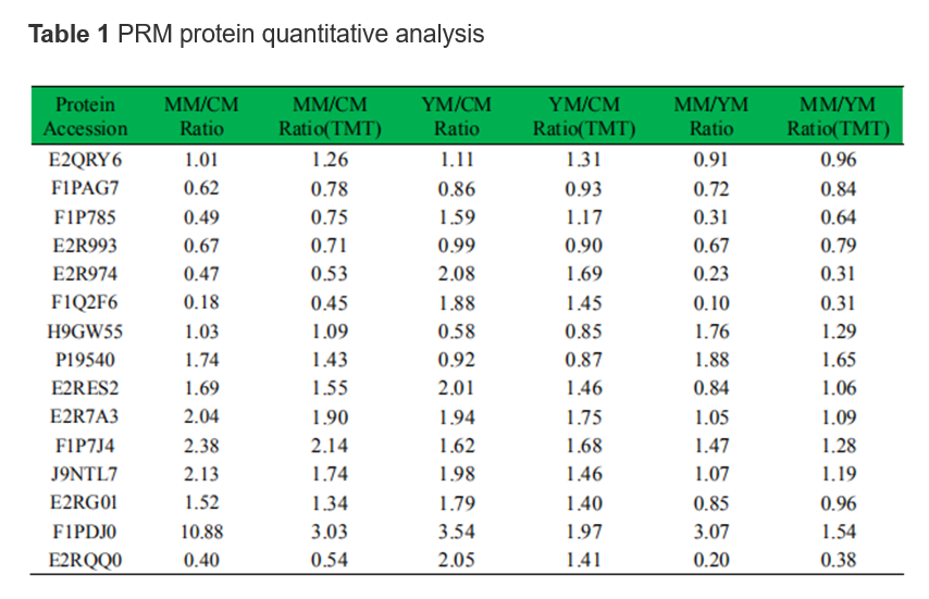
Validation of differentially expressed proteins
Parallel reaction monitoring (PRM) analyses were performed using a Q ExactiveTM Plus (Thermo) coupled online to the UPLC. The resulting MS data were processed using Skyline (v.3.6). Peptides parameters: enzyme was set to Trypsin [KR/P], max missed cleavage set as 0, the peptide length set as 7–25, and the cysteine alkylation set as a fixed modification. Transition parameters: the parent ion charges were set as 2, 3, the daughter ion charge set as 1, and the ion types set to b, y. The product ion selection started from the third to the last, with the ion match tolerance set to 0.02 Da.