Screening strain
About 400 pieces of rice straw and hay collected from all over the world such as Korea, China, Japan and the United States were used as samples to isolate microorganisms. A small amount of sterile saline is added to each sample and suspended, and the spore solution obtained by heat treatment in an 80℃ water bath for 20 minutes is used in an LB plate medium containing 2% agar (1% tryptone, 0.2% sucrose, 0.5% yeast extract and 0.5% NaCl, pH 7.0) and anaerobically cultured in an incubator at 55℃. for two days to isolate colony-forming microorganisms. Single colonies obtained by separating from hay were inoculated in 5 mL of 5% soy flour suspension medium and cultured with shaking at 37℃ for 24 hours, and then 60 species with high AGI activity in the supernatant were selected. The selected 60 strains were capable of growing for anaerobic growth at 50℃ and three strains with high AGI activity were selected as strains that were capable of using propionic acid. The selected three strains were identified as Bacillus licheniformis as a result of classification by taxonomic characteristics (Table 2) and analysis method according to Manual of Systematic Bacteriology (Bergey 2001).
Table 2. Bacillus licheniformis NY1505 physiological specificity.
|
Gram staining
|
+
|
Acid from
D-Glucose
L-Arabinose
|
+
+
|
|
55℃ growth
|
+
|
D-Xylose
|
+
|
|
Anarobic growth
|
+
|
D-mannitol
|
+
|
|
Propionate utilization (3%)
|
+
|
Hydrolysis of
|
|
|
Citrate utilization
|
+
|
Casein
|
+
|
|
Nitrate redued to nitrite
|
+
|
Gelatin
|
+
|
|
Voges-Proskauer test
|
+
|
Starch
|
+
|
The microorganism with the highest AGI production capacity was finally selected and to increase the AGI production capacity of the strain after inducing mutation by treatment with NTG (100μg/mL) to reach 99.9% kill rate, spread to 200-300 per sheet on L-broth plate medium and incubate anaerobically for two days at 37℃. The resulting colonies were randomly inoculated in 5 percent soybean flour medium and cultured with shaking at 40℃ for two days, and then the centrifuged supernatant was measured for the AGI activity. By repeating the mutation twice in the same way, a strain having high AGI activity was selected and named B. licheniformis NY1505. As a result of analyzing the 16s RNA nucleotide sequence of B. licheniformis NY1505 strain, it showed high homology with B. licheniformis. The dendrogram shows that B. licheniformis NY1505 (the accession number KCTC13021BP) is allied species with the B. licheniformis type strain (Fig. 1).
Analysis of AGI
5 x 10⁵ spores of NY 1505 were inoculated into 500g of steaming soybeans and covered the film, incubated at 37℃ for 24 hours, added 2.5L of 70% (v/v) ethanol, extract twice, and evaporated under reduced pressure. To 150 mL of the concentrated extract, 300 mL of hexane, dichloromethane, and Ethyl acetate were sequentially added twice, stirred for 2 hours, allowed to stand for 2 hours, and fractionated to wash the aqueous layer. After drying the aqueous layer, 100 mL of 90% ethanol was added to dissolve it, followed by silica gel column (100 mL) chromatography. The mobile phase was stepwise gradient from 1:1 solution of acetonitrile and methanol to 3 : 2 solution (Total 1000 mL). The AGI activity of each fraction was measured to obtain two fractions, AGI 1 and AGI 2. AGI 1 and AGI 2 each appeared as a single spot in thin layer chromatography, and the structure was determined through NMR analysis.
The chemical shift of AGI 1 is as follows.
1H-NMR (400 MHz C5D5N) δH, J (Hz)α = 4.71 (d, 1H, J = 1.8 Hz, H29a); 4.54 (d, 1H, J = 1.8 Hz, H-29b); 3.88 (d, 1H, J = 11.2, 4.4 Hz, H-11); 1.60 (s, 3H, H-30); 1.06 (s, 3H, H-25); 1.03 (s, 3H, H-23); 1.00 (s, 3H, H-24); 0.98 (s, 3H, H-27); 0.97 (s, 3H, H-26).
13C-NMR (100 MHz, C5D5N) δC = 216.1 (C-3); 169.4 (C-28); 148.1 (C-20); 108.1 (C-29); 68.2 (C-11); 59.7 (C-5); 59.4 (C-17); 52.7 (C-9); 42.7 (C-18); 41.8 (C-14); 41.7 (C-19); 40.8 (C-8); 39.5 (C-4); 38.5 (C-1); 34.1 (C-10,13,22); 37.1 (C-7); 33.8 (C-16); 30.1 (C-15); 28.5 (C-21); 26.9 (C-12); 26.7 (C-2); 24.7 (C-23); 19.4 (C-6); 17.8 (C-25); 19.5(C-26); 15.4 (C-24,30); 12.4 (C-27).
AGI 1 is a triterpene-type substance in which five rings are connected and has a structure similar to betulinic acid. The chemical structure was found to be 3-oxo-11α-hydroxy-lup-20(29)-en-28-oic acid.
The inhibition pattern of AGI 1 through the Lineweaver-Burk plot was confirmed as non-competitive inhibition (Fig. 2).
AGI 2 was presumed to be 1-deoxynojirimycin (DNJ), and as a result of running NMR, DNJ and NMR results were confirmed to be consistent. The chemical shift of AGI 2 is as follows.
1H-NMR (400 MHz D2O) δH, J (Hz)α = 3.96(d, 1H, J = 12.7, 3.5 Hz, H-6a); 3.89 (d, 1H, J = 12.8, 5.5 Hz, H-6b); 3.78 (m, 1H, H-2); 3.65 (1H, t, J =10 Hz, H-4); 3.52 (t, 1H, J = 9.5 Hz H-3,); 3.52 (s, 1H, J = 12.7, H-1a); 3.19 (m, H, H-5) 3.15 (s, 1H, J = 12.8, H-1b).
13C-NMR (100 MHz, D2O) δC = 78.1 (C-3); 70.2 (C-4); 69.4 (C-2); 62.2 (C-5); 59.8 (C-6); 48.5 (C-1).
AGI 2 showed a tendency to inhibit competitive inhibition (Fig. 3).
Animal experiment
High carbohydrate diet
After starting the diet and measuring the weight of each group every week, the lowest and highest values were excluded and statistically processed. In addition, more than 3g of fresh feces were collected for each cage to examine the number of microorganism.
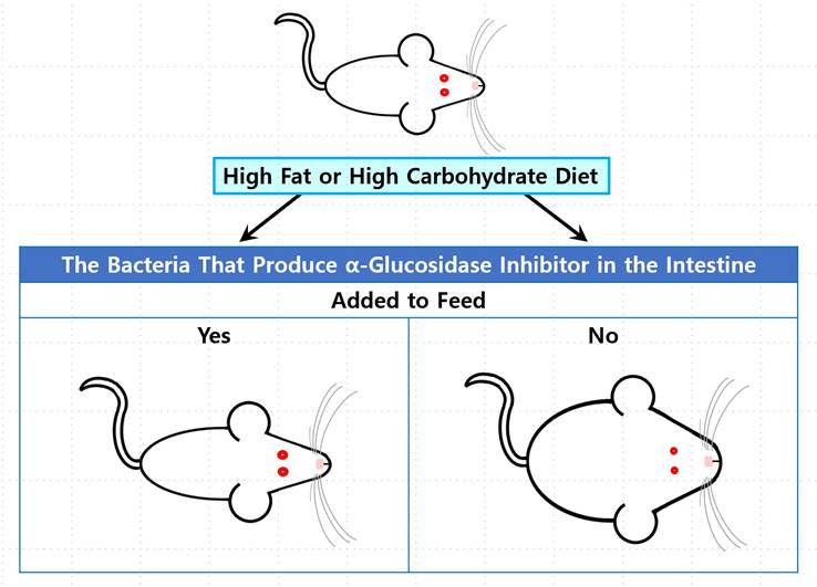
The high carbohydrate diet group showed a weight gain rate about 130% compared to the standard diet group in week 4, but the high carbohydrate diet group with spores of the B. licheniformis NY1505 strain showed the weight gain rate of about 90% compared to the standard diet group( Fig 4).
High fat diet
The high fat diet group showed a weight gain rate about 150% compared to the standard diet group in week 6, but when the high fat diet group with spores of the B. licheniformis NY1505 strain showed the weight gain rate about 120% compared to the standard diet group( Fig. 5).
Comparing the results in Fig. 4 and 5 the high fat diet group was more sensitive than the high carbohydrate group to the administration of B. licheniformis NY1505 spores at the week 3 or 4. In particular, it has been reported that when DNJ, a component of AGI 2, is administered for a long period of 12 weeks or longer, it activates β-oxidation, which decomposes fatty acids in mitochondria, and inhibits liver fat formation (Tsuduki et al. 2013; Tsuduki et al. 2009). Therefore, this is expected to be because B. licheniformis NY1505 proliferates in the intestine and produces AGI that activates β-oxidation, which is the catabolic action of fatty acids. It is known that betulinic acid, which has a similar structure to AGI 1, also inhibits adipogenesis by inhibiting differentiation in adipocyte growth (Mohsen et al. 2019).
The number of B. licheniformis NY1505 microorganism excreted in feces
When fecal extract samples with sterile saline are anaerobically cultured at 55℃ in a medium containing 3 percent propionate, only B. licheniformis grows selectively. So the surviving strain in this culture condition were determined as B. licheniformis NY1505. A mouse ingested about 5 x 10⁵ spores per day from the feed. Three weeks after spore administration, about 107 cells of B. licheniformis NY1505 were detected per 1g of feces. This strain proliferates vigorously in the intestine and is excreted (Fig. 6). Spores were administered for seven weeks and the number of microorganism rapidly decreased at 8th week when not administered for one week. It can be judged that it inhabits temporarily without adhering to the intestine.
The number of α-glucosidase inhibitor NY1505 microorganism excreted in feces
The B. licheniformis NY1505 strain proliferates vigorously in the intestine and produces AGI. From the 2nd week when the B. licheniformis NY1505 strain which had proliferated in the intestine was being detected in the feces, AGI that produced in the intestine was excreted into the feces equally (Fig. 7). As shown in Fig. 6, the amount of B. licheniformis NY1505 excreted is stabilized, and the amount of AGI excreted is also constant from 4thweek to 7th week. In other words, AGI is always continuously produced in the intestine, its concentration is maintained and a constant amount of the produced AGI is excreted. Considering that the AGI activity of natto produced from the B. licheniformis NY1505 strain is 90-95 units/g (data not shown), it can be seen that a significant amount of AGI is produced in the intestine and some of it is excreted.