Materials and reagents
Ampicillin sodium salt (# 69-52-3), vancomycin hydrochloride from Streptomyces orientalis (# 1404-93-9), neomycin trisulfate salt hydrate (# 1405-10-3), gentamycin sulfate (# 1405-41-0), erythromycin (# 114-07-8), metronidazole (# 443-48-1), Sucrose (# WXBC7932V) and TritonTM X-100 (#SLBV4122) were obtained from Sigma-Aldrich (St. Louis, MO). Phosphate buffer solution (PBS, # BF001) powder was purchased from Heart Biological Technology Co., Ltd (Xian, China). QIAamp Powerfecal DNA Kit (# 12830-50) was obtained from QIAGEN (Duesseldorf, Germany). Rabbit polyclonal antibody against GFAP (# Z0334) was purchased from Agilent Technologies, Inc (Santa Clara, CA). Mouse anti-β-Amyloid, 1-16 antibodies (# 9300-02) was obtained from BioLegend, Inc (San Diego, CA). Rabbit-anti-p2ry12 (# AS-55043A) was purchased from AnaSpec, Inc (Fremont, CA). Alexa FlourTM 555 donkey anti-mouse IgG (H+L) (# A31570) was obtained from ThermoFisher Scientific, Inc (Waltham, MA) and goat anti-rabbit IgG H&L (Alexa Flour® 488) (# ab150077) was purchased from Abcam, Inc (1:250, Abcam). 4’6-diamidine-2-phenylindole (DAPI) (# 10236276001) was obtained from Hoffmann-La Roche Ltd (Basel, Switzerland). Glycerol (# G8190) was purchased from Beijing Solarbio Science & Technology Co., Ltd (Beijing, China). Paraformaldehyde (PFA) (# 30525-89-4) was obtained from Shanghai Aladdin Biochemical Technology Co., Ltd (Shanghai, China). The identification and purification of peptide WN5 was previously reported elsewhere [44]. In this study, WN5 was chemically synthesized by GL Biochem (Shanghai) Co., Ltd (Shanghai, China) using L-isomers of each amino acid by solid-phase synthesis via the fluorenylmethoxycabonyl (Fmoc) method. It was then stored at -20 ℃ for subsequent usage.
APP/PS1 transgenic mice
APP/PS1 transgenic mice were obtained from Nanjing Biomedical Research Institute of Nanjing University (SCXK 2017-0174). APP/PS1 transgenic mice contains the KM670/671NL Swedish mutation of human amyloid precursor protein (APP) and the L166P mutation of human presenilin 1 (PS1) under the control of the Thy-1 promoter that lead to greater aggregation of amyloid-β in the cerebral brain. Mice were on the C57BL/6J background and maintained in the specific pathogen free (SPF) facility at the Institute of Laboratory Animal, Jinan University. Mice were allowed access to autoclaved food and water ad libitum. Wild-type female C57BL/6J mice were crossed with APP/PS1 male double transgenic mice to generate the APP/PS1 double transgenic mice and WT littermates. The animal experiments were conducted following the guidelines established by the Chinese Committee on Experimental Animal Supervision.
HEK-293-E22G cells
HEK-293-E22G cells, a cell line constructed by Aβ42-mCherry plasmid, were provided by Prof. Alan Tunnacliffe (Department of Chemical Engineering and Biotechnology, University of Cambridge, Cambridge, UK) and were used as the model. Cells were cultured in DMEM supplemented with 10 % heat-inactivated FBS containing 100 U/mL of penicillin, 100 μg/mL of streptomycin, 50 μg/mL of hygromycin B, 5 μg/mL of blasticidin S and 5 mM of L-glutamine at 37 ℃ under humidified air with 5 % CO2. Cells in exponential growth phase were used for the experiment.
In vitro treatments
Cell viability measurement
The effects of WN5 on the viability of HEK-293-E22G cells were determined by MTT assay. In brief, cells were seeded at a density of 5 × 10³ cells/mL in a 96-well plated for 24 h and then were cultured with different concentrations of WN5 (0.05 and 0.5 mM) for 48 h, respectively. After incubation, 20 μL of MTT was added to each well and the plates were further incubated for 4 h. The absorbance at 490 nm was measured by microplate ELISA reader.
Imaging and analysis
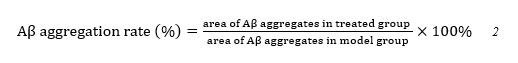
IncuCyte ZOOM live cell imaging system (Essen BioScience, MI, USA) was used to observe the Aβ-42 aggregates inside the cells. Cells pre-treated with WN5 (0.05 and 0.5 mM) were routinely propagated in culture medium. After 48 h, a solution of tetracycline was added to the cells and incubated at 37 ℃ for 72 h in the IncuCyte ZOOM apparatus and images of cells were recorded every 4 hours. We performed three identically prepared experimental replicates (n = 3) and 9-fields of vision were photographed in each hole. The Aβ aggregation rate was calculated according to formula 1. (see Formula 1 in the Supplementary Files)
Flow cytometry
In order to validate the above results, the Aβ aggregation in cells was quantified by ImageStreamx MKII imaging cytometer. The detection parameters were set up as follows: Channels 01 (bright field) and 04 (fluorescence channels). Magnification was 60x, providing a pixel size of 0.3 μm3 and the laser 561 nm activated for fluorescence of mCherry. For simple enumeration of pre-prepared microparticle samples, the acquision cut-off was set to 10000. The aggregation rate was estimated using formula 2. (see Formula 2 in the Supplementary Files)
In vivo treatments
FMT protocols
During the antibiotics phase, four APP/PS1 mice (3 month old) received antibiotic cocktail for 2 weeks by gavage. Mice assigned to the FMT (n = 5) study also received 2 weeks antibiotic cocktail prior to FMT. Mice then received oral gavage of 200 uL of donor fecal matter for 7 consecutive days. Fecal pellets were collected prior to antibiotic treatment (C1), following antibiotic exposure at days 7 and 15 (C2 and C3), during and after FMT at days 0, 4, 8, 15, 22, 30, 37 and 45 (C4, C5, C6, C7, C8, C9, C10, and C11). Gavage needles were cleaned with 70% ethanol and autoclaved after each experimental day.
Candidate treatments
After 1 week acclimatization, 6-month-old APP/PS1 mice were divided into four groups: vehicle treated wild-type mice group (n = 5, WT), vehicle treated APP/PS1 transgenic mice group (n = 5, AD), WN5-treated APP/PS1 mice group (400 mg/kg, n = 6, WN5), and Metformin-treated APP/PS1 group (500 mg/kg, n = 6, Metformin). Normal saline, peptide WN5 and Metformin were accordingly by oral administration for 12 weeks from the age of 6-months. Mice were sacrificed at the age of 9 months.
Behavior test
Cognitive function was assessed by Morris water maze and shuttle box tests, as previously described [19] in mice after 12 weeks of WN5 and Metformin intervention.
Immunostaining
5 months old APP/PS1 mice after FMT were sedated with pentobarbital and well-perfused with PBS and 4% PFA. Brain matter was dissected and hemispheres fixed in 4% PFA for 24 h, followed by 10%, 20%, and 30% sucrose gradient dehydrated for 24 h, until the brain completely sank to the bottom. Brain tissues from prefrontal lobe to hippocampus were processed into 25 μm per sections for immunostaining. There were 12 brain tissue sections on each slide that covered different brain areas (3750 μm). Sections were stained with anti-Aβ42 (1:200), anti-p2ry12 (1:200) and anti-GFAP (1:200) over night at 4 ℃. Subsequently, these sections were stained with Alexa FlourTM 555 donkey anti-mouse IgG (H+L) (1:250) or Goat anti-rabbit IgG H&L (Alexa Flour® 488) (1:250) at room temperature for 2 h. Then sections were stained with DAPI for 5 min and then mounted with 60% glycerol, and imaged with a 4x/20x objective on a Zeiss fluorescence microscope. Aβ plaques were analyzed with Fiji software and the morphology of microglia and astrocytes were analyzed with Fiji software using Sholl analysis.
Immunohistochemistry
After the behavior tests, WN5 and Metformin treated APP/PS1 mice were sacrificed and brain tissue was processed into sections (4-μm) for immunohistochemistry. Aβ immunohistochemical staining was performed on coronal slices by rabbit anti-Aβ42 antibody (1:1500, Abcam). Primary antibodies were detected with biotinylated goat anti-rabbit IgG (1:200) in conjunction with the DAB kit coupled with diaminobenzidine substrate. The stained sections of the β-amyloid plaques were observed and captured with an Olympus IX-73 microscope. Quantification of the β-amyloid plaque was performed on the hippocampus and sub-regions of the hippocampus using ImageJ software.
DNA extraction and 16S rRNA gene sequencing
Bacterial genomic DNA was extracted from mouse fecal pellets using the QIAamp DNA Stool Mini Kit. The library was generated according to a previously reported method. The V3-V4 regions of the 16S rRNA gene were PCR amplified using Forword primer 5’-ACTCCTACGG GAGGCAGCA-3’ and Reverse primer 5’-GGACTACHVGGGTWTCTAAT-3’, purified and then sequenced using the Illumina MiSeq platform according to the standard protocols. The 16S rRNA genes of the gut microbiota were analyzed using QIIME (version 1.17). Raw FASTQ files were processed to remain high-quality sequences and further clustered into operational taxonomic units (OTU) at 97% similarity using the Silva reference data base (Release128, http://www.arb-silva.de). OTUs were further classified into six taxonomic ranks of phylum, order, class, family, genus, and species. Principal component analysis (PCA) was performed to visually evaluate the differences and similarities in bacterial communities between groups (β-diversity) using Bray-Curtis method. Alpha-diversity was assessed using the species diversity indices (Inverse Simpson, Shannon, or Simpson).
Statistics analysis
Excluding the microbiome population statistics mentioned above, data from other experiments were presented as mean ± SEM (standard error of the mean). Differences between two groups were determined using two-tailed, unpaired Student t-test with Welch’s correction. Statistical significance among > 2 groups with only one variable was assessed using ANOVA followed by Bonferroni’s post hoc test. Krustal-Wallis test on ranks was performed for data that failed the normality test. Differences were considered significant at P < 0.05.