Bacterial Strains, cells and culture
S Typhimurium SL1344, the deletion of sseK3 Typhimurium SL1344 (∆sseK3 mutant) and its complemented (sseK3-complemented) were used in this study and conserved in the laboratory. The ∆sseK3 mutant was constructed through use of counterselectable suicide vectors. The sseK3 gene was cloned into the pBR322 plasmid for complementation studies. Macrophages RAW264.7 cells were obtained from the American Type Culture Collection (ATCC, Manassas, VA), and cells were cultured in Dulbecco's Modified Eagle Medium (DMEM)/high-glucose medium (HyClone, USA) containing 10% fetal calf serum (FCS) in an incubator at 37 ℃ and 5% CO2.
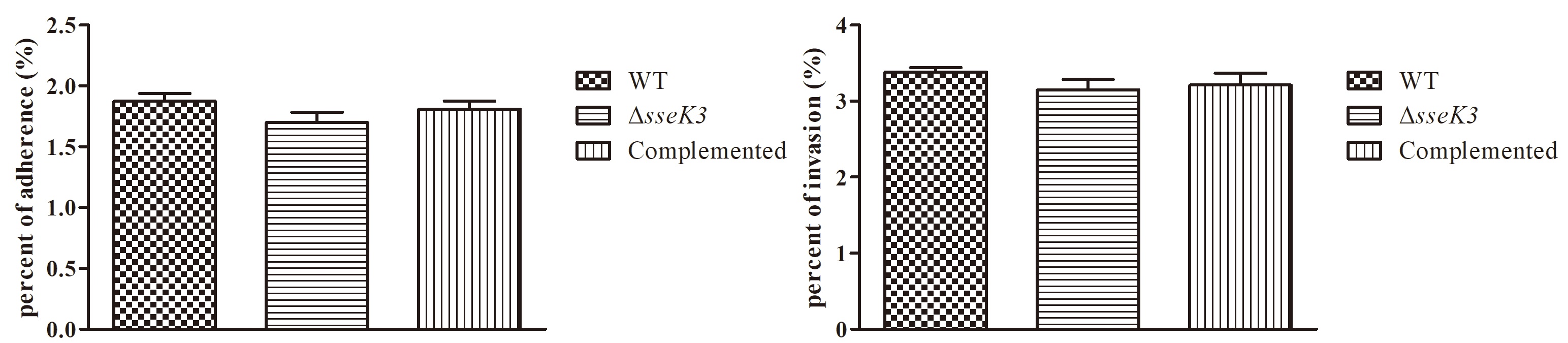
Adherence and invasion assay
Adhesion and invasion of RAW264.7 cells was assessed as previously described [53, 54]. A 24-well cell culture plate was inoculated with 1×10⁵ RAW264.7 cells per well and incubated for 16 h. WT, ΔsseK3 mutant and sseK3-complemented strains were coincubated with RAW264.7 cells at a multiplicity of infection (MOI) of 100:1, with three replicate wells per strain. To allow the bacteria to fully contact the RAW264.7 cells, the plates were centrifuged and incubated with 5% CO2 for 2 h at 37 ℃. For the adherence assay, the supernatants were aspirated, and the cells were washed three times with PBS. Subsequently, the cells were digested with 0.25% trypsin and plated in a gradient dilution and counted. For the invasion assay, the supernatants were aspirated, the cells were washed three times with PBS, and gentamicin-containing medium (100 μg/mL) was added and incubated at 37 ℃ with 5% CO2. After incubation, the supernatants were aspirated, and the cells were washed three times with PBS. Subsequently, the cells were lysed using 0.1% Triton X-100 and plated with a gradient dilution and counted.
Flow cytometry assay
A 6-well cell culture plate was inoculated with 1×10⁶ RAW264.7 cells per well and incubated for 16 h. WT, ΔsseK3 mutant and sseK3-complemented strains were coincubated with RAW264.7 cells at a multiplicity of infection (MOI) of 100:1, with three replicate wells per strain. To allow the bacteria to fully contact the RAW264.7 cells, the plates were centrifuged with 1000 rpm/min. Then, gentamicin-containing medium (100 μg/mL) was added and incubated at 37 ℃ with 5% CO2. After incubation, the supernatants were aspirated, and the cells were washed three times with PBS. The percent of cells undergoing apoptosis was detected by flow cytometry using Annexin V-FITC/PI apoptosis detection kit (KeyGEN BioTECH Jiangsu China). The cells of infected and mock groups were digested with 0.25% trypsin and washed three times with ice-cold phosphate buffered saline (PBS) and suspended in Binding Buffer with 500 μL, followed by adding 5 μL Annexin V-FITC and 5 μL Propidium Iodide (PI). Then the solution was placed in the dark room for 15 min at room temperature followed by immediately analysis using flow cytometry (Beckman Coulter, Inc., Fullerton, CA, US).
Caspase-3, caspase-8 and caspase-9 activity assay
The activitiy of caspase-3, caspase-8, and caspase-9 was measured by Caspase-3 Assay Kit, Caspase-8 Assay Kit, Caspase-9 Assay Kit (Beyotime, Shanghai, China), respectively. The 6-well cell culture plate was inoculated with 1×10⁶ RAW264.7 cells per well and incubated for 16 h. WT, ΔsseK3 mutant and sseK3-complemented strains were co-incubated with RAW264.7 cells at a MOI of 100:1, with three replicate wells per strain. The plates were centrifuged with 1000 rpm/min and gentamicin-containing medium (100 μg/mL) was added and incubated at 37 ℃ with 5% CO2. After that, the supernatants were aspirated, and the cells were washed three times with PBS. Subsequently, the cells of infected and mock groups were digested by trypsinization without EDTA and washed three times with ice-cold lysis buffer 3 times, followed by adding 100μL lysis buffer on ice. After incubated for 15 minutes, the concentration of protein was detected using the Bradford protein assay kit (Beyotime, Shanghai, China). Subsequently, after the cell lysates were incubated with Ac-DEVD-pNA for 4 h at 37℃, the samples were read at 405 nm.
Glycolysis level assay
The glycolysis levels were measured using pyruvic acid analysis kit, lactic acid analysis kit and ATP analysis kit respectively, which were purchased from Nanjing Jiancheng Bioengineering Institute (Nanjing, China). WT, ΔsseK3 mutant and sseK3-complemented groups were treated as above similar methods. After incubation with gentamicin-containing medium (100 μg/mL), the supernatants were aspirated, and the cells were washed three times with PBS. Then they were processed based on manufacturer’s instruction at 2 h, 4 h, 6 h and 8 h, respectively. The concentration of protein each group was detected using the Bradford protein assay kit (Beyotime, Shanghai, China). Finally, the absorbance values of pyruvic acid analysis kit, lactic acid analysis kit and ATP analysis kit were read at 505 nm, 530 nm and 636 nm in a microplate spectrophotometer, respectively.
Statistical analysis
The data are presented as the mean ± standard deviation (SD) of three independent experiments, as based on triplicates assays. Two-way analysis of variance (ANOVA) with a post-hoc test (Bonferroni’s multiple-comparison test) was used to compare and assess significance of the differences among all groups. The value of *P<0.05, **P<0.01 or ***P<0.001 was considered significant.