Chemicals And Reagents
Dulbecco’s modified Eagle’s medium (DMEM) and fetal bovine serum (FBS) were purchased from Gibco (Grand Island, NY, USA). Lineage negative selection cocktail kit, Sca-l positive selection cell isolation kit and StemSpan serum-free expansion medium were purchased from StemCell Technologies (Vancouver, BC, Canada). Stem-cell factor (SCF), thrombopoietin (TPO) and Flt3 ligand (Flt-3) were purchased form R&D Systems (Minneapolis, MN, USA). Anti-mouse lineage-Percp-cy5.5, Sca1-PE, CD34-FITC, CD48-APC-cy7, CD45-FITC, CD29-PE, CD44-PE, CD34-PE antibodies were obtained from BD Biosciences (San Jose, CA, USA). Primary antibodies against RFP, EGFP, α-SMA, K19 and desmin were purchased from Abcam(Cambridge, United Kingdom).
Animals
Eight-week-old enhanced green fluorescent protein(EGFP) C57BL/6 mice were purchased from Model Animal Research Center of Nanjing University. Eight-week-old red fluorescent protein(RFP) C57BL/6 mice were purchased from Institute of Laboratory Animal Sciences, Chinese Academy of Medical Sciences. All C57BL/6 mice were purchased from Guangdong Medical laboratory Animal Center. Experimental procedures were approved by the Bioethics Committee of General Hospital of Guangzhou Military Command. ALL protocols were approved by the Bioethics Committee of General Hospital of Guangzhou Military Command.
Isolation of bone-marrow-derived HSCs
To get bone-marrow-derived HSCs, six weeks old to eight weeks old C57BL/6 mice(n=10) were isolated as before[14]. Briefly, bone marrow-derived cells were collected by flushing the femurs and tibias. Then the red blood cells of bone marrow-derived cells were removed by red blood cell lysis buffer. Lineage-negative(Lin-) bone marrow cells were obtained through lineage positive cells depletion using Lineage negative selection cocktail kit by Dynabeads. Sca-l positive(Sca-1+) lin- bone marrow cells were obtained through Sca-1 negative cells depletion using Sca-l positive selection cell isolation kit by magnetic cell sorting again. After that we got lin-Sca+ HSCs. To obtain HSCs marked fluorescent protein, the EGFP or RFP mice were used.
In vitro culture of HSCs
Lin-Sca+ HSCs were cultivated in 6-well plates or 25 cm2 flasks (Costar, Cambridge, MA) at a concentration of 106/mL nucleated cells in StemSpan serum-free expansion medium supplemented with 50 ng/ ml SCF, 50 ng /ml TPO and 50ng/ml Flt-3. Cultures were incubated at 37°C in a 5% CO2 atmosphere[15, 16]. The cells grew in suspension. When the cells grew to 3-4 layers, suspending cells were harvested and expanded in more flasks. Three to four passages of Lin-Sca+ HSCs were used for follow-up studies.
Isolate and culture bone-marrow-derived MSCs
MSCs were isolated as before[17]. Briefly, bone marrow cells were collected by flushing the femurs and tibias of 4 weeks old mice(n=10). These cells were cultivated in 6-well plates or 25 cm2 flasks (Costar, Cambridge, MA) at a concentration of 106/mL nucleated cells in DMEM, with low glucose (4.5 mM), GLUTAMAX I (Gibco), 10% heat-inactivated FBS, 100 U/mL penicillin, and 100 μg/mL streptomycin (Gibco). No cytokines were added at any stage. Cultures were incubated at 37°C in a 5% CO2 atmosphere. After 72 hours, nonadherent cells were removed. When 70% to 80% confluent, adherent cells were trypsinized, harvested, and expanded in larger flasks. The adherent spindle-shaped cells were further propagated for three passages. To obtain MSCs marked fluorescent protein, the EGFP or RFP mice were used.
HSCs and MSCs were identified byFlow Cytometry
Bone marrow-derived HSCs of the 3rd-4th passage were harvested directly, and rinsed with PBS containing 2%FBS 3 times. Then they were incubated with anti- lineage-Percp-cy5.5, Sca1-PE, CD34-FITC, CD48-APC-cy7 antibodies for 30 min according to the manufacturers’ instructions. After that, the cells were rinsed with PBS containing 2%FBS 2 times and analyzed by flow cytometry (BD Biosciences, San Jose, CA, USA).
The bone marrow derived MSCs at the 2rd–3rd passage were trypsinized, centrifuged, and rinsed with PBS containing 2%FBS 3 times. Then they were incubated with anti- CD34-PE, CD45-PE, CD29-FITC, CD44-FITC antibodies for 30 min according to the manufacturers’ instructions. After that, the cells were rinsed with PBS containing 2%FBS 2 times and analyzed by flow cytometry (BD Biosciences, San Jose, CA, USA).
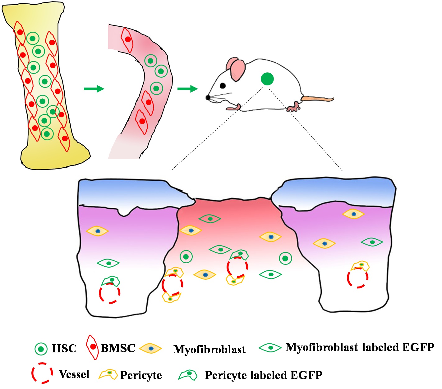
Normal mice with bone marrow derived HSCs marked EGFP and MSCs marked RFP models were prepared.
Forty C57BL/6 mice, weighting 20-25g, 6-8 weeks old, were randomly divided into irradiated group(n=20) and control group(n=20). Mice in irradiated group(IR) received a split dose of 8 Gy of g irradiation[18]. Then bone marrow derived HSCs labeled EGFP and MSCs labeled RFP were injected into blood though tail vein. Mice in irradiated group randomly received tail veil injections of 106 HSCs and 106 MSCs (IR+TP group) or equal amounts of solvent (sterile PBS), each group have 10 mice. Mice in control group randomly received tail veil injections of 106 HSCs and 106 MSCs (TP group) or equal amounts of solvent (sterile PBS, Ctr group), each group have 10 mice.
The mice bone marrow function was evaluated by clone culture
Bone marrow cells of mice in irradiated group were used for clonal culture. The mice were divided into transplantation group (IR+TR group) and PBS group according to receiving stem cells injection or PBS injection. The clonal culture was executed according to the instruction manual. Briefly, bone marrow-derived cells were collected by flushing the femurs and tibias. Then the red blood cells of bone marrow-derived cells were removed by red blood cell lysis buffer. Resuspend cells to 105 cells/ml using IMEM containing 2% FBS. 100 μl of cells were added to 1 ml of clonal medium and implanted in a 35mm culture dish for culture. Cultures were incubated for 12 days at 37°C, 5% CO2 and 95% humidity atmosphere. We observed clone formation under an inverted microscope.
Preparation of wound model
To investigate the effect of bone marrow derived HSCs and MSCs on would healing, three group mice were prepared as would healing models on the second day. The would healing models were prepared as before[19]. Briefly, three groups mice were euthanized by 10% chloral hydrate (0.002 ml/g body mass). Full-thickness skin wounds of 6mm in diameter were created on each mouse on the dorsal-right or -left shaved skin with sterile puncher.
Traces of HSCs and MSCs on would healing were tracked by live imaging system
The fluorescent protein of mice were detected by live imaging system(Roper Scientific, Martinsried, Germany), anesthetized with chloral hydrate. The pictures were taken on day1, 3, 5, 7 after the wound formation.
Traces and differentiation of HSCs and MSCs on would healing were detected by immunohistochemistry
The skin were harvested on day 5 after the wound formation. Two mice were randomly equally selected and killed each time in one group. The dorsal skins around the wound (area 1.5 × 1.5 cm) were carefully dissected and fixed in 4% paraformaldehyde and residual samples were immediately frozen in liquid nitrogen, and further processed for the morphometric analyses as described below[20].
The paraffin specimens were serial sectioned. The serial sections were immunostained with EGFP, RFP, desmin, α-SMA and K19. Briefly, sections were deparaffinized with immersion in dimethylbenzene and rehydrated, then heated in citrate buffer (0.01 M, pH 6.0) for 5 min at 100 and were then treated with endogenous peroxidase (3% hydrogen peroxide solution) for 5 min at room temperature. After blocking in 10% goat serum for another 30 min at room temperature, sections were immunostained with primary antibodies for EGFP (1:500; Abcam, Cambridge, UK) or RFP, desmin, α-SMA , K19 containing 0.1% Tween-20 and 5% bovine serum albumin (BSA) overnight at 4℃. After washing three times with PBST (PBS supplemented with 0.1% Tween-20), sections were incubated with secondary antibodies, avidin-biotin-peroxidase complex and DAB reagent [19, 21]. Subsequently, all sections were double stained with hematoxylin and visualized under the microscope (BX51, Olympus, Japan).