Animal experiments
All animal experiment protocols were approved by the Ethical Committee of Dalian Medical University, China (approval number: CXK (Liao) 2015 − 2003) and performed following the Institutional Animal Care and Use Committees (IACUC) of Dalian Medical University and following the international guidelines for the animal care and use (ARRIVE guidelines and NIH guidelines). The HD-intoxication experiment was as being described in our earlier publication [21]. Briefly, adult male Sprague Dawley rats (200–230 g body weight) were maintained in the Experimental Animal Center of Dalian Medical University. Routine husbandry procedure was provided with 22°C room temperature, 50 % relative humidity, a 12 h light-dark cycle and supply of food and drinking water ad libitum. To induce HD-mediated neurotoxicity, rats were intraperitoneally injected with HD (Solarbio, China), 400 mg/kg/day, five times per week, for 5 consecutive weeks. After the development of characteristic phenotypes of peripheral nerve degeneration, rats were engrafted with 5×10⁷ cells/kg BMSCs (in a single bolus dose) [22] by tail vein injection and observed to follow-up functional recovery for 5 weeks until necropsy. The control rats received no BMSCs treatment but only normal saline (NS). At the end of week 10, rats were necropsied and the sciatic nerves were quickly dissected, soaked in fixation solution or snap-frozen in liquid nitrogen for further analyses.
Neurobehavioral and nerve electrophysiological tests
To assess the phenotypical effect of axon degeneration/regeneration, gait score test, distal latency test and motor nerve conduction velocity test were carried out[23, 24]. A well-trained, test-blinded neurologist who was not involved in the project was asked to perform the measurements.
A gait score was assigned from 1 to 4, with 1 = normal, unaffected gait; 2 = slightly affected gait (tip-toe walking, slight ataxia, and hindlimb weakness); 3 = moderately affected gait (obvious movement abnormalities characterized by dropped hocks and tail dragging); 4 = severely affected gait (frank hindlimb weakness and inability to rear). Three successive measurements were taken and averaged for each animal [25].
The distal latency and conduction velocity were measured once a week, using a method published in literature [26]. The rats were anesthetized with ether and placed in a self-made rat fixation at the prone position. Three testing points, A, B and C, were marked at 3 cm, 10 cm and 15 cm from the base of the tail, respectively. Stimulation electrode was placed at Point A, and recording electrodes were placed at Points B and C. The nerve conduction velocities between A and B and between A and C were recorded. One hundred impulses were recorded and summated to calculate the conduction velocity: motor nerve conduction velocity (MCV) = Length BC / Latency BC (Length BC is the distance between B and C, Latency BC is the latency time between B and C). Distal latency was expressed using the latency BC time.
Electron microscope (SEM) analysis
Sciatic nerves were immersed in 2% glutaraldehyde overnight at 4°C, rinsed in PBS, fixed with 1% acetic acid for 3hrs, subjected to gradient dehydration, embedded and sliced with an ultramicrotome[27–29]. The slides were then stained with uranyl acetate and lead citrate and observed under a transmission electron microscope (H-7500, Hitachi, Japan).
BMSCs isolation and culture
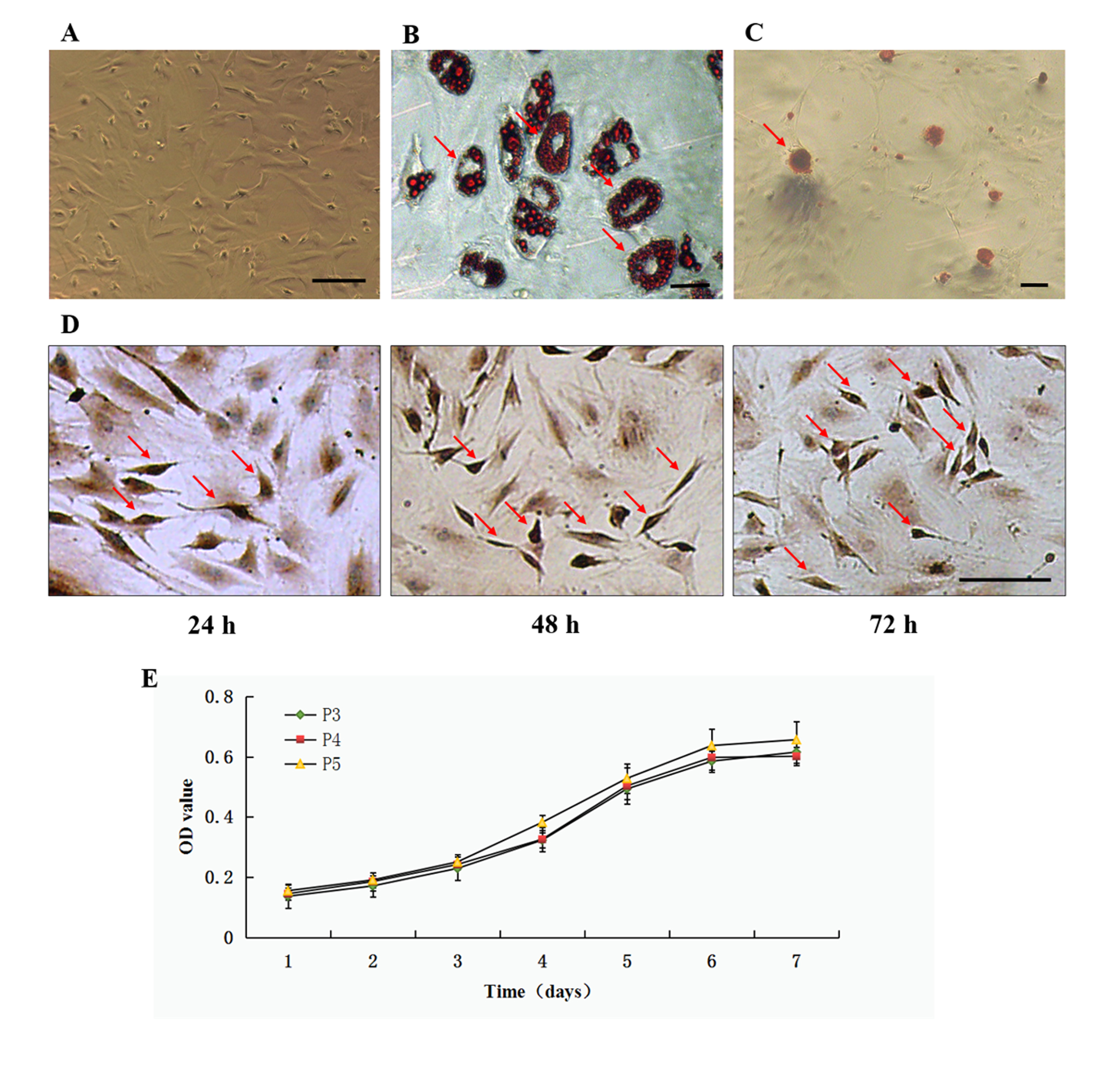
BMSCs were prepared as being described [30]. Briefly, Sprague Dawley rats (5–6 weeks old, 60–90 g, Laboratory Animal center, Dalian Medical University, China) were euthanatized and bone marrow was flushed from the femur and tibia bones with PBS. A mixture of single-cells was obtained by passing the cells through a sieve, seeded in Dulbecco’s modified eagle medium-low glucose (Gibco, USA) supplemented with 10% fetal bovine serum (FBS, HyClone, USA), 100U/mL penicillin and 100 µg/mL streptomycin (Beyotime, China) and culture at 37°C with 95% humidified air. Twenty-four hours later, the medium was replaced. The cells were cultured and used in in vivo or in vitro experiments within 5 passages. To validate the correct harvest and culture of these BMSCs, they were immunocytochemically stained using neuron specific enolase (NSE) antibody 24, 48 and 72 h after seeding. Adipogenic and osteogenic differentiation of BMSCs was evaluated by specific media Oil Red O and Alizarin Red S staining (Cyagen, China) [31].
Preparation of BMSCs-derived conditional Medium (BMSC-CM)
BMSC-CM was prepared following an established method[32]. Briefly, confluent BMSCs were washed, supplemented with fresh DMEM-10% FBS and cultured for 24 h. The culture media were collected, centrifuged at 200 g for 10 minutes at 4°C, applied to a filtration column (Millipore, Billerica, MA, USA), centrifuged at 3, 500 g for 45 minutes at 4°C, desalted and filter-sterilized.
Culture of primary Schwann cells and DRG neurons
To harvest DRG neurons, 2 day-old SD rats were sacrificed and lumbar dorsal root ganglia were dissected under a 16x anatomical microscope. The tissue was washed, cut and digested using 0.5% Type II Collagenase (Sigma, USA) for 45 minutes at 37°C and then with 0.25 % trypsin for 10 minutes at 37°C. The dissociated DRG neurons were centrifuged, resuspended, filtered, and cultured with D-MEM/F-12 supplemented with 10% FBS, 2% B-27 (Gibco, USA), 0.25 mg/ml insulin (Sigma, USA), 0.1 mg/ml L-glutamine (Sigma), 100 U/ml penicillin and 100mg/ml streptomycin at 37 ℃ with 5% CO2.
To harvest Schwann cells, 5-day-old SD rats were sacrificed and sciatic nerves were cut and digested with 0.125% trypsin and 0.03% type IV collagenase (Beyotime, China) for 30–40 min and centrifuged at 1,500 g for 5 min. The Schwann cells were cultured in DMEM with 10% FBS and with 1uM cytarabine at 37 ℃, 5% CO2 for 24 h. For co-culture, Schwann cells were seeded on a density of 1 x 10⁶/mL and DRG neurons were seeded on 5 x 10⁴/mL onto two poly-L-lysine-coated 0.5x0.5 cm coverslips, respectively. After 3–4 days of culture, the two coverslips with Schwann cells and DRG neurons were placed together with a distance of 2 mm and continued to grow for 3 days.
Cell transfection
Lipofectamine 3000 transfection reagent (Invitrogen, USA) was used to deliver NGF siRNA, scramble control siRNA, let-7e-5p mimics, let-7e-5p anti-miRNA and control miRNA into cells. All the siRNAs and miRNAs were purchased for RiboBio (China).
Treatment of DRG neurons with BMSC-CM, NGF, anti-NGF, and K252a
DRG neurons were cultured and added with HD (8 mM), HD (8 mM) plus 20% (v/v) BMSC-CM, HD (8 mM) plus anti-NGF (1:500) plus 20% BMSC-CM, HD (8 mM) plus non-specific immunoglobulin (1:500) plus 20 % BMSC-CM, HD (8 mM) plus recombinant NGF (200 µg/mL), or HD (8mM) plus K252a (200 nM) plus 20 % BMSC-CM. The cells were grown for additional 12hrs before harvest.
ELISA analysis of NGF
The concentrations of NGF in sciatic nerve tissue, BMSC-CM and cell culture medium were detected using an enzyme linked immunosorbent assay (ELISA) kit (USCN, USA) following manufacturer’s instruction. Medium of cells were collected and filtered through a 0.22 µm filter (Millipore) for removing cell debris. Each sample were measured three times using a spectrophotometer with absorbance at 490 nm wavelength.
Western Blot
Tissues and cells were homogenized using ice-cold RIPA buffer (Beyotime, China). Protein lysates were separated on SDS-PAGE gel and transferred to PVDF membrane (Millipore, France). The membrane was blocked and incubated with primary antibodies as following: NGF (Cell Signaling Technology, USA), TrkA, p-TrkA, Akt, p-Akt, mTOR, p-mTOR, CREB, p-CREB, GAP-43, GAPDH (Abcam, USA), and β-actin (ZS-Bio, China). The membranes were incubated with horseradish peroxidase-conjugated secondary antibody (Sigma, USA) and immunoreactivity was visualized enhanced chemoluminescence (Beyotime, China). For quantification, each sample was subjected to three independent experiments (n = 3) with triplicate loading per experiment.
Immunofluorescence
For immunofluorescence, frozen tissue sections (10 µm) or cold methanol-fixed DRG neurons were blocked with 10% donkey serum, incubated with either anti-MBP (Cell signaling, USA) or anti-MAP2 antibody (Cell signaling, USA) overnight at 4°C and fluorophore-conjugated secondary antibody, then incubated with anti-SMI312 (Cell signaling) or anti-GAP-43 antibody (Cell signaling) and secondary antibody. The stained slides were observed and imaged under a confocal microscope (Olympus, Japan). Image analysis was performed using Image-Pro Plus.
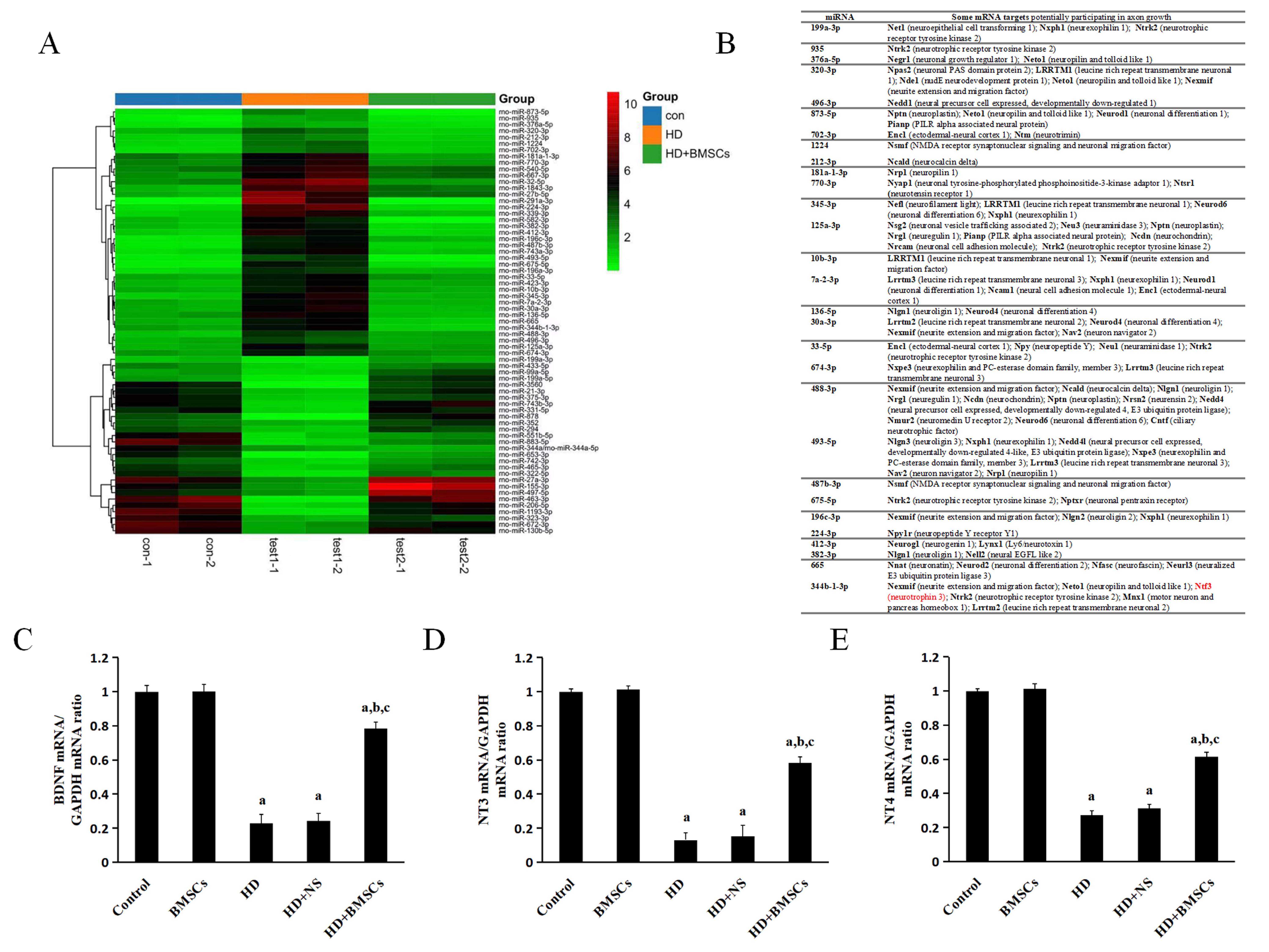
Microarray analysis of miRNAs
The expression profile of miRNAs in sciatic nerves of rats receiving no treatment (control), HD, and HD + BMSC was investigated using a miRNA microarray chip (KangChen Bio-Tec, China). The differential expression of miRNAs was analyzed using fold change (FC) > 2 and p-value < 0.05, respectively, in comparison between control and HD groups and between HD and HD + BMSC groups. Heatmap and clustering from sequencing were used to show the differential expression of miRNAs.
Real Time-PCR
Total RNA from sciatic nerve tissues and culture cells was extracted using RNAiso Plus (Takara, Japan). Real Time PCR was performed to analyze expressional change of Let-7 miRNAs and NGF mRNA using the PrimeScript RT Reagent Kit and SYBR Premix Ex TaqⅡ (Reagents from Takara, Japan) on a Thermal Cycle Dice Real-Time system. The Bulge-Loop miRNA primers for let-7 miRNAs and U6 miRNA were from RiboBio (China). The primers (NGF Forward primer 5’-3’: TGC CCC TGC TGA ACC AA, Reverse primer 5’-3’: GCT TGC TCC TGT GAG TCC TGT; GAPDH Forward primer 5’-3’: GGC ACA GTC AAG GCT GAG AAT G; Reverse primer 5’-3’: ATG GTG GTG AAG ACG CCA GTA) were also from RiboBio (China). All reactions were run in triplicate. The relative expression was calculated using the comparative 2−ΔΔCt method.
Statistical Analysis
All results were expressed as mean ± SD, and the statistical analysis was performed with one-way analysis of variance (ANOVA), followed by LSD test, which was performed using SPSS 13.0 statistical software. The p-values less than 0.05 were considered to be significant.