FDC tumors are very rare neoplasms that may mainly arise in lymph nodes and extranodal sites, which have a heterogeneous histology with spindle tumor cells and variable degrees of pleomorphism and arrange in fascicular or storiform patterns. The FDC tumors received increasing attention since the first case of a tumor with follicular dendritic cell differentiation was reported by Monda et al. in 1986 [2]. By now, there are less than 100 cases of follicular dendritic cell tumors have been reported in the English literature [2, 9]. The differential diagnosis includes Hodgkin's disease, sarcoma, leiomyosarcoma, gastrointestinal stromal tumor and inflammatory pseudotumor [10–12]. According to immunohistochemical analyses, the most commonly used FDC tumor markers include CD21 (C3d receptor, positive in 93% of cases) and CD35 (C3b receptor, positive in 89% of cases). Other quite specific markers used are: R4/23 (63%), Ki-67 (5%-50%), EMA (41%), vimentin (61%), HLA-DR (57%), CD45 (21%) and S-100 protein (31%). The tumor cells typically lack expression of CD1a, CD 68, and desmin [9, 13].
IPT-like FDC tumor of the liver is an extremely rare tumor and only 20 cases have been reported in the English literature, including ours [1, 6, 9, 10, 13–22]. (Table 1). In 1996, Shek and colleagues reported the first case of hepatic FDC sarcoma.[14] The most common main complaint of the patient was epigastric pain and weight loss. Malaise, anemia and fever were also part of the initial presentation in some patients. In five cases, patients were completely asymptomatic and the mass was found incidentally by using CT scan and abdominal ultrasound. The age of all the patients at initial presentation ranged from 19 to 82 years (mean 49.4 years), the mean tumor diameter was 11.9 cm (3-20cm), and the mean reported survival was more than 30 months (follow-up ranging from 6 to 108 months). The histology is similar to that of the conventional hepatic FDC tumor and it is generally considered to be a distinctive variant. This variant is characteristically restricted to the abdomen and seems to be a separate clinicopathologic entity. Comparing with conventional FDC sarcoma, it has a marked female predominance (female to male ratio, 4:1), whereas conventional FDC sarcomas are not more prevalent in only one sex [23]. IPT-like FDC sarcomas have prominent inflammatory component, which makes it difficult to distinguish them from inflammatory pseudotumors. IPT-like FDC sarcomas are strongly associated with the presence of EBV (80%), which is rare for conventional FDC sarcomas. Both IPT-like FDC sarcomas and conventional FDC sarcomas show generally a indolent clinical behavior, nevertheless, conventional FDC sarcomas of the liver can be more aggressive than IPT-like FDC ones, and may recur or metastasize and even lead to death.
Table 1
Characteristics of patients with hepatic FDC sarcoma of the liver.
| Case | Sex | Age (year) | Main complaint | Diameter (cm) | EBV | Treatment | Recurrence/Survival | Published year |
| 1 | F | 68 | Malaise, weight loss, anemia | 11 | + | SR | No/>30 m | 1996[15] |
| 2 | F | 35 | Epigastric discomfort, fever,weight loss | 20 | + | SR | Yes/>30 m | 1996[14] |
| 3 | M | 37 | Malaise, weight loss, anemia | 15 | + | SR | No/>24 m | 1998[16] |
| 4 | F | 19 | Right upper quadrant pain, weight loss, palpable mass | 12 | + | SR | No/40 m | 2001[6] |
| 5 | F | 56 | Gastrointestinal discomfort | 15 | + | SR | Yes/NA | 2001[6] |
| 6 | F | 40 | Epigastric pain, weight loss | 12.5 | + | SR | No/108 m | 2001[6] |
| 7 | F | 49 | Incidental at ultrasound | 4.2 | + | SR | No/9 m | 2001[6] |
| 8 | F | 31 | Abdominal distention, weight loss | 15 | + | SR | No/60 m | 2001[6] |
| 9 | F | 57 | Epigastric pain, weight loss | 9.5 | + | SR | No/36 m | 2001[10] |
| 10 | F | 51 | Epigastric pain, weight loss | 12 | + | SR | No/12 m | 2001[10] |
| 11 | M | 82 | Incidental on a CT abdomen | 15 | - | SR | No/18 m | 2005[1] |
| 12 | F | 30 | Incidental at ultrasound | 5.5 | + | SR | No/12 m | 2006[17] |
| 13 | F | 57 | Abdominal pain, vomiting, dizziness, liver dysfunction | 13 | + | SR | No/24 m | 2008[13] |
| 14 | F | 78 | Incidental at ultrasound | 3 | + | TACE | 27 m | 2010[18] |
| 15 | F | 59 | Asymptomatic | 6 | + | SR | NA | 2010[19] |
| 16 | F | 53 | Right upper quadrant pain, fever, anemia, jaundice | 11.5 | - | SR | No/6 m | 2011[9] |
| 17 | M | 56 | Right upper quadrant abdominal pain | 11 | NA | SR | No/12 m | 2011[20] |
| 18 | F | 31 | Palpable abdominal mass | 20 | + | SR | NA | 2016[21] |
| 19 | M | 19 | Painless swellings around several joints | 6 | - | SR | NA | 2016[22] |
| SR, surgical resection; TACE, Transcatheter Arterial Chemoembolization |
The initial diagnosis of FDCS is based on clinical examination, imaging and pathologic assessment. The role of imaging is mainly in describing the extent of the mass and staging. When suspected a mass in the liver, the diagnosis of IPT-like FDC sarcoma is very difficult without pathological findings. It is noteworthy that the diagnosis of IPT-like FDC sarcoma should be based on the recognition of FDCs from microscopic appearance. Actually, the distinction of hepatic IPT-like FDC sarcoma from other liver tumors is usually impossible without immunohistochemistry. However, sometimes IPT-like FDC sarcoma can be difficult to consider the disease because the tumor shows bland spindle cells without nuclear atypia and the overshadowing chronic inflammatory infiltrate. Shek et al. [14] reported a case of primary IPT-FDC tumor of the liver that was initially misdiagnosed as an inflammatory pseudotumor. The tumor recurred 30 months after complete resection of the "inflammatory pseudotumor "[14]. CD21 and CD35 have been widely used as the preferred FDC markers, which were expressed in almost IPT-like FDCs. EBV infection is identified as a key role in the genesis of IPT-like FDC sarcoma. Almost all IPT-like FDC sarcomas exhibited positive EBER by in situ hybridization. LMP-1 gene, the major oncogene of EBV, was identified in several cases of IPT-like FDC sarcomas [15, 23]. However, the pathogenic mechanism of EBV in IPTFDC sarcoma remains unclear and further investigation is required.
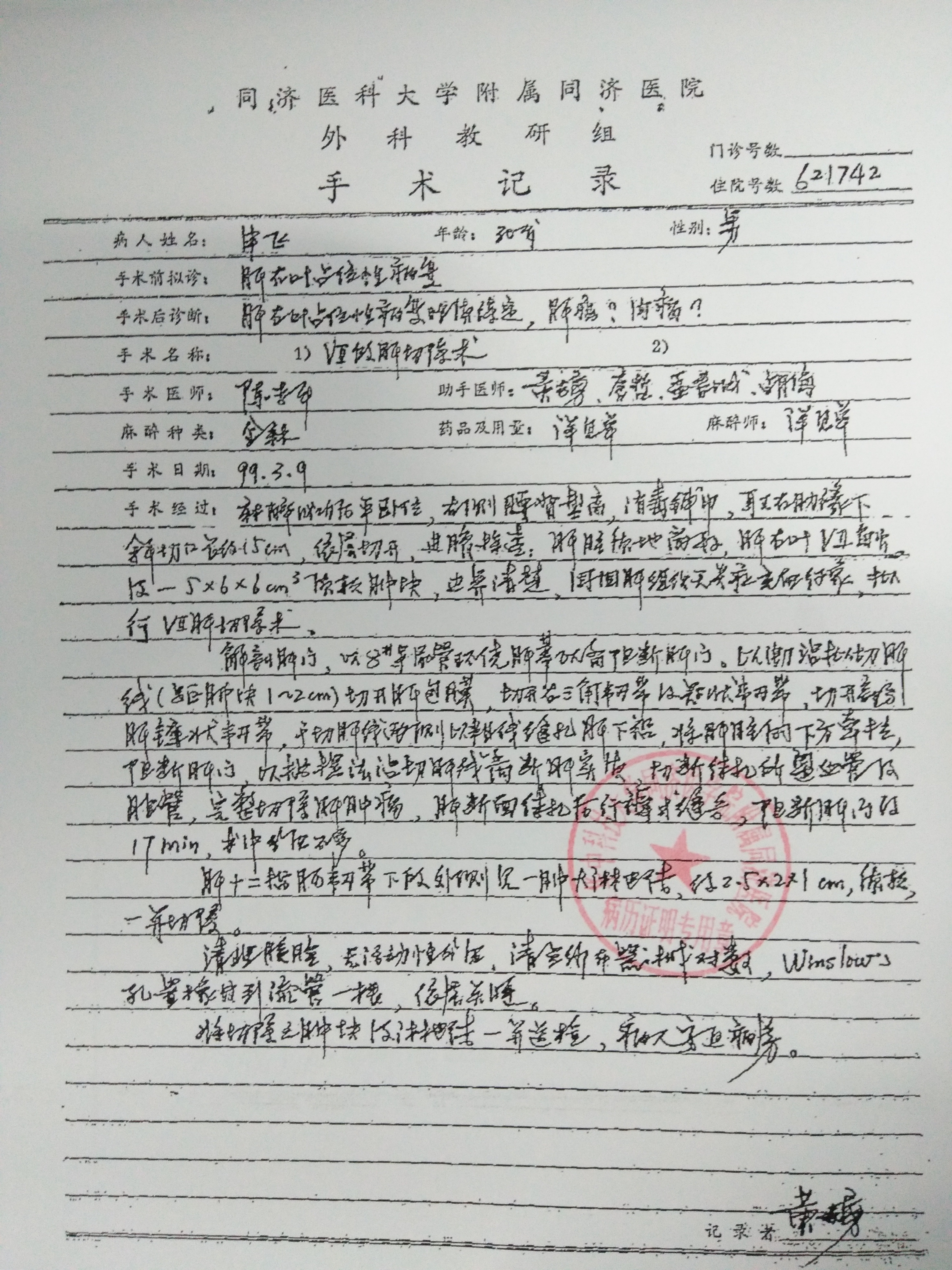
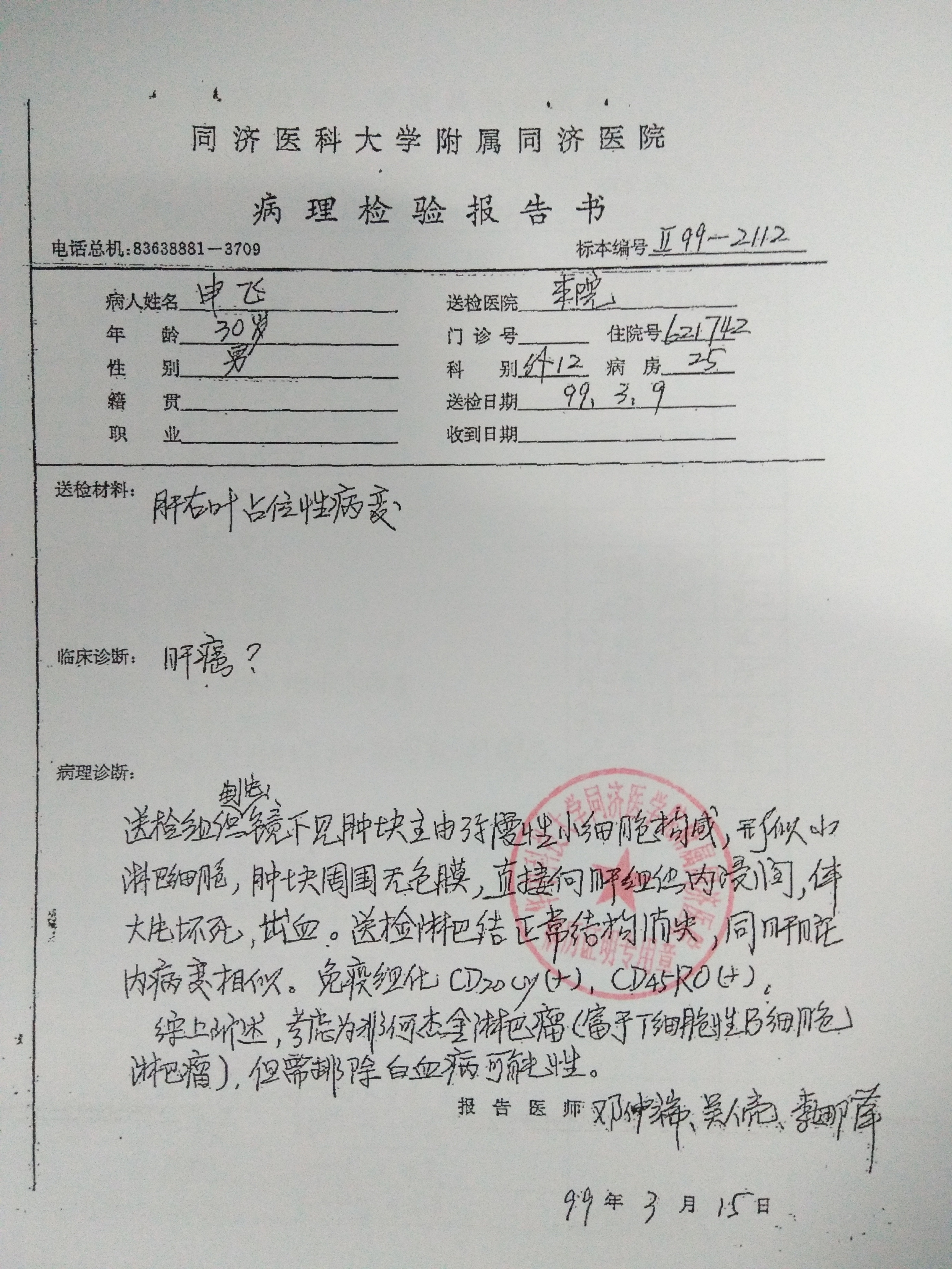
Primary NHL of the liver has rarely been reported and because there are no typical laboratory or image diagnostic findings, and pathological analysis is the standard diagnostic method[24]. The coexistence of hepatic NHL and hepatic IPT-like FDC sarcoma is extremely rare. The patient we reported in this article received hepatic segmentectomy in our hospital due to non-hodgkin lymphoma (B-cell lymphoma) of the liver in 1999 (no data available because of long time). Immunohistochemical studies demonstrated that all the tumor cells were strongly positive staining with CD20 and CD45. A diagnosis of hepatic NHL was rendered (Supplemental Figs. 1 and 2). Several studies indicated that chronic HCV infection may be associated with the pathogenesis of NHL, however, the present case has no HCV infection. The detailed mechanism of HCV-mediated lymphomagenesis remains unclear[25]. In the present case, it was misdiagnosed with HCC or non-hodgkin lymphoma at initial evaluation. Our search of the literature found no such cases. This is the first report to demonstrate hepatic IPT-like FDC sarcoma in a patient with primary hepatic NHL history.
Surgical resection is the treatment of choice for primary hepatic IPT-like FDC sarcomas and hepatic lymphoma whenever possible. The efficacy of chemotherapy and radiotherapy is unclear. Daniel et al.[26] reported that even if complete resection has been achieved for hepatic NHL, postoperative chemotherapy is mandatory. The patient did not receive chemotherapy after the first hepatectomy. Tsunemine et al. 18suggested that TACE was useful for the management of hepatic FDC sarcoma, and the patient was still alive more than 2 years after the diagnosis with hepatic tumors favorably controlled by repeated TACE. Shinagare et al.[20] reported a case which the patient was not determined to be a surgical candidate because of large tumor mass and small residual liver volume. The patient received four cycles of standard-dose CHOP and portal vein embolization in an attempt to cause hypertrophy of the residual liver. Seven months later, the patient underwent a successful resection of the mass [20]. Eighteen of the patients with hepatic IPT-FDC sarcoma have undergone partial hepatectomy.