CD70 expression in human pancreatic cancer cells
In total, 166 patients who underwent PDAC resection were included in our study. To evaluate the significance of CD70 expression in human pancreatic cancer, we first performed IHC analysis on FFPE tissues. Figure 1 shows the staining pattern for CD70 expression in different tissues. CD70 was expressed mainly in the plasma membrane and cytoplasm of cancer cells (Fig. 1a, b). In several cases, tumor-infiltrating leukocytes that expressed CD70 were observed and used as internal controls (Fig. 1d). In normal tissues, positive staining was restricted to lymphoid tissues (tonsils, thymus, and spleen) and gut-associated lymphoid cells. Positively stained tissues (tonsils) were used as a positive control (Fig. 1e). CD70 was not detected in the normal pancreas tissues, including acinar cells and normal glands (Fig. 1f).
Patients characteristics
Patient demographic, clinical, and histopathological variables are summarized in Table 1. Among the whole cohort, CD70 protein expression was confirmed in 42 patients (25%). We found no significant association between neoadjuvant treatment and CD70 expression. A tumor with CD70 high expression tended to be located in the pancreatic body or tail. None of the other clinicopathological variables, such as age, sex, lymph node metastasis, and tumor grade differentiation, showed a significant relationship with CD70 expression.
Survival analysis
In the whole cohort, high CD70 expression was not associated with OS (33.1 vs. 40.8 months, P = 0.256; Fig. 2a). We also found no differences in OS according to CD70 expression, neither in the upfront-surgery subgroup, nor in the neoadjuvant chemoradiation therapy subgroup (data not shown). As previously described, completion of adjuvant chemotherapy is one of the strongest prognostic factors associated with pancreatic cancer treatment [24]. To test whether CD70 expression could interact with the efficacy of adjuvant chemotherapy, we compared survival and recurrence data according to CD70 expression in patients that either did or did not complete adjuvant chemotherapy. High CD70 expression was significantly associated with inferior OS in the population of patients that completed adjuvant chemotherapy (N = 110, OS: 45.4 vs. 63.8 months, P = 0.027; Fig. 2b), whereas no such association was detected with the population of patients that did not complete adjuvant chemotherapy (N = 56, OS: 20.7 vs. 14.9 months, P = 0.969; Fig. 2c).
Moreover, the incidence of hematogenous metastasis was significantly higher in patients with high CD70 expression than in those with low CD70 expression (P = 0.018; Fig. 3a). In contrast, high CD70 expression was not associated with local recurrence (P = 0.218; Fig. 3b).
Multivariate analysis revealed that high CD70 expression, high T stage, para-aortic lymph node invasion, and incompletion of adjuvant chemotherapy were significant risk factors for hematogenous metastasis (Table 2). Moreover, significant prognostic factors for the entire adjuvant chemotherapy-completed population were carbohydrate antigen 19-9, a high T stage, the lymph node status, and CD70 expression (P = 0.011, Supplementary Table 1).
Relative mRNA expression of CD70 in pancreatic cancer cell lines
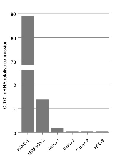
We next assessed relative CD70-expression levels in pancreatic cancer cell lines by real-time PCR. CD70 was relatively highly expressed in PANC-1, MIAPaCa-2, and AsPC-1 cells (Supplementary Fig. 1). With the exception of these cell lines, three out of the six pancreatic cell lines analyzed were almost negative for CD70 expression. Therefore, we conducted in vitro assays using the PANC-1 and MIAPaCa-2 pancreatic cancer cell lines.
CD70 knockdown suppressed pancreatic cancer cell proliferation independently of gemcitabine
Transient CD70 knockdown and gemcitabine therapy significantly reduced the growth of PANC-1 and MIAPaCa-2 cells (Fig. 4). Importantly, CD70 knockdown also removed enhanced proliferation in gemcitabine-treated cells. Overall, these results suggest that CD70 is involved in cell proliferation, and that blocking CD70 has the potential to suppress cell proliferation independently of gemcitabine treatment in pancreatic cancer cell lines.
CD70 knockdown decreased migration in pancreatic cancer cell lines
To elucidate the mechanism involved in the CD70-mediated metastatic increase, CD70 knockdown cells were used to study in vitro migration. Migration and transwell chamber assays showed that CD70 knockdown reduced PANC-1 and MIAPaCa-2 migration (Fig. 5). Using a migration assay, we found that CD70 induced pancreatic cancer cell migration in vitro.
Correlation between CD70 and epithelial-to-mesenchymal transition (EMT)-related gene expression in human pancreatic cancer tissues
To investigate how CD70 expression may be associated with a negative impact on prognosis, we focused on genes whose expression are associated with the EMT transition because of its broad role in metastasis and treatment resistance. We analyzed the association between CD70 expression and EMT markers in pancreatic cancer tissue by real-time PCR. In 20 available tissues from patients with positive IHC staining for CD70, CD70 expression significantly correlated with Vimentin (r = 0.640, P = 0.004), Snail (r = 0.466, P = 0.038), and Twist (r = 0.495, P = 0.027) mRNA expression (Fig. 6).
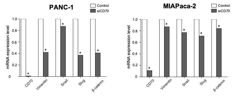
Effect of CD70 knockdown on EMT-related gene expression in pancreatic cancer cell lines
To investigate the possible association between CD70 and the EMT, and whether CD70 regulates EMT in pancreatic cancer cell lines, we knocked down CD70 expression in PANC-1 and MIAPaCa-2 cells using siRNA. The mRNA-expression levels of Vimentin, Snail, and Slug were significantly reduced by CD70 knockdown (Supplementary Fig. 2). Moreover, mRNA expression of β-catenin, the key molecule in the canonical Wnt-signaling pathway, was also significantly reduced by CD70 knockdown.
Association between CD70 expression and tumor-infiltrating lymphocytes (TILs)
Next, we investigated the immunomodulatory function of CD70 in pancreatic cancer. We performed IHC analysis of TILs, including CD4, CD8, and FoxP3. Our results showed that the numbers of tumor-infiltrating CD8+ and FoxP3+ T cells tended to be lower or higher in patients with high CD70 expression than in those with low CD70 expression (P = 0.094 and 0.075, respectively; data not shown). However, these differences were not statistically significant.