Histopathological Study
Informed written consent was obtained from relatives of a 45-year-old male to perform clinical autopsy. The use of brain sample was carried out according to the current guidelines of Spanish legislation (Real Decreto 2011/1716) and the Declaration of Helsinki and all assessments. The autopsy confirmed peritonitis with intestinal perforation, infiltrated by ring cell adenocarcinoma and disseminated metastasis. After the brain removal from the skull, forty-two fresh samples (21 brain areas derived from both cerebral hemispheres) were extracted in parallel without delay, immediately frozen and stored at −80 °C for proteomic analysis. The areas assessed in this study were: Olfactory bulb (OB), Substantia nigra (SN), Spinal cord (SpC), Brainstem (BS), Pons (Prot/Pons), Cerebellar vermis (CV), Dentate gyrus (DG), Cingulum (CING), Motor cortex (MCx), Putamen (PUT), Caudate nucleus (CN), Lenticular extern (LE), lenticular intern (LI), Frontal cortex (FC), Temporal cortex (TC), Thalamus (TH), Hippocampus (HIP), Amygdala (AMY), Parietal cortex (PC), Occipital cortex (OC), and Frontal white matter (FWM). The remaining brain areas were fixed with 4% buffered formalin for 4 weeks and some other selected regions were embedded in paraffin and stained with haematoxylin and eosin following the BrainNet Europe guidelines35. Protein expression of Tau, β-amyloid, TDP-43, α-synuclein, ubiquitin, and α–β crystalline was analysed by immunohistochemistry using specific antibodies across different brain regions. The microscopic study demonstrated minor neuronal hypoxic changes in the temporal and hippocampal area. No neuropathological signs of neurodegenerative disorders were found on pathological examination of the subject.
Sample preparation for LFQ analysis
Protein extraction was performed as previously described36. Brain samples were homogenized in lysis buffer containing 7 M urea, 2 M thiourea and 50 mM DTT supplemented with protease and phosphatase inhibitors. Then, samples were spun down at 100,000×g for one hour at 15 °C. After protein precipitation, protein concentration in the supernatant was measured with the Bradford assay kit (Biorad). Protein enzymatic cleavage was carried out with trypsin (Promega; 1:20, w/w) at 37 °C for 16 h. Peptides were subjected to desalting (C18-sep packs column, Merck Milipore) and vacuum dried. Reconstitution of peptides was undertaken using 0.1% Formic acid, and peptide quantification was performed using Scopes method37.
Liquid chromatography tandem mass-spectrometry (LC-MS/MS)
1µg of digested and desalted peptides were loaded onto the column at 5µl/min flow rate. Peptides were resolved on an analytical column at a flow rate of 300 nl/min over 180 min gradient in solvent B (80% ACN in 0.1% FA). Mass spectrometric acquisition was performed in the DDA (Data Dependent Acquisition) mode in full scan range of 375-1700 m/z with Orbitrap fusion mass analyser at a mass resolution of 60,000 in Mass Spectrometry Facility at IIT Bombay (MASSFIITB). The mass window was set to be 10 ppm with a dynamic exclusion duration of 40s. All MS/MS spectra were acquired by HCD, i.e. High energy Collision Dissociation method of fragmentation. Each sample was run in duplicates in a randomized manner to prevent a run-to-run bias and remove prevent batch variation.
LFQ data analysis in MaxQuant
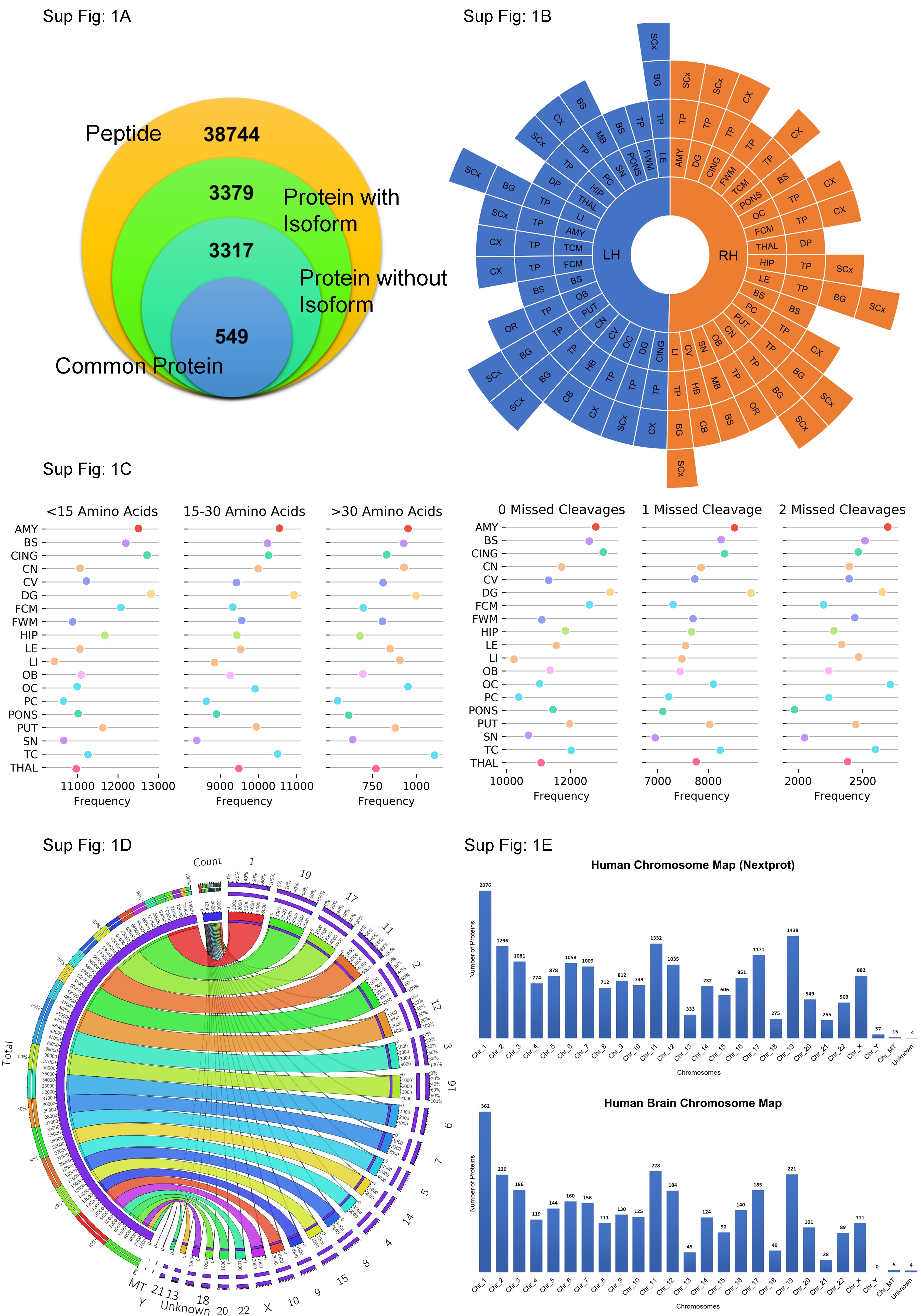
The raw datasets were processed with MaxQuant (v1.6.5.0) against UniProt Human Proteome Database (Proteome ID: UP000005640) and searched with the built-in Andromeda Search Engine of MaxQuant38. Raw files were processed within Label-Free-Quantification (LFQ) parameters setting label-type as "standard" with a multiplicity of 1. The Orbitrap was set to Orbitrap Fusion mode. Trypsin was used for digestion with a maximum of 2 missed cleavages. Carbamidomethylation of Cysteine (+57.021464 Da) was set as the fixed modification, whereas oxidation of Methionine (+15.994915 Da) was set as the variable modification. The False-Discovery-Rate (FDR) was set to 1% for the protein and peptide levels to ensure high reliability of the protein detection/identification. The minimum length for a peptide was set to 7AA. Decoy mode was set to "randomize", and the type of identified peptides was set to "unique+razor".
Data quality check and analysis
The analysis of 21 samples showed that the left hemisphere of Motor Cortex (MCx) sample were not giving appropriate number of proteins. The sample was re-run for confirmation and MCx sample was excluded from the study. The max quant analysis was performed with 20 samples and the data was taken forward for further analysis.
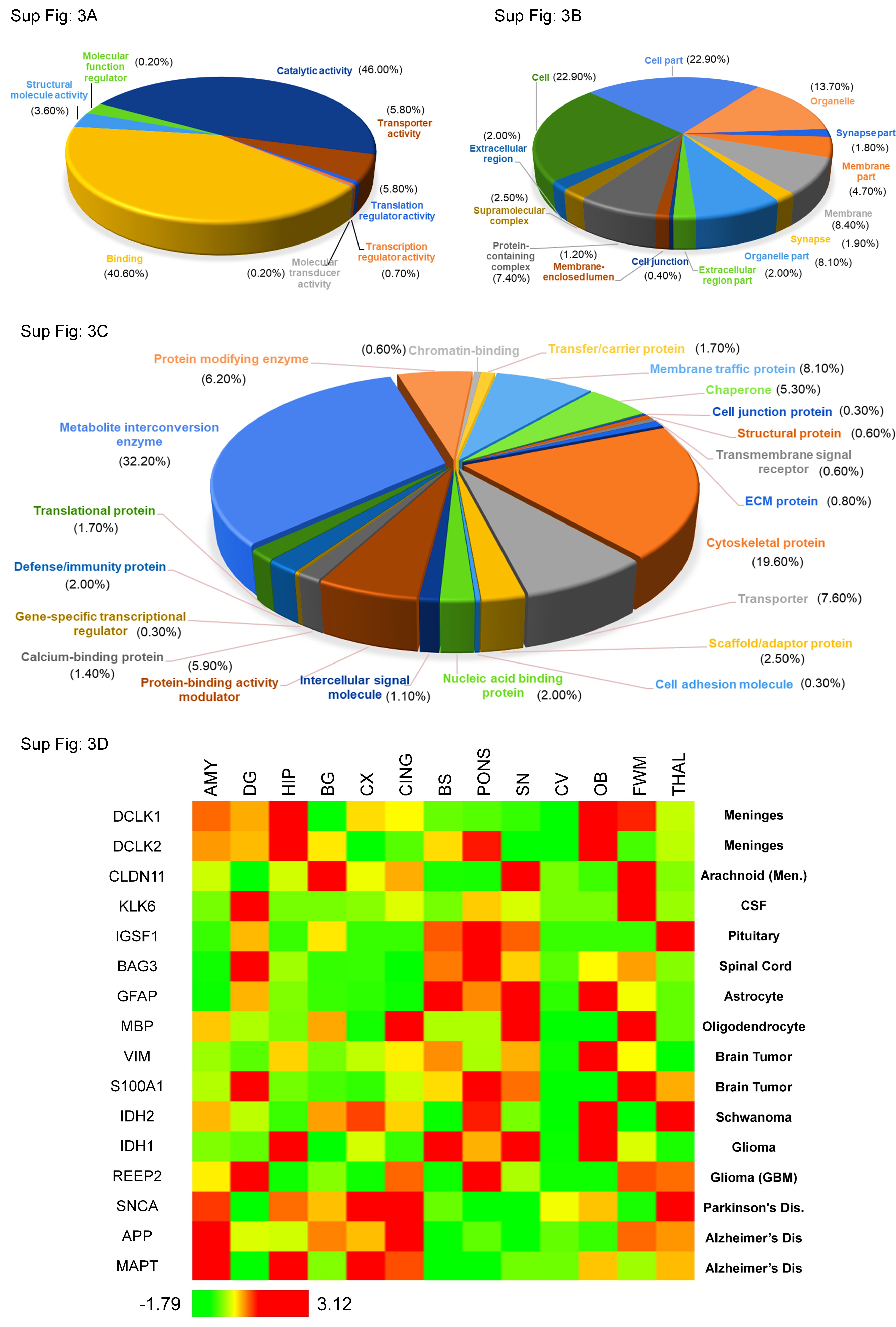
Statistical and bioinformatics analysis of spectra and proteomics data was performed using Metaboanalyst39, Microsoft Excel, Python, and R statistical software. No missing value imputation has been done. The hierarchical clustering was performed using z-score transformed LFQ intensities and was further clustered using Euclidean as a distance in Metaboanalyst39 and Hierarchical Clustering Explorer40 (Version 3.5). PANTHER41, WebGestalt42 and SynGO were used to perform some pathway enrichment and synapse biology-based on Gene Ontology (GO). STRING and NetworkAnalyst were used for protein-protein interaction and enrichment analysis.
Regression/Residual Analysis of inter-hemispheric data
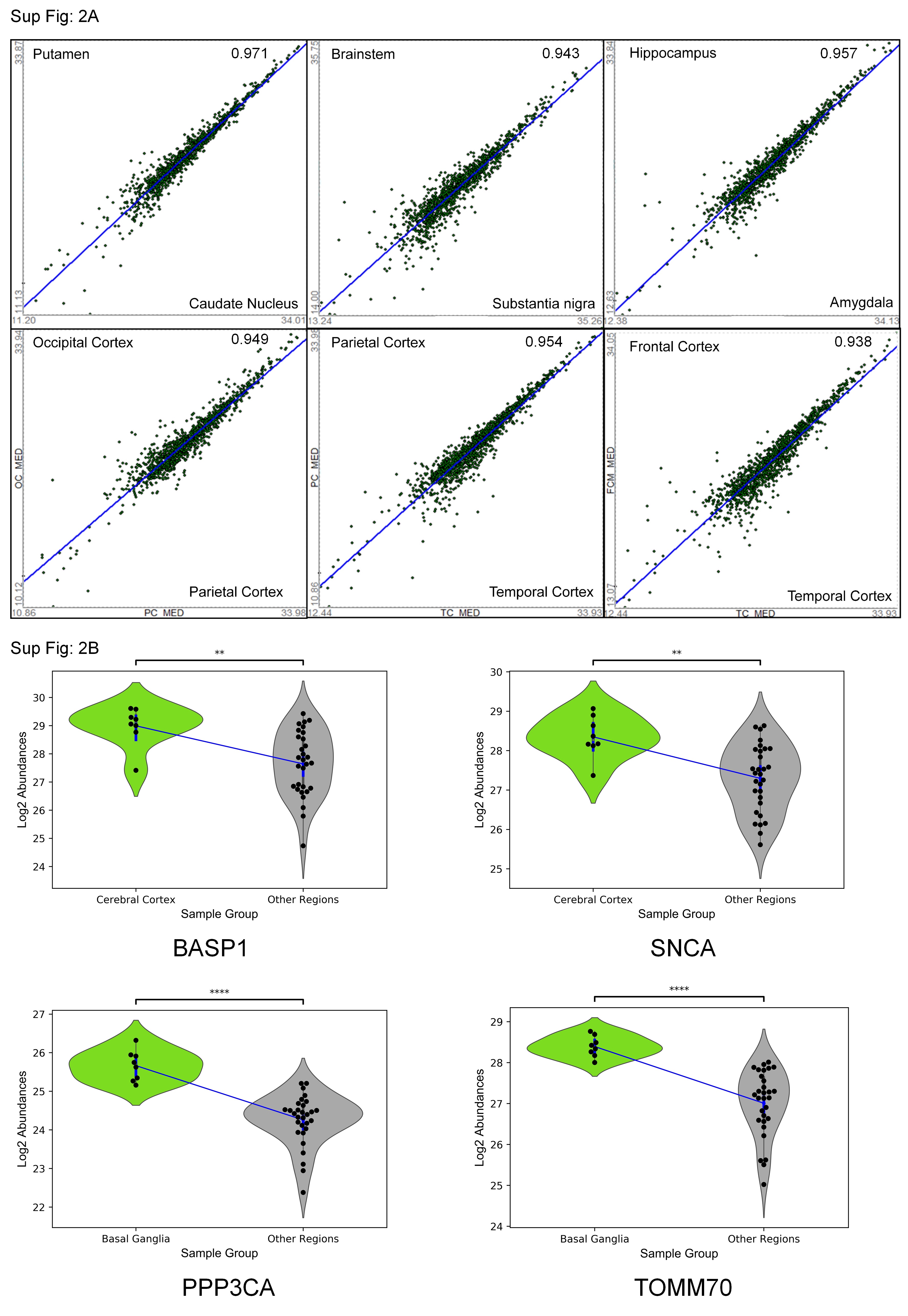
A simple bivariate linear regression analysis, used to identify significantly over or under-expressed proteins associated with a particular sample was performed. In this approach, the protein expression levels were regressed for pairs of individuals. For example, the expression levels for a replicate of AMY were put on the Y-axis, and the expression levels for another non-AMY sample were put on the X-axis. A regression analysis was conducted in MS Excel and standard residual values were calculated for each protein for the regression. This approach was repeated for all replicates in a pair-wise fashion, regressing all of the sample profiles in the region against all sample profiles out of the region. The mean residual value was calculated for a given region. The advantage of this approach is that it combines the power of a volcano plot with the ability to examine replicate-to-replicate variation and region-to-region variation. For each regression, the distribution of residuals and the correlation coefficients were examined. Raw regression results are presented in the supplementary data. This approach was not only used to look at the region-specific proteins but also used to examine the Left versus Right expression for each region. In this way, the Left v/s Right differential expression could be examined across all regions.
Data mining and data extraction for comparative analysis
Proteomics study with keywords related to the human brain and different neuro-anatomical regions of the human brain, according to the Allen Brain Atlas Classification table were searched in Proteome Exchange and PRIDE repository. All the studies available in the repository up to 10.02.2020 with the following filter criteria - Species: Human (Homo sapiens) and Method: LFQ were downloaded. The manuscript of the papers and sample metadata files were thoroughly studied to understand the experimental design and which raw files belong to the healthy subjects. In most cases, authors were contacted to verify the raw files. Additionally, in-house generated mass spectrometry-based data of few other regions from our previous study were also taken. All these curated raw mass spectrometry files were taken forward for MaxQuant data analysis with Human Proteome Database (Isoform). Further, brain-related databases and repositories were also considered, which included Brain Atlas of Human Protein Atlas (HPA), CSF Proteome Resource (CSF-PR)43, and Harmonizome44.
Inter-hemispheric brain proteome map (IBPM) portal
The IBPM portal under www.brainprot.org is designed on the Django framework with robust features for visualization of data in the study and it will act as an interface for the researchers in the field of brain proteomics for data integration and region-specific information. The portal also fetches general information like the chromosome localization, function, and peptide information about the protein from various existing repositories. The portal is designed with scalability at its core; the database in the portal is highly dynamic and can be extended to incorporate public databases.
Data availability
All MS raw files could be accessed from Proteome-Xchange using the identifier PXD019936.
References
- Bell, J. E. et al. Management of a twenty-first century brain bank: experience in the BrainNet Europe consortium. Acta Neuropathologica 115, 497–507 (2008).
- Ferrer, I. et al. Familial globular glial tauopathy linked to MAPT mutations: molecular neuropathology and seeding capacity of a prototypical mixed neuronal and glial tauopathy. Acta Neuropathologica 139, 735–771 (2020).
- Scopes, R. K. Measurement of protein by spectrophotometry at 205 nm. Analytical Biochemistry 59, 277–282 (1974).
- Tyanova, S., Temu, T. & Cox, J. The MaxQuant computational platform for mass spectrometry-based shotgun proteomics. Nature Protocols 11, 2301–2319 (2016).
- Chong, J. et al. MetaboAnalyst 4.0: towards more transparent and integrative metabolomics analysis. Nucleic Acids Research 46, W486–W494 (2018).
- Seo, J. & Shneiderman, B. Interactively exploring hierarchical clustering results [gene identification]. Computer 35, 80–86 (2002).
- Mi, H., Muruganujan, A., Ebert, D., Huang, X. & Thomas, P. D. PANTHER version 14: more genomes, a new PANTHER GO-slim and improvements in enrichment analysis tools. Nucleic Acids Research 47, D419–D426 (2018).
- Liao, Y., Wang, J., Jaehnig, E. J., Shi, Z. & Zhang, B. WebGestalt 2019: gene set analysis toolkit with revamped UIs and APIs. Nucleic Acids Research 47, W199–W205 (2019).
- Guldbrandsen, A. et al. CSF-PR 2.0: An Interactive Literature Guide to Quantitative Cerebrospinal Fluid Mass Spectrometry Data from Neurodegenerative Disorders. Molecular & Cellular Proteomics 16, 300 (2017).
- Rouillard, A. D. et al. The harmonizome: a collection of processed datasets gathered to serve and mine knowledge about genes and proteins. Database 2016, (2016)