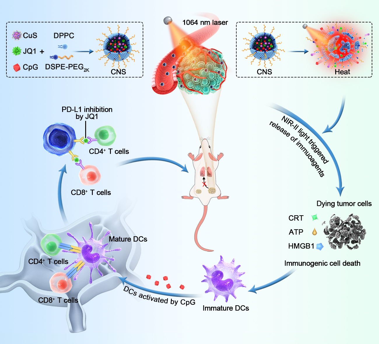
Synthesis and characterization of CNS
Various photoactivated CNS loading agents were prepared using a film hydration method[49]. In brief, a thin film composed of DPPC (MW = 734.0) and DSPE–PEG2k (MW = 2805.5) with a feeding mass ratio of 25:5 was synthesized and then hydrated with ultrapure water including CuS, JQ1, and CpG. Hydrophobic JQ1 and hydrophilic CuS and CpG were encapsulated into temperature-responsive liposomes through hydrophobic and π–π stacking interactions. The EE of CNSD was 71.8% for CuS, 57.2% for JQ1, and 69.8% for CpG, and the corresponding LC values were 4.3%, 1.7%, and 0.1%, respectively. Three control counterparts (CNS0, CNSC, and CNSJ) were simultaneously prepared using a similar process and used as a comparison.
As shown in the transmission electron microscopy (TEM) images, CNSD and the three control counterparts (CNS0, CNSC, and CNSJ) were quasi-spherical and showed a uniform size distribution (Figure 1a). Dynamic light scattering (DLS) revealed that the hydrodynamic size of CNSD was approximately 31.3 ± 1.4 nm, which was slightly larger than that of CNS0 (25.1 ± 1.1 nm), CNSC (27.0 ± 2.7 nm), and CNSJ (26.2 ± 3.7 nm) (Figure 1b). Moreover, the polydispersity indexes (PDIs) of CNS0, CNSC, CNSJ, and CNSD were 0.23, 0.22, 0.24, and 0.23, respectively, suggesting good mono-dispersity of the four types of nanoparticles. The zeta potential was measured to investigate the surface charge of various prepared nanoparticles. Compared with CNSC (-28.9 mV), CNSD (-23.4 mV) in 1× phosphate-buffered saline (PBS) showed an elevated surface charge, indicating successful JQ1 loading. Compared with CNSJ (-20.9 mV), CNSD (-23.4 mV) in 1× PBS showed a reduced surface charge, indicating successful CpG ONDs encapsulation (Figure 1c). CNSD and the three control counterparts (CNS0, CNSC, and CNSJ) showed similar peaks in the ultraviolet-visible (UV-vis) absorption spectrum, indicating the inappreciable affection of loaded two drugs on the optical property of the prepared nanoparticles (Figure 1d). Furthermore, the prepared CNSD nanoparticles showed excellent colloidal stability due to their stable hydrodynamic size and PDI over 10 days (Additional file 1: Figure S1).
Photothermal properties and photothermal-induced drug release performance
The NIR-II photothermal properties and photothermal-induced release of immune agents from CNS were further investigated. Under conditions of photoirradiation (1064 nm) at a power intensity of 1 W cm−2, the temperatures of CNS0, CNSC, CNSJ, and CNSD solution showed an obvious increase up to around 55°C in 7 min (Figure 1e, Additional file 1: Figure S2). The photothermal temperature curves exhibited minimal differences after five cycles of heating and natural cooling (Figure 1f), showing good photothermal stability of the nanoparticles. Furthermore, the photothermal conversion efficiency of CNSD was ≈ 32.6%, which was similar to that of CNS0 (32.4%), CNSC (31.8%), and CNSJ (31.5%) (Additional file 1: Figure S3).
The photo-triggered release properties of the loaded immune agents were further analyzed. Under conditions of photoirradiation (1064 nm) at a power intensity of 1 W cm−2 for 5 min, the cumulative release of JQ1 from CNSD was 42.3% in 1 h, which was 3.6-fold higher than that without laser irradiation (Figure 1g). The underlying mechanism can be explained as follows: CNSD mediated the photothermal effect and induced the dissociation of temperature-responsive liposomes with a phase-transition temperature of around 42 °C, resulting in the on-demand release of JQ1.
CNS-mediated intracellular photothermal-activated immune response
The cellular uptake of CNS was first analyzed in Panc02 cells. The cellular uptake of CNS was confirmed by loading 2.5% (w/w) CNS with indocyanine green (ICG), which is a cyanine dye approved by the FDA, to construct four loaded CNS nanoparticles (CNS0@ICG, CNSC@ICG, CNSJ@ICG, and CNSD@ICG) (Additional file 1: Figure S4). The ICG-loaded CNS nanoparticles exhibited identical peaks in the UV-vis absorption spectrum, indicating their successful preparation (Additional file 1: Figure S5). After a 24-h incubation, the fluorescence intensity of CNSD-cultured Panc02 cells was 105-fold greater than that of the control group, suggesting the effective internalization of nanoparticles into Panc02 cells. There was no obvious difference in fluorescence intensity in Panc02 cells cultured with the four nanoparticles (Figure 2a and b). The cytotoxicity of these CNS nanoparticles was analyzed. The viability of the Panc02 cells was >90% after incubation with four CNS nanoparticles for 24 h, indicating the nontoxicity and good biocompatibility of CNS in vitro (Figure 2c). The capacity for CNS to suppress tumor cells was further assessed. After incubation with CNS0, CNSC, CNSJ, or CNSD for 24 h, Panc02 cells were exposed to a 1064 nm NIR laser at a power density of 1 W cm−2 for 5 min. The results showed that cells treated with diverse CNS nanoparticles plus NIR-II light all decreased with elevating CuS concentrations (Figure 2c). Using 100 μg/mL CuS, the viabilities of the Panc02 cells treated with CNS0 + L, CNSC + L, CNSJ + L, and CNSD + L decreased to 24.6%, 38.7%, 17.3%, and 11.9%, respectively, suggesting excellent CNS-mediated photothermal effectsagainst cell growth.
Exposure of the tumor cells to calreticulin (CRT) as a specific signal for triggering APCs was a characteristic sign of ICD induction. Thus, CRT exposure of the Panc02 cells was further evaluated to validate the performance of CNS-mediated ICD under conditions of NIR-II irradiation in vitro. After NIR-II laser irradiation for 24 h, the mean fluorescence intensity (MFI) of CRT in Panc02 cells treated with the four CNS nanoparticles was elevated. Of note, the MFI of CRT in the CNSD + L group was 3.2 and 2.5-fold higher than that in the control and CNSD groups, respectively (Additional file 1: Figure S6). These data indicate that CNS nanoparticles were capable of efficiently triggering ICD under conditions of NIR-II laser irradiation.
As a key role in human immunity, immature DCs can engulf antigens and transfer them to neighboring lymph nodes, where they are processed into peptides for T cell activation. Mature DCs can be detected by the co-stimulatory molecules, CD80/CD86, which are representative markers that signify DC maturation. Murine bone marrow-derived dendritic cells (BMDCs) extracted from C57 mice received different treatments and the corresponding percentages of matured DCs (CD11c+CD80+CD86+) were then measured by flow cytometry (Figure 2d). NIR-II laser irradiation significantly elevated the maturation of various CNS-treated DCs compared with the non-laser irradiation group (Figure 2e). DC maturation in the CNSD + L group was 26.4-fold higher than in the PBS group. Notably, DC maturation in the CNSD + L group was 3.7, 2.0, and 1.8-fold higher than that of the CNSD, CNS0 + L, and CNSJ + L groups, respectively, suggesting that the NIR-mediated photothermal effect can be attributed to the laser-induced release of tumor antigens and loaded immune-agents (Figure 2f). However, DC maturation was similar in the CNSD +L and CNSC + L groups, which may be explained by the fact that JQ1 does not directly facilitate DC maturation.
Immune-related cytokines, such as interleukin-6 (IL-6) and tumor necrosis factor-α (TNF-α), can be secreted by mature DCs. Thus, we measured the concentration of cytokines in the medium using ELISA kits. The secretion levels of TNF-α and IL-6 in the NIR-II laser-treated CNS groups were all increased (Figure 2g and h). In particular, levels of IL-6 and TNF-α in the CNSD + L group were 2.0- and 1.6-fold, respectively, higher than that of the CNSD group.
CNS-mediated synergistic NIR-II photothermal immunotherapy in Panc02 tumors
Panc02 tumor-bearing C57BL/6 mice with primary and distant tumors were used to evaluate the curative effect of CNS-mediated NIR-II photothermal immunotherapy in living mice. The mice were intravenously administered with various CNS nanoparticles and the primary tumor was then exposed to NIR-II laser treatment. The growth of primary and distant tumors was subsequently monitored for two weeks (Figure 3a). The ICG labeled CNS nanoparticles were intravenously injected into the Panc02 tumor-bearing mice to investigate the optimal tumor accumulation of CNS. The fluorescence intensity in tumors of CNS0, CNSC, CNSJ, and CNSD-treated mice gradually increased and peaked at 24 h post-injection (Additional file 1: Figure S7a). At this time-point, the intensity of the tumors of CNS-injected mice was at least 5.9-fold higher relative to the background, demonstrating the accumulation of nanoparticles into tumors (Additional file 1: Figure S7b).
Thephotothermal properties of CNS nanoparticles were further investigated in both Panc02- and 4T1tumor-bearing mice to examine the effects of PTT in vivo. At 24 h post-treatment using various CNS nanoparticles, the primary tumors of the mice were exposed to the NIR-II laser (1064 nm) at a power intensity of 1 W cm−2 for 5 min. The temperatures of the region of Panc02 tumors in all CNS-treated groups were gradually elevated and peaked at least ≈54.8°C for 5 min post-irradiation (Figure 3b and c). Similarly, the temperatures of the region of 4T1 tumors in all treated groups were at least ≈54.2°C for 5 min post-irradiation (Additional file 1: Figure S8). These results showed that the CNS nanoparticles have good photothermal properties in vivo. Furthermore, the tumor temperatures in all treated groups were similar at the same time points, showing that all CNS nanoparticles possessed similar photothermal conversion efficacies and intratumoral aggregation.
Due to the good photothermal conversion efficiency and excellent intratumoral aggregation properties of CNS nanoparticles, Panc02 tumor-bearing mice were then randomly divided into six groups to investigate the antitumor capacities of various CNS nanoparticles. After treatment, the curative effects of CNS in vivo were assessed by monitoring the growth of the primary and distant tumors. In the absence of photoirradiation, the growth of primary and distant tumors in CNSD and PBS-treated mice showed negligible inhibition (Figure 3d and e). Following photoirradiation, the primary tumor volume in the CNSD-treated group was effectively suppressed and was 6.7, 4.1, and 3.2-fold lower than CNS0, CNSC, and CNSJ-treated mice, respectively. The distant tumor volume in the CNSD-treated group was also effectively inhibited and was 3.0, 2.4, and 3.1-fold lower than CNS0, CNSC, and CNSJ-treated mice, respectively. Furthermore, the tumor weights of the primary and distant tumors in the CNSD + L group were 0.06 and 0.2 g, which was 25.7 and 6.6-fold lower than that in the CNSD group, respectively (Figure 3f). The pathological data from the CNSD + L group exhibited larger regions of cell apoptosis and necrosis [hematoxylin and eosin (H&E) and terminal deoxynucleotidyl transferase dUTP nick end labeling (TUNEL)] in the primary tumors compared with the CNS0 + L, CNSC + L, and CNSJ + L groups (Figure 3g). The PBS and CNSD groups showed no obvious areas of apoptosis in these masses. Similarly, Ki-67 staining images showed the greatest inhibition of cancer cell proliferation in the CNSD + L group.
CNS-mediated synergistic NIR-II photothermal immunotherapy on 4T1 tumors
4T1 tumor-bearing Balb/c mice with primary and distant tumors were given various treatments and monitored to investigate the efficacy of CNS nanoparticles-mediated photothermal immunotherapy in inhibiting lung metastasis (Figure 4a). The inhibition effects of both primary and distant tumors were investigated due to the desirable photothermal effects of CNS nanoparticles in 4T1 tumor-bearing mice. In contrast to the results of the Panc02 tumor-bearing mouse model, the growth of primary and distant tumors was not suppressed in the control and CNSD groups. However, photoirradiation showed the greatest suppressive effects of both primary and distant tumors in CNSD-treated mice compared with CNS0, CNSC, and CNSJ-treated mice (Figure 4b and c). Compared with the other groups, the CNSD + L group exhibited the most apoptosis of tumor cells, as demonstrated by the H&E staining of the tumor biopsy (Figure 4e). The lungs of the 4T1 tumor-bearing mice were extracted and pathologically examined 30 days after various treatments to investigate the therapeutic effects of CNS-mediated inhibition of lung metastasis (Figure 4d and f). The greatest inhibitory effect of tumor lung metastasis was observed in CNSD-treated mice. Furthermore, the average number of pulmonary metastasis in the CNSD + L group was at least six times lower than that in the other groups. These data showed that CNSD nanoparticles possessed the potential to inhibit diverse malignant tumors and prevent tumor metastasis.
CNS-mediated synergistic immune response in vivo
The mechanisms of CNS-mediated photothermal immune responsein vivowere further evaluated due to the superior suppression of primary tumors and metastatic tumors.
An in-depth analysis of the key processes of the immune response in vivo, including ICD induction, DC maturation, and T cell infiltration, was performed to investigate the CNS-based photothermal immune-activation and the synergic effects. Since the induction of ICD is the first step in photothermal immune activation, activation of ICD triggered by CNS nanoparticles under laser irradiation was analyzed first. The representative biomarkers of ICD including adenosine triphosphate (ATP), CRT, and expression of high mobility group box 1 protein (HMGB1) were used to facilitate the uptake, processing, and presentation of tumor antigens in DCs. One day after treatment, the intratumoral ATP levels of various CNS nanoparticles plus laser irradiation were elevated and compared with the single PBS-treated and CNSD-treated groups. The ATP levels of the CNSD + L group were 2.2 and 1.8-fold higher than those of the control and CNSD groups, respectively (Figure 5a). Moreover, there was a negligible difference in intratumoral ATP levels between the four CNS nanoparticles with photoirradiation, showing that ATP was mainly influenced by NIR-irradiated CNS nanoparticles. Immunohistochemical staining of the tumor section revealed CRT in the tumors as a brown signal. Almost no obvious brown signal was shown in the tumors after treatment with CNSD alone or PBS (Figure 5b). In contrast, tumors treated with various CNS nanoparticles with photoirradiation exhibited an obvious enhanced brown signal. HMGB1 was shown as a red fluorescent signal in the immunofluorescence staining images. Similarly, compared with the CNSD alone and PBS-treated groups, tumors treated with diverse CNS nanoparticles with photoirradiation exhibited significantly enhanced red fluorescence signals (Figure 5c). These data suggested that CNS-mediated NIR-II PTT was able to induce intratumoral ICD.
The tumor-draining lymph nodes in C57BL/6 mice were collected after treatment and measured using flow cytometry to evaluate DC maturation (CD11c+CD80+CD86+) (Additional file 1: Figure S9). The percentage of matured DCs in CNSD-treated mice with laser irradiation was the highest, and was 2.4, 2.1, and 1.6-fold higher than that in the control, CNSD, and CNSJ + L groups, respectively, suggesting that CNS-mediated hyperthermia could activate DCs maturation via the synergic action of PTT-induced ICD and the photothermal-induced release of immune-agents (Figure 5d and e). Since JQ1 facilitates the downregulation of PD-L1 expression, we examined whether NIR-II laser-induced JQ1 released from CNSD could suppress PD-L1 expression in Panc02 tumor xenografts in vivo by examining immunohistochemical staining of the tumor section. As shown in the representative immunohistochemistry staining images, the brown signal intensity in the CNSD + L and CDSJ + L groups was significantly weaken compared with those in the other treatment groups (Figure 5f). The brown signal intensity in the CNSD group without photoirradiation did not decrease compared with that in the CNSD group with photoirradiation, implying that tumor PD-L1 expression decreased due to NIR laser-induced JQ1 release from CNSD nanoparticles.
Cytotoxic T lymphocytes, such as CD8+ T cells, can identify and kill tumor cells[50]. Hence, tumor tissues were collected after various treatments to assess the percentage of cytotoxic T lymphocytes cells (CD8+/CD3+) via flow cytometric analysis to investigate the CNS-mediated synergized antitumor immune response (Additional file 1: Figure S10). Levels of CD8+ T cells in various CNS nanoparticles-treated mice were all increased after photoirradiation compared with those without photoirradiation. In particular, the percentages of T cells (CD8+/CD3+) in the CNSD + L group were 1.6, 1.2, and 1.2-fold higher than those in the CNS0 + L, CNSC + L, and CNSJ + L groups, respectively (Figure 5g and h). Similarly, immunofluorescence analysis revealed a greater red fluorescence intensity in the CNSD + L group for CD8 than that in other groups, which is in keeping with the flow cytometry results (Figure 5i). These results indicated that CNSD-mediated photothermal immunotherapy enabled the acquisition of synergetic antitumor immune response bycombining the photothermally released immunoregulators and photothermal-induced immune response. Furthermore, the secretion of cytokines, such as IL-6, TNF-α, and interferon-gamma (IFN-γ), is a key indicator of an antitumor immune response[22]. Therefore, the levels of these cytokines in the serum of Panc02 tumor-bearing C57BL/6 mice were also investigated byELISA. The levels of IL-6, TNF-α, and IFN-γ in the CNSD + L group were the highest and were 1.3, 1.2, and 1.2-fold higher than those in other groups (Figure 5j–l).
Gene expression analysis in vivo
Transcriptomics analysis of CNSD and PBS-treated mice was conducted to thoroughly investigate the mechanisms of immune activation at the genetic levelin vivo. Analysis of immune-related genes across the whole genome revealed a total of 75 differentially expressed immune-related genes, among which, 57 were upregulated and 18 were downregulated (Additional file 1: Figure S11). These differentially expressed immune-related genes were depicted in the heat map (Figure 6a). Among the upregulated immune-related expressed genes, gene ontology (GO) process analyses and Kyoto Encyclopedia of Genes and Genomes (KEGG) pathway analyses were further performed to determine biological information, such as regulation pathway and biomolecular function. GO process analyses showed that the most enriched pathways included immune response, immune system response, cytokine-mediated signaling pathway, and regulation of immune response, which are closely related to immune activation (Figure 6b). KEGG pathway analyses also indicated that several immune activation-related pathways, including Toll-like receptor signaling pathway and IL-17 signaling pathway, were enriched in CNSD-treated mice (Figure 6c). In the CNSD-treated groups, several upregulated genes, including Tnf, Irf7, Jun, Ccl3, and Ccl4, were associated with the Toll-like receptor signaling pathway, which plays a major role in proinflammatory cytokine-induced DC maturation, implying that combined CNSD and laser irradiation treatment may reawaken the antitumor immune response in the immunosuppressive tumor microenvironment[51, 52].
Biocompatibility of CNS nanoparticles in vivo
The biocompatibility and biotoxicity of CNS nanoparticles were evaluated in vivo. No obvious behavioral abnormalities or weight loss were observed during the treatment of both 4T1 tumor-bearing Balb/c mice and Panc02 tumor-bearing mice (Additional file 1: Figure S12, 13). Moreover, after treatment, H&E staining images showed that regions with necrosis or apoptosis were rarely detected in the murine major organs, including heart, liver, spleen, lung, and kidney in the different treatment groups (Additional file 1: Figure S14, 15). Additionally, analysis of long-term biotoxicity in C57BL/6 mice revealed no significant differences in diversely vital blood parameters, and liver and kidney function indexes among the Control, D15, and D30 groups (Additional file 1: Figure S16). The corresponding histological morphology in the H&E staining images remained normal at different treatment points (Control, D15, and D30). These results indicated the perfect biocompatibility of CNS nanoparticles for tumor immunotherapyin vivo.