Exosomes isolation and identification
Exosomes were isolated from EC-derived conditioned media or serum of EC patients according to the protocol of exoRNeasy serum kit (Qiagen, Hilden, Germany) with a differential centrifugation method. EC cells were cultured in DMEM/F12 medium supplemented with 10% exosome-depleted fetal bovine serum. The pelleted exosomes were subjected to transmission electron microscopy, nanoparticle tracking analysis, RNA extraction, protein assay, in vitro cell treatment, or in vivo administration. The amount of exosomes was measured by the BCA Protein Assay kit (Qiagen). For RNA extraction from exosomes, we used miRNeasy Mini Kit (Qiagen). For exosomes labeling, exosomes were fluorescently labeled with PKH67 membrane dye (Sigma). Labeled exosomes were resuspended in 10 ml phosphate-buffered saline (PBS), collected by ultracentrifugation. For in vitro cell treatment, 2 μg of exosomes were added to 2 × 10⁵ recipient cells for 48 h. For in vivo administration, 10 μg of exosomes resuspended in 20 μl PBS were injected into the center of the xenograft tumors once every three days.
Next-generation sequencing exosomal miRNA analysis
We collected 8 serum samples from 4 cases of EC patients and 4 cases of healthy donors, and then exosomes were extracted from serum. Then, total RNA, containing miRNA, was extracted from serum exosomes using the miRNeasy Mini kit (Qiagen, Hilden, Germany). RNA samples were sent to Guangzhou Gminix, Biotechnology Co, Ltd. (Shanghai, China) for miRNA next-generation sequencing on an Illumina HiSeq 2500 sequencing platform with 10 M reads (Illumina, San Diego, CA).
Cell lines and human tissue specimens
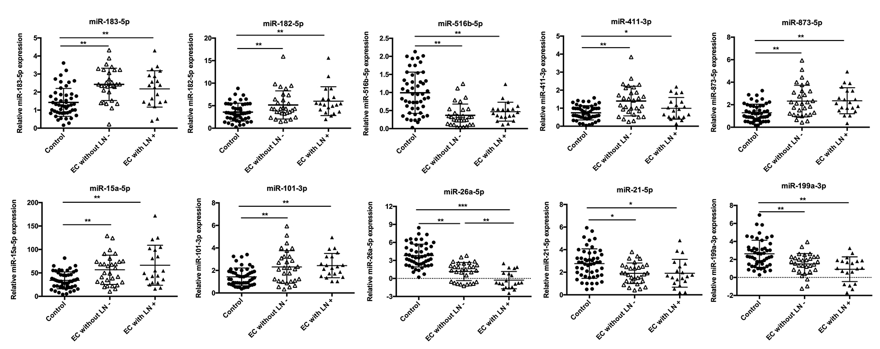
Human lymphatic endothelial cell (HLEC), normal endometrial epithetial cell (EEC), human endometrial cancer cell lines Ishikawa (ISK), HEC-1B, HEC-1A, KLE, AN3CA were all purchased from the American Type Culture Collection (ATCC). HLEC was cultured in ECM medium (ScienCell, CA, USA) supplemented with 5% fetal bovine serum (HyClone, Logan, USA). EEC, ISK, HEC-1B, HEC-1A, KLE, and AN3CA cell lines were cultured in DMEM/F12 medium (Gibco, NY, USA) supplemented with 10% fetal bovine serum (HyClone, Logan, USA). A total of 50 cases of EC tissue specimens, corresponding to 30 cases without LN metastasis and 20 cases with LN metastasis, were collected from EC patients who underwent radical surgery without prior radiotherapy and chemotherapy between 2018 and 2019 at the International Peace Maternity and Child Health Hospital. A total of 50 cases of serum samples were collected from EC patients and 50 cases of serum samples were collected from healthy donors. Informed consent was obtained from all patients, and the research was approved by the Ethics Committee of International Peace Maternity and Child Health Hospital.
Stable transfection with lentiviral vector
Lentivirus vectors expressing miR-26a-5p and repressing miR-26a-5p were constructed and generated by Genechem Inc. HEC-1B was transfected with LV-Fluc-miR-26a-5p-up and its negative control vector (LV-Fluc-miR-26a-5p-up-NC). ISK was transfected with LV-Fluc-miR-26a-5p-down and its negative control vector (LV-Fluc-miR-26a-5p-down-NC).
RNA interference and plasmids
Inhibitor and mimic of miR-26a-5p were purchased from RiboBio Inc. The LEF1-coding sequence (without 3’-UTR) was cloned into pCDNA3.1(+)-vector. The empty vector was used as a blank control. In the rescue experiments, cells that stably expressed miR-26a-5p or incubated with exosomal miR-26a-5p were transfected with the human LEF1 expressing plasmids (OBiO Technology, Shanghai). TFEB expressing plasmids and siTFEB were designed and synthesized by OBiO Technology. Lipofectamine 3000 Reagent (Invitrogen) was then used to transfect siTFEB, human TFEB expressing plasmids, and human LEF1 expressing plasmids according to the manufacturer’s protocol.
Fluorescence in situ hybridization and Immunofluorescence
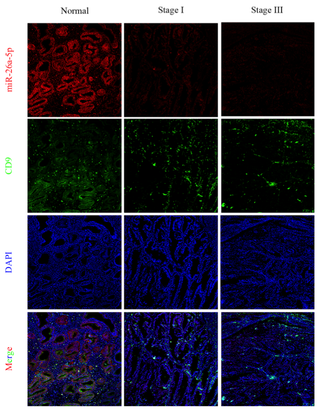
Formalin-fixed paraffin-embedded sections were cut into 4.0-μm sections of tumor specimens. FISH was performed in tumor sections using fluorescence in situ hybridization (FISH) kit (Bosterbio, USA) and the miR-26a-5p detection probe (Boster, Wuhan, Chian) by following the manufacturer’s protocol. For immunofluorescence, serial 4.0-μm paraffin section from EC tissues were analyzed by immunofluorescence with the Opal 4-color kit (PerkinElmer) according to the manufacturer’s protocol.
Bioinformatic miRNAs target prediction
Two online programs TargetScan and miRDB were used to predict potential target genes for miR-26a-5p. GeneCards website (http://www.genecards.org) was used to identify relative regulators of endometrial cancer and lymphatic metastasis.
Quantitative real-time-PCR
Total RNA was extracted from cell lines and human EC tissues using the TRIzol Reagent (Invitrogen, USA), and miRNA was extracted by the RNeasy/miRNeasy Mini kit (Qiagen). Reverse transcription was performed using PrimeScriptrt reagent kit (Qiagen), and the ABI Prism 7000 Sequence Detection System (ABI 7000 SDS) was used for real-time PCR analysis. U6 was used as the endogenous control of miRNAs, and GAPDH gene was used as an internal control for other mRNAs. Each experiment was repeated three times.
Western blotting
Total protein from cells and exosomes were prepared in RIPA buffer with protease inhibitors and quantified using BCA assay kit (Thermo Fisher Scientific Inc., MA, USA). Subsequently, protein lysates were resolved by SDS-PAGE, transferred onto PVDF membranes (Millipore, Bedford, MA). The antibodies used in the experiments included anti-β-catenin monoclonal antibody (1:1000, Cell Signaling Technology), anti-LEF1 monoclonal antibody (1:1000, Cell Signaling Technology), anti-c-myc monoclonal antibody (1:1000, Cell Signaling Technology), anti-VEGFA monoclonal antibody (1:1000, Abcam), anti-TFEB monoclonal antibody (1:1000, Cell Signaling Technology), anti-PI3K monoclonal antibody (1:1000, Cell Signaling Technology), anti-p-PI3K monoclonal antibody (1:1000, Cell Signaling Technology), anti-AKT monoclonal antibody (1:1000, Cell Signaling Technology), anti-p-AKT monoclonal antibody (1:1000, Cell Signaling Technology), and anti-GAPDH (1:5000, Santa Cruz). Each experiment was repeated three times.
HLEC tube formation assay and transwell migration assay
For tube formation assay, matrigel matrix (Corning) was plated in 48-well plate and incubated at 37 °C for 30 min to allow the matrigel to form a solid structure in the bottom. The treated HLECs were seeded onto the matrigel-coated well. After 12 h incubation, the plate was observed for the tubular structure with microscope. The tube length was quantified by ImageJ software (National Institutes of Health, Bethesda, USA). Each experiment was repeated three times. For transwell migration assay, 1 × 10⁵ cells in 200 μl ECM medium without FBS were seeded on a fibronectin-coated polycarbonate membrane insert in a Transwell apparatuses (Corning), and 600 μl medium with 10% FBS was added to the lower chamber. After 24 h of incubation, the cells invaded to the bottom of the inserted membrane were fixed with methanol for 15 min and then stained with 0.1% crystalline violet solution. We counted the cell numbers for analysis under a microscope in five random fields.
Chromatin immunoprecipitation (ChIP)
Cells were fixed with 1% formalehyde for 10 min at room temperature. Then, the cells were washed twice with PBS at 4 ℃, collected and resuspended in lysis buffer and lysed on ice for 30 min. Cells were sonicated 5 times for 5 s to solubilize and shear cross-linked DNA. The chromatin (25μg) was immunoprecipitated for 12 h with 2 μg of anti-TFEB antibody or IgG. After incubation, protein G magnetic beads were then washed for 5 min with buffers. The immune complexes were eluted with elution buffer. After RNase A and proteinase K treatments and reversal of cross-linking, DNA was obtained by phenol and phenol/chloroform extractions. PCR amplifications of the precipitated DNA were carried out.
Luciferase activity assay
The putative miR-26a-5p complementary site in the 3’-UTR of LEF1 or its mutant sequence was cloned into the pMIR-REPORT Luciferase vector (OBiO Technology, Shanghai, China). 293T cells were co-transfected with the pMIR-REPORT-LEF1-3’UTR-WT or pMIR-REPORT-LEF1-3’UTR-MT vector and miR-26a-5p mimic/NC. And, cells were seeded into a 24-well plate and co-transfected with luciferase reporter constructs encoding the wild-type 3’-UTR region of miR-26a-5p or a mutated miR-26a-5p 3’-UTR region and TFEB plasmid using Lipofectamine 3000 (Invitrogen, California, USA). Luciferase activity was measured 48 h after transfection and analyzed by using a Dual-Luciferase Reporter Assay System (Promega). Firefly luciferase signal was normalized to Renilla luciferase signal.
Popliteal LN metastasis model
NOD-SCID mice (5 weeks old) were purchased from the Experimental Animal Center, Shanghai Jiaotong University School of Medicine (Shanghai, PR China) and were used for the lymphatic metastasis model. The studies were approved by the Institutional Animal Research Ethics Committee of Shanghai Jiaotong University School of Medicine. Luciferase-labeled HEC-1B cells (1 × 10⁷) were injected into the footpad of the mice. Then, the mice were randomly divided into three groups (n = 5 per group) and injected intratumorally with PBS, HEC-1B-exovector, or HEC-1B-exomiR(20 μg per dose) every 3 days. Lymphatic metastasis was analyzed using a IVIS® Spectrum In vivo Imaging System (Xenogen Corporation). The footpad tumors and popliteal LN were excised when the tumors reached a comparable size 150 mm3 (volume = length × (width)2 / 2). FISH and IHC were used to analysis the sections of primary tumors and popliteal LN.
Statistical analysis
GraphPad Prism 6 software was used for statistical analysis. Quantitative values were presented as the mean ± SD. Differences among/between groups were analyzed by one-way ANOVA or Student’s t-test. The χ2- test was used for categorical variables. The degree of linear relationship between the expression levels of exosomal miR-26a-5p and miR-26a-5p in endometrial cancer lesion was analyzed by pearson’s correlation coefficient. Significant difference was indicated by P < 0.05. Adobe Illustrator CC, Adobe Photoshop CC, and Image J software were used for figure presentation.