Fabrication of hydrogel
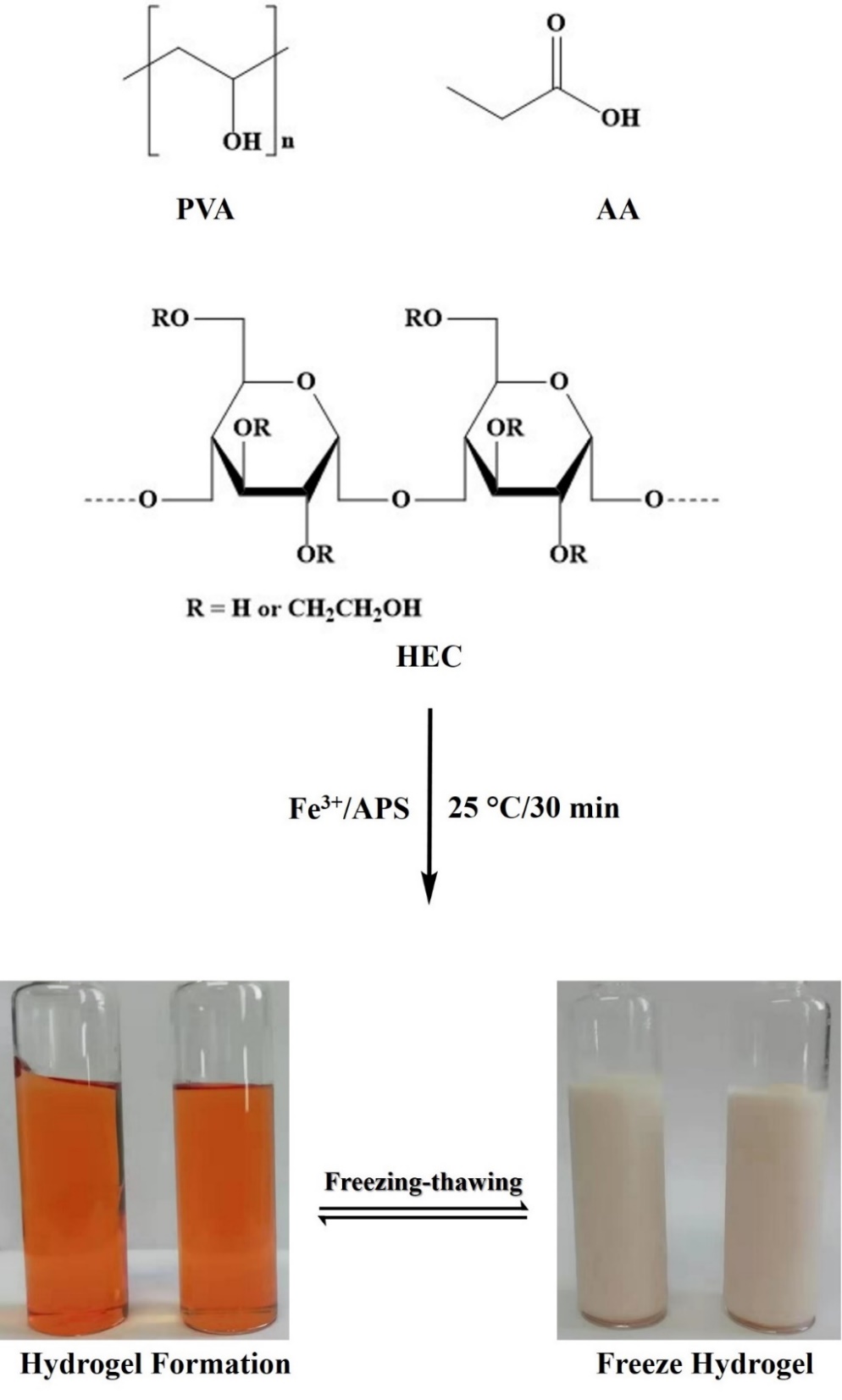
The construction of a physical double network strategy in hydrogel networks generally improves their mechanical performance and self-recovery efficiency. Based on this concept, we engineered a natural polymer-based (HEC) double network hydrogel sensor with robust mechanical, self-healing, and conductive properties. Figure 1 shows the schematic diagram for the fabrication of HEC-PVA/PAA-Fe3+ hydrogels via free radical polymerization of the first network and freezing-thawing cycles for the construction of the second network. Firstly, the HEC-PAA solutions of different concentration ratios were prepared at different temperatures, and then various concentrations of AA were added to the pre-cooled HEC-PAA solution under continuously stirring. After the formation of the homogeneous mixture, various molar concentrations of Iron (III) hexahydrated chloride were added to the obtained mixture and followed by the addition of APS solution. The formation of hydrogels was carried out at room temperature followed by the freezing-thawing cycles. Owing to the abundance of oxygen-containing groups on the polymer chain, such as carboxyl, hydroxide, and ether groups, as well as metal ions, the development of numerous non-covalent associations occurred concurrently in the hydrogel network. The majority of the hydroxyl groups in hydroxyethyl cellulose and polyvinyl alcohol form intermolecular and intramolecular hydrogen bonds, which help to keep the hydrogel network stable. The ethyl oxygen and carboxylic groups of PAA form an ionic coordination interaction with a tri-positive ferric cation (Fe3+), which plays an important role in the hydrogel network's self-healing performance. Besides, hydrogen bonding might also form in the HEC and PAA network between the –OH groups of HEC with the –COO− and –OH groups of PAA. All these binding interactions were confirmed by FTIR spectroscopic technique.
The FTIR spectra of the pure HEC, HEC/PAA-Fe3+, and the hydrogel sensor HEC-PVA/PAA-Fe3+ is shown in Fig. 1. The absorption peaks at 3450 and 2932 − 2862 cm− 1 in the HEC spectrum are due to the presence of stretching vibrations of –OH and –CH bonding in the HEC polymer. The absorption bands at 1186 cm− 1 represent the C-O bond stretching, while the band at 1049 cm− 1 shows the deformation of the O-H bond and the bands in the fingerprint region are due to C-H bond bending vibrations. The disappearance of C-H stretching and narrowing of O-H stretching in the HEC/PAA-Fe3+ spectrum confirm the involvement of these groups in metal coordination and hydrogen bond formation. The change in the fingerprint region also confirms the formation of non-covalent bond formation in the HEC/PAA-Fe3+ hydrogel materials. The spectrum of the strain sensor hydrogel HEC-PVA/PAA-Fe3+ shows clear cut differences both in the functional group region and fingerprint region. The most significant change is in the O-H stretching vibration field, which has widened, implying that more –OH groups are involved in the hydrogel materials as a result of the addition of PVA.
This broad region also confirms the formation of the metal-ligand bond between the –OH groups and metal cations. The band at 1754 cm− 1 also confirms the formation of coordination bonds among metal ions and carboxylate ions in the hydrogel network. The disappearance of certain peaks in the fingerprint region also confirms the formation of hydrogen and metal-ligand coordination bonds in the strain sensor hydrogel materials.
Compression analysis
The compression strength of the hydrogel samples with different PVA concentrations in cylindrical shape was performed at a cross-head speed of 2mm/min by using (CMT4501) universal tensile testing machine. All the hydrogel sensors were capable of enduring compression above 98% fracture strain. The fracture stress, fracture strain, fracture toughness, and compression modulus (0–30% strain) were determined from the corresponding compressive stress-strain graphs (Fig. 2a). The compressive stress-strain curves for the HEC hydrogel sensor containing PVA 5%, 10% & 15% are shown in Fig. 2a. It can be seen from Fig. 2c, that all the hydrogel sample curves exhibit a linear behavior at initial strain percent and from which we can calculate the modulus of the hydrogel sensors. The compression stress of the hydrogel sensor increases slightly with increasing deformation and achieved about 28 MPa compression stress irrespective of their different PVA concentration. These phenomena indicate that these hydrogels could be compressed completely without any fracture, which confirms their 3D tough network nature. The hydrogel sensor has a higher maximum compression stress (28 MPa) than pure hemicellulose-based hydrogels. These hydrogels, however, have shown different strain percentages and a hydrogel with a 10% PVA level is able to resist the high compression of ~ 100%. High PVA content formed a brittle hydrogel network causing a decrease in the compression strength including compression strain (%), toughness, and compression modulus. The PVA content showed a trend of the first increase and then decrease in compression strain, compression modulus, and compression toughness. This indicates that with low PVA content the number of hydrogen bonds was less as compared to the optimized concentration (10%) of PVA, while higher concentration results in a brittle network due to the formation of extra crosslinking in the hydrogel network.
In this study, we investigated the effect of PVA concentration on the compression modulus and the strain percent (0–30%) was selected for the comparison of their modulus strength. Compression modulus is an important parameter for the measurement of the strength of hydrogel materials and it was calculated by dividing stress by strain percent. As shown in Fig. 2c & d, the compression modulus of the hydrogel sensor increased as the concentration of PVA increased from 5–10%, while a further increase in PVA% content results in a decrease in the compression modulus. This indicates that optimum PVA concentration could promote a maximum number of inter and intra hydrogen bonds that stiffen the hydrogel network structure. This indicates that the incorporation of PVA polymer affects the mechanical strength of the hydrogel network.
Tensile strength.
The mechanical properties of the hydrogel sensor were characterized via a uniaxial tensile testing machine (CMT4501) with a crosshead speed of 60 mm/min at room temperature. The tensile stress-elongation tests were performed quantitatively and we found that the mechanical strength of the hydrogels was significantly influenced by HEC concentration, PVA concentration and iron (III) ions Fe3+ molar concentration. The mechanical properties of the hydrogel materials are greatly affected by the hydrogel network structure and crosslinking density. Therefore, it is important to establish a relationship between the mechanical properties and network structure of hydrogels. To investigate these phenomena, we performed tensile-elongation tests on hydrogel sensors with different HEC concentrations, and their stress-strain curves are shown in Fig. 3a. The mechanical strength of the designed hydrogel increased with the increase in HEC concentration. However, beyond a certain limit there was a decrease in the mechanical strength with the increase in HEC content due to the formation of the brittle and more dense crosslinked network. The variation in strength, such as fracture stress, fracture strain, and fracture toughness is summarized in Fig. 3b, c & d, respectively. Hydrogels with 2% HEC concentration showed fracture stress of 0.34 MPa with a fracture strain of 880% and fracture energy of 1.32 MJm− 3. Increasing the HEC content to 3% increased the tensile stress of 0.38 MPa with a fracture strain of 1100% and a fracture energy of 1.95 MJm− 3. Hydrogels with 4% HEC concentration exhibited maximal tensile stress (0.51 MPa), elongation strain (1250%), and fracture energy (2.99 MJm− 3), and a further increase in HEC content results in a decrease in the mechanical strength (0.45 MPa stress, 1010% strain & 2.12 MJm− 3). Thus, we choose 4% HEC concentration as an optimum concentration for further fabrication. The increase in tensile strength influenced by the increase in HEC content was due to a suitable number of dynamic crosslinking points, causing the formation of a compact crosslinked hydrogel network. Excess HEC content raises the viscosity of polymer solution, impeding macromolecular chain movement and free radical diffusion during the polymerization process. Thus, the creation of an unstable hydrogel network reduces the tensile strength of the engineered hydrogel.
Further, we have also investigated the PVA concentration effect on the mechanical properties of the hydrogel sensors systematically. We used three different PVA concentrations (5%, 10%, and 15%), and the stress-strain mechanical strength results are shown in Fig. 4a. The mechanical properties of the hydrogel could be tuned easily by changing the PVA concentration. At low PVA (5%) content, the hydrogel shows more stretchable, elastic, and flexible properties. The relative fracture stress, fracture strain, and fracture energy to their corresponding PVA percent concentration are given in Fig. 4b, c & d, respectively. At a low percent concentration of PVA, the hydrogel exhibited the lowest tensile stress (0.43 MPa), largest tensile strain (1350%), and moderately fracture energy (2.56 MJm− 3). By increasing the PVA concentration from 5 to 10%, the tensile stress reached 0.51 MPa, while there was a decrease in the elongation strain (1250%) and the highest fracture energy (2.99 MJm− 3). When the PVA concentration was increased to 15%, there was a decrease in fracture strain (0.42 MPa), fracture strain (1040%), and fracture energy (2.28 MJm− 3). These results indicate that the PVA polymer serves as a crystallite as a physical cross linker and could make more inter-and intra-hydrogen bonding interactions, which in turn improve the tensile strength of the hydrogels. These physical cross linkers could also act as energy dissipation bonds. However, the excess crosslinking will decrease the tractability and flexibility of the hydrogel network due to the formation of a brittle network. Thus, 10% PVA concentration was chosen as an optimum concentration for the fabrication of an optimized hydrogel sensor.
Incorporation of metal ions also acts as a physical crosslinking point, which produces high strength hydrogels with excellent self-healing ability due to the dissociation and association of metal-ligand bonding interaction. Therefore, we further investigate the key role of ferric ion (Fe3+) concentration on the mechanical strength of the prepared hydrogel sensors. We used six different (0.05, 0.10, 0.20. 0.40, 0.60 & 0.80 M) molar concentrations of iron (III) ions for the synthesis of the hydrogel sensor. Beyond a certain limit (0.40 M), the formation of hydrogels did not take place and the four samples with 0.05––0.4 molar concentrations of iron (III) ions were subjected to tensile stress-strain tests to find their comparative mechanical strength as shown in Fig. 5a. As depicted in Fig. 5b, the tensile fracture stress increases as a function of an increase in F3+ ions molar concentration from 0.05 to 0.2 M and it showed the highest fracture stress (0.51 MPa) at 0.20 M concentration while further increase in metal ions content results in a decrease in tensile stress (0.40 MPa) at 0.4 M concentration. There was a dramatic change in the tensile fracture strain as a function of various metal ions concentrations. It can be seen from Fig. 5c, that at low Fe3+ ions concentration (0.05 & 0.10 M), the hydrogel showed higher elongation strain of 1290 & 1350%, respectively. These results indicate that a flexible, stretchable, and elastic hydrogel could be formed at low iron (III) concentration. However, at 0.20 M, the hydrogel showed the maximum tensile fracture stress (0.51 MPa), and a further increase in ferric ion content showed a decrease in both tensile fracture stress, strain, and toughness. The trends in toughness as a function of ferric ions concentration follow the same trends as in tensile trends, which are depicted in Fig. 5d. The metal ions (Fe3+) serve as physical connection points for building hydrogels with a high mechanical strength, forming coordinated bonds with PAA and HEC ligands. These bonds will dissociate and reassemble quickly and reversibly to dissipate energy and contribute to the enhancement of the hydrogels' characteristics and gradual recovery of the hydrogels' mechanical properties. The drastic shift in mechanical strength versus ferric ion content may be due to a particular phenomenon, such as the formation of mono-, di-or tridentate coordination’s or retardation of radical polymerization.
The ionic coordination among the ferric ions and functional groups of ligands also affects the strength of the hydrogel network. At very low iron (III) concentration, usually bidentate (even monodentate) coordination is formed, causing lower crosslinking density in the hydrogel network, which results in worse mechanical properties. At moderate ferric ion concentration, the formation of tridentate coordination occurs, which increases the tensile stress and results in shrinkage of the hydrogel network that decreases the tensile strain of the hydrogels. Therefore, 0.20 M Fe3+ concentration was chosen as an appropriate concentration, which forms a considerable secondary cross-linking degree and strengthens the hydrogel materials. Additionally, the excess content of ferric ions may impede the radical polymerization of AA, causing a decrease in molecular weight of PAA polymer, which in turn lowers the mechanical strength of the designed hydrogels. We compared the mechanical properties of hydrogels to investigate the effect of the HEC, PVA, and iron (III) ions on the performance of the hydrogels. The compressive strength, tensile strength, and fracture energy of the HEC4PVA10/PAA3-Fe3 + 0.2 hydrogels show the best performance in all samples.
Self-healing efficiency
The self-healing ability of materials not only prolongs their lifetime but also retains the materials’ original properties. Autonomously self-healing in hydrogel at room temperature and without the mediation of a peripheral stimulus broadens their applications. The prepared hydrogel sensors exhibited an excellent self-healing capability, which was studied macroscopically and measured the tensile strength of healed hydrogel sensors by using (CMT4501) universal tensile testing machine. The comparative self-healing study was carried out as a function of the hydrogel composition to find their self-healing abilities and quantify the self-healing efficiencies (SHE) as a proportion of the tensile strength (TS) of the healed hydrogel to the TS of the original sample. We have also investigated the comparative percent growth ratios as a function of change in various constituents of the hydrogel sensors in original and healed samples, as summarized in Table S2. For tensile testing, the rectangular hydrogel samples were cut with a blade in the middle and rejoined the cut pieces at the cut interfaces without any external intervention. The hydrogel sensors were heald for 24 h at room temperature, before being subjected to a tensile stress-strain test to determine their mechanical healing strength. The tensile stress-strain graphs of original and healed hydrogel sensors with different HEC concentrations (2, 3, 4, & 5%) are shown in Fig. 6a. It was found that HEC concentration not only affects the mechanical strength of the hydrogels but also affects their self-healing efficiency. The trend of first increase and then decrease in stress, strain, and toughness was also found in SHE of hydrogel sensors with different HEC concentrations as depicted in Fig. 6b. The hydrogel with 2% HEC concentration showed about 89% self-healing efficiency (SHE) in stress and with 4% HEC content SHE reached 97%. Similarly, the SHE in a strain of hydrogel sensor with HEC (2%) was 95%, which increased with the increase in HEC concentration (4%) and achieved 99% recovery in tensile strain. The recovery in toughness with 2 & 4% HEC concentration hydrogels was 83% & 94%, respectively. Further increase in HEC content results in a lower SHE in stress (85%), strain (92%), and toughness (76%).
Furthermore, we also quantified the SHE as a function of PVA concentration as shown in Fig. 6c & 6d. The hydroxyl moieties in the PVA chain play a key role in the self-healing process of hydrogel materials. At moderate concentration, PVA also shows the excellent self-recovery ability of hydrogels. The hydrogel with 5% PVA concentration got recovery of 86% in stress, 96% in strain, and 84% in toughness. On doubling the PVA concentration (PVA 10%), the SHE in stress, strain, and toughness was reached to 97%, 99%, and 94% respectively, which could be considered as an excellent recovery in hydrogel materials. A further 5% increase in PVA concentration caused a decrease in the self-healing efficiency as a whole, which may be attributed to the difficult rearrangement of hydroxyl moieties to form inter-and intrachain hydrogen bonds.
The influence of key factor Fe3+ ions were also studied systematically on the SHE of the hydrogel sensor and the tensile stress-strain curves of original and healed hydrogels with different Fe3+ ions molar concentrations are shown in Fig. 6e. All the curves of healed hydrogel obey the order of their respective original hydrogel to confirm their recovery and mechanical strength. The self-healing efficiency in stress, strain, and toughness as a function of various concentrations of iron (III) ions is shown in Fig. 6f. The trend of first increase and then decrease was also found in SHE as a function of metal ions concentration. The hydrogel containing 0.05 M Fe3+ ions concentration achieved about 90% recovery in stress, 93% in strain, and 83% in toughness. On increasing the concentration of metal ions from 0.05 M to 0.20 M, the SHE in stress, strain, and toughness reached 97%, 99%, and 93%, respectively. Further increase in the metal ions concentration causes a decrease in SHE in stress, strain, and toughness to 90%, 93%, and 83%, respectively. The mobilized Fe3+ ions and polymers chain plays a critical role in the process of self-healing, which diffuse towards the cut interfaces and interact with the functional groups of polymer chain to re-establish the ionic coordination. However, at higher Fe3+ ions concentration, a rigid hydrogel network is formed that hinders the free diffusion of metal ions and polymer chain, and therefore the self-healing ability of the hydrogel gradually decreases.
The mechanical healing efficiency was also studied as a function of healing time on the optimized hydrogel sensors. The two freshly cut pieces were healed for different time intervals (3, 6, 12 & 24 h) without any external intervention at room temperature. The healed specimen was subjected to the stress-strain tests and their relative was compared strength with the original hydrogel sample, as shown in Fig. 7a. Hydrogel recovered 73% in stress, 84% in strain, and 60% in toughness, even at the shortest healing time of 3 hours. With an increasing in healing time, the self-healing efficiency also increases and achieved 91% SHE in strain, 99% SHE in strain, and 94% SHE in toughness after a healing time of 24 h, as represented in Fig. 7b. The comparative fracture stress, strain, and toughness of original and healed hydrogel sensors as a function of the healing time are depicted in Fig. 7c.
The self-healing ability was also studied macroscopically as shown in Fig. 8. The freshly prepared hydrogel was cut into small pieces with a sharp cutter. The cut pieces were placed together in a self-made dumbbell-shaped mold, pressed between two glass plates, and were healed at room temperature for 24 h, as shown in Fig. 8a. It was interesting to find that the cut pieces were healed together and formed a desired shaped hydrogel without any visible fracture and scratch (Fig. 8b). The healed hydrogel showed strong mechanical strength that can be bent and twist without any fracture (Fig. 8c). Thus, we can say that the designed hydrogel has excellent self-healing ability.
The supreme self-healing capability of the hydrogel sensor is due to two types of interactions, i.e. ionic and hydrogen bonding interactions. The hydroxyl moieties in the poly (vinyl alcohol) (PVA) chain form hydrogen bonding with the adjacent chains on the contact of two cut pieces. The polymer chains diffuse transversely through the interface and the reformation of the new hydrogen bonding occurs. However, the rearrangement of the hydroxyl moieties in the PVA hydrogel to reform intra and inter-chain hydrogen bonds limits the predominant self-healing ability of PVA hydrogels. The self-healing of hydrogel sensor is endorsed by the natural transmission of PVA; HEC; and PAA polymer chains, ferric ions and water at the cut edges, and the successive formation of hydrogen bonding. The simultaneous diffusion and subsequent entanglement of Fe3+ ions and PAA chain across the cut interfaces help in the self-healing ability of hydrogel due to the Fe3+ ions mediated ionic interaction and hydrogen bonding association.
Electrochemical and sensing performance
Electrochemical impedance spectroscopy was used to measure the ionic conductivity of the hydrogel sensor and its Nyquist plot is represented in Fig. 9a. The initial semicircular pattern of the Nyquist plot indicates the charge transfer and then the linear curve shows the mass transfer electrical conductivity. The starting point observed at the start of the high frequency is the electrolyte resistance (Rs) and the charge-transfer resistance (Rp) can be determined by the diameter of the semicircle. The hydrogel showed about 2.22 × 10− 1 S m− 1 electrical conductivity at room temperature, which is sufficient for use in the fabrication of biomedical e-skins, soft robotics, health monitoring, and biomedical diagnostic devices. The mobile ferric ions and free electrons on carboxylate ions in the hydrogel network greatly contributed to the electrical conductivity of hydrogels. To investigate the probability of the hydrogel as a strain sensor, the hydrogels were subjected to relative electrical resistance change tests as a function of assorted strain. The sensitivity of the strain sensor was measured by finding their linearity as a change in the relative electrical signals as a function of applied strain (%), as depicted in Fig. 9b. The relative change in resistance (ΔR/R₀ (%) increases linearly with an increase in strain from 0 to 400% and reached to 340%, indicating superior sensitivity of the prepared hydrogel sensor. The stability of the sensor was measured by holding-loading a stepwise test as a change in ΔR/R₀ (%) versus time (s) and sustained for a while at some specific strain. The hydrogel exhibited steps of stair-like trends as displayed in Fig. 9c, in which the ΔR/R₀ (%) shows direct proportion with the change in strain (%) and maintains a specific strain for some time without any change in the (ΔR/R₀ (%), indicating outstanding stability in resistance as a function of various stains. The reliability and durability of the hydrogel sensor were further investigated through the electromechanical (EM) cyclic test as a relative change in resistance as a function of time (s) and the results are depicted in Fig. 9d. The EM loading-unloading signals in Fig. 9d almost show similar trends in their resistance change versus time, which confirm their excellent durability and reliability in EM behavior. These results indicate that the designed hydrogel sensor is a stable, sensitive, reliable, and durable sensor, which can boost its long life expectancy.
To investigate the performance of hydrogel as a wearable strain sensor in a real-time application, the pressure-based strain sensors were directly attached to the numerous junction points (index finger and wrist joints) of a human body for monitoring various human motions. The relative change in resistance at various strains was measured by monitoring the movements of these muscular joints.
The sensitivity, ionic conductivity, and feasibility of the hydrogel-based sensor were recorded via a bending-holding process by bending the index finger and holding for some time at fixed strain, as shown in Fig. 10a. The hydrogel demonstrated a relative change in resistance as a function of time with an increase in finger bending angle as a steps of stair trend. Bending the finger causes the hydrogel to stretch, which increases the relative resistance, and at a constant angle, the ΔR/R₀ (%) remains constant, indicating accurate control of the finger moment. Several consecutive electromechanical loading-unloading cycles were used for the analysis of the reliability of electrical conductance on the bending figure as shown in Fig. 10b. The cyclical bending of the hydrogel sensor on the forefinger produced nearly identical signals, indicating that the sensor's responsive activity is repeatable and consistent.
The hydrogel sensor was further subjected to 100 cycles at 50% strain in the EM loading-unloading test and the results are displayed in Fig. 10c. The cyclic EM loading-unloading curves (inset Fig. 10C) showed almost identical intensity at a 50% strain, indicating their stability and long lifespan as a strain sensor. This was further confirmed by a cyclic bending-relaxing test of the wrist joints and a stable ΔR/R₀ (%) as a function of time was obtained as displayed in Fig. 10d that further confirms their durability, stability, and recoverability. Based on this performance, the prepared hydrogel has the potential for e-skin applications that could be used as a wearable strain sensor to monitor and quantify various human motions in real-time.