2.1. Materials
DSPE and DSPE-PEG-2000 were obtained from Lipoid (Germany), D L-lactide and glycolide were purchased from Corbion Purac (Netherland),Ir obtained Generous gift from Emcure (Pune),1,1'-Dioctadecyl-3,3,3',3' Tetramethylindotricarbocyanine Iodide (DiR), 1,1'-Dioctadecyl-3,3,3',3'-Tetramethylindocarbocyanine Perchlorate-4′ (Dil),6-Diamidino-2-phenylindole (DAPI), dialysis tubing (MWCO 2000 Da), SP-Sephadex C-25 resin were purchased from Sigma (UK). Snake Skin® dialysis tubing (MWCO 10000 Da) was a gift sample from Thermo-fisher (USA). Soybean lecithin (Epikuron 140 V) was a kind gift from Cargill Pharmaceuticals. Methylene chloride, acetone, n-hexane, absolute ethanol and diethyl ether (ultra-pure grades) were obtained from Sigma Aldrich (India). RPMI-1640 media,fetal bovine serum (FBS), penicillin/streptomycin, Trypsin/EDTA, and phosphate buffered saline (PBS) were obtained from Gibco, Invitrogen (India). BD flow cytometry tubes were purchased from VWR (UK). Vectashield®mounting media was from Vector Labs (UK); 16% Formaldehyde, methanol-free, was from Thermo Scientific Pierce. All reagents were used without further purification.
2.2 Synthesis of PLGA using ring opening polymerization
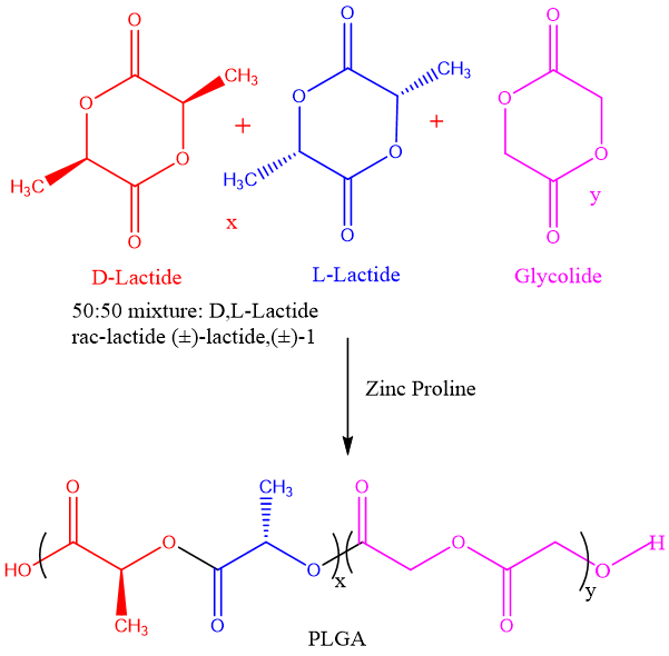
The ring opening polymerization of monomers of D, L-Lactide and Glycolide performed using Zinc proline complex [9]. Polymerization’s reaction was carried out in bulk (without any solvent used in reaction medium) in sealed glass ampoules of (inner diameter of 2 cm and 10 cm in height). Glass ampoules were passivated using 20% TMSCl (Trimethyl silyl chloride in acetone solvent) before polymerization and dried properly in the oven. The addition of monomer and initiator was carried out in MBRAUN UNilab™ glove box to prevent moisture absorption in the monomers of D, L-Lactide and Glycolide. Ampoules were subjected to 3-4 Freeze-Pump-Thaw cycles in vacuo to remove moisture content from the monomers of D, L-Lactide and Glycolide. Ampoules containing D, L-lactide 50% mole (500 mg, 3.472 mmol), glycolide 50% mole (395 mg, 3.472 mmol) ratio initiator Zinc Proline (5 mg, 0.03472 mmol) respectively used to carry out polymerization reaction. The initiator zinc proline to monomer ratio 200 were used in the reaction sealed under vacuum. Ampoules were then immersed in Techne SBL-2D™ Fluidized sand bath previously set at the temperature of 200 ˚C for 4h. Ampoules were cooled and broken so the solid material containing Poly (D, L-Lactide-co-Glycolide) polymer, and unreacted D, L-lactide, glycolide monomer was dissolved in a small amount of dichloromethane and precipitated in excess n-hexane to obtain a Poly (D, L-Lactide-co-Glycolide) polymer. Polymer samples were dried at 40 °C under vacuum for 48 h. Further characterized performed successfully. The polymerization reaction was reported with our research group in previous study for PLA repeated with some modification carried for PLGA [38].
2.3 Characterization of PLGA copolymer
The Synthesized PLGA copolymers were characterized by 1H NMR, MALDI-TOF MS, Differential Scanning Calorimetry (DSC), Gel Permeation Chromatography (GPC) and ATR-FTIR etc.
2. 3.1 1H Nuclear magnetic resonance (NMR)
1H-NMR spectra were recorded using 400 MHz Bruker 400 spectrometer using CDCl3 as the solvent containing a small amount of the TMS as an internal standard. The sample preparation was carried out by dissolving 10 mg of PLGA polymers in CDCl3 at room temperature.
2.3.2 Matrix assisted laser desorption ionization -Time of flight mass spectroscopy (MALDI-TOF MS)
MALDI-TOF MS analysis was performed on an AB SCIEX4800 plus MALDI TOF/TOFTM Analyzer. The PLGA copolymer samples were dissolved in tetrahydrofuran (1mg/mL) and mixed with the matrix (15mg/mL of tetrahydrofuran) and dried on the sample plate. 2,5-dihydroxybenzoic acid and dithranol were used as the matrix.
2.3.3 Differential scanning calorimetery (DSC)
Differential Scanning Calorimetry (DSC) measurements were performed on TA Instrument DSC Q10 in a nitrogen atmosphere. The heating rate was 10°C /min and cooling rate was 100°C/min for every run from 0°C to 200°C. The glass transition temperature data were recorded from second heating curves.
2.3.4 Gel permeation chromatography (GPC)
The molecular weights such as number-average molecular weight [Mn], weight-average molecular weight [Mw], and polydispersity [Mw/Mn] were determined with respect to polystyrene standards by size-exclusion chromatography on an Agilent Technologies, Polymer Laboratories Gel permeation chromatography (PL-GPC) 220 machine (Santa Clara, CA, USA) at 25 °C, with eluting PLGA solutions (10 mg/mL of CHCl3) and toluene as an internal standard, and through a series of five 30 cm long Styragel columns with pore sizes of 500, 105, 104, 103, and 100Å. CHCl3 was used as the mobile phase (flow rate: 1 mL/min), and a refractive index detector was used for the detection of different molecular weight fractions.
2.3.5 Attenuated total reflectance - fourier transform infrared (ATR-FTIR)
ATR-FTIR spectra of PLGA copolymer samples were recorded using a (Perkin Elmer IRFTIR spectrometer USA) in 500–4000 cm‐2 wavelength range in attenuated total reflectance (ATR) mode. The instrument was calibrated with an indium standard before measurements.
2.4 Formulation of the LPs
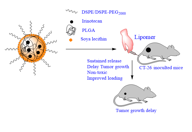
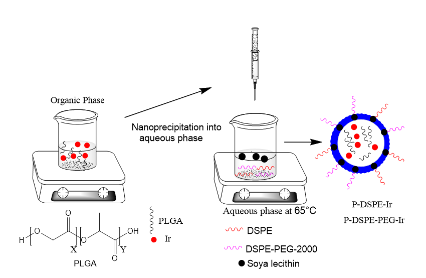
LPs were formulated from PLGA, soybean lecithin with varied DSPE and DSPE–PEG-2000 shell using a modified nanoprecipitation technique [39]. PLGA was first dissolved in acetonitrile with concentration 1 mg/mL. Lecithin and DSPE/DSPE– PEG–2000 (7:3 molar ratio) were dissolved in 4% ethanol aqueous solution (aqueous phase) and 20 % of the PLGA (in acetonitrile as organic phase) weight with respect to lipid weight and heated to 65 0C temperature. The PLGA solution was then added slowly into the previously preheated lipid aqueous solution (DSPE/DSPE-PEG-2000 and Soyabean Lecithin) drop-wise (1 mL/min) under gentle stirring followed by vortexing for 3 min. The LPs were allowed to self-assemble for 2 h with continuous stirring while the organic solvent was allowed to evaporate. The remaining organic solvent and free molecules were removed by washing the LPs solution 3 times using an Amicon Ultra-4 centrifugal filter (Millipore, Billerica, MA) with a molecular weight cut-off of 10 kDa and then resuspended in water to obtain a final desired concentration. The LPs were freeze dried in liquid nitrogen and lyophilized for storage at -80 0C for later use [40-42]. P-DSPE-Ir LPs is referred to PLGA core, DSPE shell LPs loaded with Ir whereas P-DSPE-PEG-Ir LPs was referred to PLGA core DSPE-PEG2000 shell LPs loaded with Ir.
2.5 Characterization of the formulated LPs
Formulated nanoparticles were characterized for the following characterization as follows
2.5.1 Determination of percentage encapsulation efficiency
The encapsulation efficiency (EE %) of Ir loaded LPs was determined by measuring the concentration of the free drug in the LPs suspension. The unencapsulated Ir was separated by filtration followed by centrifugation. A definite volume of the LPs suspension was transferred to the upper chamber of the Nanosep and centrifuged at 6000 rpm for 60 min. The amount of free drug in the filtrate was measured using UV-vis spectrophotometer (Model UV- 1601 PC; Shimadzu, Kyoto, Japan) by measuring the absorbance at 360 nm as previously described [43-44]. The EE (%) was calculated by
EE (%) = ([Drug] total -[Drug] Supernatant)/[Drug] total) * 100
The percentage of drug loading efficiency (% LE) was calculated using the following equation:
LE (%) = Amount of Ir encapsulated (mg)/ weight of all excipients (polymer, Lipid and Soya lecithin) *100.
2.5.2 Size and Zeta potential measurements
The Polydispersity Index (PDI) and Zeta potential of LPs were determined by using zetasizer (PCS, Nano ZS90 zetasizer, Malvern Instruments Corp, U.K). Charge on the LPs surface was determined using zetasizer 300HSA (Malvern Instruments, Malvern, UK). Disposable polystyrene cells and disposable plain folded capillary Zeta cells were used. LPs suspensions were diluted in deionized water and measurements were performed at 25 °C. Electrophoretic mobility was used to calculate the zeta-potential using the Helmholtz-Smoluchowski equation. Analysis time was kept for 3 min and hydrodynamic size was presented as the average value of 20 runs, with triplicate measurements within each run.
2.5.3 Transmission Electron Microscopy (TEM)
Transmission electron microscopic (TEM) imaging of LPs was carried out using (FEI Technai G2 T20) instrument with an acceleration voltage of 200 keV. The TEM sample was prepared by transferring the LPs suspension (4 mg/mL) onto a 200-mesh carbon-coated copper grid. Samples were blotted away after 30 min incubation and grids were negatively stained for 10 min at room temperature with freshly prepared, 2% (w/v) phosphotungstic acid aqueous solution. The grids were then washed twice with distilled water and air-dried before imaging.
2.5.4 Atomic force microcopy (AFM)
The atomic force microscopy (AFM) measurements were performed on silicon wafer using a Multimode scanning probe microscope equipped with a Nanoscope IV controller from Veeco Instrument Inc., Santa Barbara, CA. All the AFM measurements were done under ambient conditions using the tapping-mode AFM probes model - Tap190Al purchased from Budget Sensors. The radii of tips used in this study were less than 10 nm, and their height was ~ 17 μm. The cantilever used had a resonant frequency of ca. 162 kHz and nominal spring constant of ca. 48 N/m with a 30 nm thick aluminium reflex coating on the back side of the cantilever of the length 225 μm. For each sample, three locations with a surface area of 1 × 1 µm2 and 500 × 500 nm2, each were imaged at a rate of 1 Hz and at a resolution of 512× 512.
2.5.5 Wide angle X-ray Diffractometer (WAXD)
The lyophilized LPs were analyzed using wide angle X-ray Diffractometer (WAXD) in the range of 2θ=5‐55° at room temperature (25 °C). The WXRD patterns of LPs sample were analyzed by Philips 1830 X-ray diffractometer (Philips, Almelo, The Netherlands) using a CuKα source at a (λ=1.5406 Å) to get more insights about nature of the sample.
2.6 In-vitro release profile
In vitro release from Ir loaded P-DSPE-Ir and P-DSPE-PEG-Ir LPs were determined by the previously reported dialysis method. 20 mg of LPs suspended in 10 ml of PBS buffer, was sealed in a dialysis bag (M.W. cut off: 10, 000, Sigma Aldrich). The dialysis bag was incubated in 100 ml of PBS buffer medium (pH 7.4 and 50% FBS in pH PBS) at 37 °C. The release amount was analyzed at 360 nm using Uv-visible spectroscopy Perkin-Elmer calculated using a calibration curve for Ir dissolved in the same media as the dialysate (R2 = 0.99995, range1-50 μg/mL) [45-46]. A control experiment was set up alongside in which the same amount of Ir was dissolved in DMSO and dialyzed for comparison. This control was set up to eliminate nonspecific adsorption of the drug to the dialysis membrane. The same conditions were used for P-DSPE-Ir and P-DSPE-PEG-Ir.
2.7 In-vitro anti-tumor efficacy studies
The In-vitro anti-tumor efficacy of the blank P-DSPE, P-DSPE-PEG, P-DSPE-Ir and P-DSPE-PEG-Ir LPs was examined in CT-26 murine carcinoma cells by MTT assay. Cells were cultured (2 × 10⁴ cells per well) in 96 well plate in RPMI media supplemented with 10% FBS, 1% penicillin, and 1% streptomycin in 5% CO2 atmosphere at 37 °C. After 24 h incubation, different concentrations (5-100 μg/mL dispersed in media) of Ir, blank P-DSPE, P-DSPE-PEG, P-DSPE-Ir and P-DSPE-PEG- Ir LPs were added into wells. Following 24 hrs incubation, wells are washed with PBS, and 200 μL of MTT (0.5mg/ml) was added. Formazan crystals formed after 4 h were dissolved in 200 μL of DMSO. Optical absorbance was recorded at 570 using a microplate reader (Epoch2, BoiTek). The results were expressed as the percentage cell survival (mean ± SE) and calculated using the following equation:
% Cell survival = (A570 nm of treated cells/A570 nm of untreated control cells) x 100.
2.8 Fluorescence Labeling of LPs
Fluorescent labeled Dil and DiR dye loaded LPs were prepared for In-vitro uptake and In -vivo tumor targeting and biodistribution studies In-vivo imaging system (IVIS). Dil, emitting in the red region, and DiR, a fluorophore with near-infrared emission, are hydrophobic fluorescent markers that were incorporated into the organic phase at 0.5 % (w/v) (dye/organic phase) for in vitro and in vivo studies, respectively. Formulations were protected from light covered with aluminum foil and utilized for in vitro (P-DSPE-Dil and P-DSPE-PEG-Dil) and in vivo (P-DSPE-DiR and P-DSPE-PEG-DiR) studies.
2.9 Cellular uptake
CT-26 cells were seeded on coverslips at a density of 1×10⁵ cells /well. To check the uptake efficiency of nanoparticles, cells were treated with Dil tagged LPs (P-DSPE-Dil and P-DSPE-PEG-Dil). After 4 hrs, media containing nanoparticles were removed and cells were fixed with 4% paraformaldehyde for 20 min. The coverslips, were mounted onto slides and uptake was visualized using Leica sp5 confocal microscope. In separate experiments, cells were treated with cold temp (40C) or sodium azide (0.1%) or sucrose (0.5M) or amphotericin B (20mM) for 1hr as inhibitor of uptake pathways prior to treatment with LPs followed by confocal analysis.
2.10 Cell cycle analysis
Effect of Ir loaded P-DSPE-Ir and P-DSPE-PEG-Ir LPs on cell cycle progression in CT-26 cells was analyzed by FACS. Briefly, CT-26 cells were seeded in a 60 mm dish at a density of 2×10⁵ cells /dish and treated with NPs (50 µg/ml) for 24 h. After treatment, cells were fixed overnight in 95% ethanol at 40C and were washed in chilled PBS for 2 times, the pellet was re-suspended in 50 μl of RNase A (0.5 mg/ml) and incubated for 20 minutes at 37 °C. After that, 450 μl of propidium iodide (50μg/ml) was added. The proportion of cells at different phases of the cell cycle was monitored by FACS Calibur (BD Biosciences).
2.11 In-vivo Biodistribution study
All animal procedures were performed in accordance with the Guidelines approved by ‘Committee for the Purpose of Control and Supervision of Experiments on Animals’ (CPCSEA), Government of India and experiments were approved by Institutional Animal Care and Use Committee (IACUC) of National Centre for Cell Science (NCCS), Pune, India.6−8 week’s old female Balb/C mice were used to perform the in vivo studies. CT-26 colon cancer cells (1×10⁶) mixed with Matrigel were injected subcutaneously into the right flanke of mouse, and tumor growth was observed. After tumor generation, DiR loaded P-DSPE-DiR and P-DSPE-PEG-DiR loaded LPs were injected intravaneously through tail vein of the mice. The biodistribution of DiR loaded nanoparticles monitored at various time points upto 7 days by In-vivo imaging. The biodistribution of the DiR in major organs (heart, lung, liver, spleen, intestine, and kidney) and tumor was examined by ex-vivo imaging after sacrifice of mice using the IVIS system (IVIS spectrum, Xenogen). In separate experiments, CT-26 tumor bearing mice were injected with (P-DSPE-DiL and P-DSPE-PEG-DiL) and tumor retention of NPs was analyzed for 9 days using IVIS.
2.12 In-vivo anti-tumor efficacy studies
All animal procedures were performed in accordance with the Guidelines approved by ‘Committee for the Purpose of Control and Supervision of Experiments on Animals’ (CPCSEA), Government of India and experiments were approved by Institutional Animal Care and Use Committee (IACUC) of National Centre for Cell Science (NCCS), Pune, India.CT-26 colon cancer cells (1×10⁶) mixed with Matrigel were injected subcutaneously into the right flanke of mouse, and tumor growth was observed. After tumor initiation, mice were treated with Ir, P-DSPE-Ir and P-DSPE-PEG-Ir nanoparticles (10 mg/kg body wt Ir corresponding conc.) intravenous twice a week for two weeks. The tumor size monitored with the help of Vernier caliper during the experiment. After two weeks, mice were sacrificed, tumors were harvested and weighed. Further, tumors sections were prepared for histopathological analysis.
2.13 In-vivo acute toxicity studies
All animal procedures were performed in accordance with the Guidelines approved by ‘Committee for the Purpose of Control and Supervision of Experiments on Animals’ (CPCSEA),Government of India and experiments were approved by Institutional Animal Care and Use Committee (IACUC) of National Centre for Cell Science (NCCS), Pune, India.6-8 week old healthy female Balb/c mice were treated with Ir, P-DSPE-Ir and P-DSPE-PEG-Ir LPs (20 mg/kg body wt of Ir corresponding conc) twice a week for two weeks. After one week, blood was drawn from mice through retro-orbital sinus and hematological and biochemical analysis was performed to examine the toxicity.
2.14 Stastical analysis
For all experiments, data were presented as mean ± SD. Significant differences were examined using one-way ANOVA. p<0.05 was considered statistically significant in all studies.