3.1. Characteristics of the PEs
The particle size of the PEs was controlled between 119 and 131.2 nm, the polydispersity index (PDI) of each nanomedicine less than 0.2, and the zeta potential below -20 mV, indicating that the nanomedicines showed good stability (Table S1).
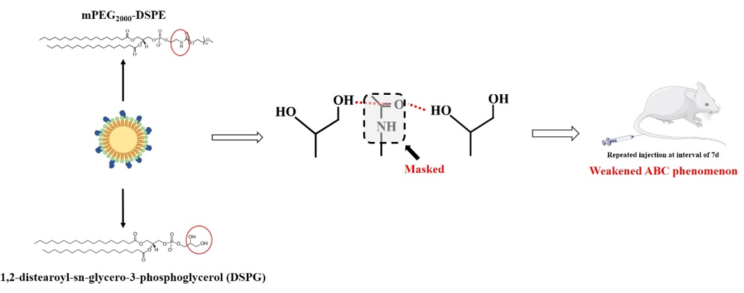
As everyone knows, the phase transition temperature of SPC is -20 oC, while that of DSPG is 55 oC. In order to eliminate the influence of the two emulsifiers on the release of emulsions in blood, we compared the release behavior of different emulsions in vitro simulated in vivo environment.
1.0 mL PEs was precisely aspirate and added to a dialysis bag (with a molecular weight cut off of 10kDa), clamped the two ends and placed it in 200 mL of PBS buffer (500 mmol/L, pH = 7.4, containing appropriate penicillin), stirred under constant temperature at 37.0 ± 0.5 oC and away from light (100rpm). At 0.5, 1, 2, 4, 8, 12, 24, and 48 h, drew 3.0 mL of dialysate, and added an equal amount of release medium. The dialysate was filtered with a 0.45µm microporous filter membrane and the fluorescence intensity was measured at λex = 750nm, λem = 790nm, and the concentration was calculated by substituting it into the standard curve equation; the cumulative release Rn of the drug is calculated, the formula is as follows, and the results are as follows Fig. 1.
$$\mathbf{R}\mathbf{n}=\frac{\mathbf{C}\mathbf{n}\mathbf{V}0+\sum _{\mathbf{n}-1}^{\mathbf{n}}\mathbf{C}(\mathbf{n}-1)\mathbf{V}}{\mathbf{M}\mathbf{t}}\times 100\mathbf{\%}$$
Among them, Cn is the concentration at the nth sampling, V0 is the volume of the release medium, V is the volume of each sampling, and Mt is the total drug concentration. Results showed that there was no significant difference in the release behavior among different PE.
3.2. Effect of the PEG modified densities on the pharmacokinetics of n mol % PEs and PE-DSPGs of single and repeated intravenous injection
The 8-hour pharmacokinetic behavior of a single injection of n mol % PEs and PE-DSPGs was evaluated by measuring the plasma concentration of the DiR. As indicated by the area under the curve (AUC) and T1/2, the circulation time of n mol % PEs and PE-DSPGs in the Wistar rats increased gradually with the increase in PEG density on the surface of n mol % PEs and PE-DSPGs (Table S2). Seven days after the ଁrst injection, the rats were injected repeatedly.
The strength of the ABC phenomenon was evaluated using the ABCindex, which indicated the ratio of AUC of the second dose to that of the first dose; that is, the ABCindex = AUC(0−60 min) of the second injection/AUC(0−60 min) of the first injection. The higher the ABCindex value, the smaller the difference in pharmacokinetic behavior between the second and first injections, which meant that a weaker ABC phenomenon is occurring. Here, the ABCindex(0−60 min) was used as the criterion to evaluate the strength of the ABC phenomenon.
The repeated administration of n mol % PEs in each group induced the ABC phenomenon. When the modification density of PEG increased from 10 mol % to 30 mol %, the ABC phenomenon decreased; however, when the PEG modification density continued to increase, the ABC phenomenon was enhanced. Compared with n mol % PEs, there were no significant differences in the level of anti-PEG IgM induced by n mol % PE-DSPGs with the same PEG modification density (Fig. 2), but the ABCindex of each group was significantly higher than that of n mol % PEs, with the same PEG modification density, indicating that the ABC phenomenon induced by each group was remarkably weakened. When the ABCindex of 30 mol % PE-DSPG reached 0.89, almost no ABC phenomenon was observed (Fig. 3). These results suggest that both the modification density of PEG and the addition of DSPG could affect the intensity of the ABC phenomenon. The modification density of PEG affected the secretion levels of anti-PEG IgM, whereas while DSPG did not affect the secretion level of antibodies, it weakened the ABC phenomenon.
3.3 Effect of DSPG concentration on the pharmacokinetics of single and repeated injections of PE-DSPG-ns
To further investigate the influence of DSPG on the ABC phenomenon, we prepared a series of PEs with different DSPG concentrations, PE-DSPG-ns, using the same procedure as that for n mol % PEs and PE-DSPGs (Fig. 4). No significant differences were observed in the pharmacokinetic behavior among the five groups after a single injection of PE-DSPG-ns, as shown by the AUCs and T1/2 values (Table S2). Seven days after the first injection, the PE-DSPE-ns containing DiR were injected repeatedly with the same phospholipid dose. The ABCindex showed that the addition of DSPG could indeed weaken the ABC phenomenon. When the modification concentration of PEG was fixed, i.e., PE-DSPG-ns groups, the ABC phenomenon gradually weakened with the gradual increase of DSPG concentration and when the molar ratio of the DSPG to PEG increased to 30:1 in the PE-DSPG-30 group, the ABCindex reached approximately 0.94, and the ABC phenomenon almost disappeared.
No significant differences were observed in the level of anti-PEG IgM induced by the first injection of PEs with the same PEG modification density; however, the addition of DSPG weakened the ABC phenomenon in each group, significantly. Therefore, we selected 5 µmol phospholipid/kg 10 mol % PE with the smallest ABCindex for the first injection, and the rats with high levels of anti-PEG IgM after the first injection of 10 mol % PE were termed as ABC (+) rats. As shown in Fig. 5(A–C), the ABC (+) rats produced a high level of anti-PEG-IgM 7 days after the first injection while the second 30 mol % PE injection produced a strong ABC phenomenon, with the ABCindex reaching 0.25, while ABCindex of repeated 30 mol % PE injection was 0.7. It was suggested that the reason for the weak ABC phenomenon after the repeated injection of 30 mol % PE may be that the higher density of PEG modification inhibited the production of anti-PEG antibodies. When the level of anti-PEG IgM reached a higher level, the 30 mol % PE would still be cleared quickly with an accumulation in the liver and spleen (Fig. 5D).
The ABCindex of the second injection of 30 mol % PE-DSPG in the ABC (+) rats reached 0.82, which was slightly lower than the repeated injection of 30 mol % PE-DSPG and further demonstrated the attenuation effect of DSPG on the ABC phenomenon; although there was a high level of anti-PEG IgM in rats, a strong ABC phenomenon did not occur. The ABC (+) rats injected with PE-DSPG-30 did not cause the ABC phenomenon. This also showed that higher DSPG concentrations could effectively inhibit the occurrence of the ABC phenomenon.
3.4 IgM binding of 30 mol % PE and 30 mol % PE-DSPG in the ABC (+) rats
To determine the influence of DSPG on the intensity of the ABC phenomenon of PEs, we measured the changes in anti-PEG IgM before and 6 h after the second injection of 30 mol % PE, 30 mol % PE-DSPG, and PE-DSPG-30 in the ABC (+) rats. As shown in Fig. 6, no significant change was observed in the plasma anti-PEG IgM levels of the ABC (+) rats 6 h after the injection of 30 mol % PE-DSPG and PE-DSPG-30, while the level of anti-PEG IgM in the ABC (+) rats decreased significantly after the intravenous injection of 30 mol % PE, indicating that the second injection of 30 mol % PEs could be combined with anti-PEG IgM of the ABC (+) rats to produce the ABC phenomenon. In contrast, 30 mol % PE-DSPG and PE-DSPG-30 combination did not enable the anti-PEG IgM recognition in the ABC (+) rats; therefore, the ABC phenomenon did not occur.
3.5 Co-injection of 10 mol % PE with 30 mol % PE, 30 mol % PE-DSPG and DSPG-PE-30
Here, we investigated the effect of the co-injection of 10 mol % PE containing DiR with blank 30 mol % PE or DSPG-PE-30 into the ABC (+) rats. As shown in Fig. 7, the co-injection of 30 mol % PE and DSPG-PE-30 and 10 mol % PE did not affect the ABC phenomenon of 10 mol % PE.