Animals and ethics statement
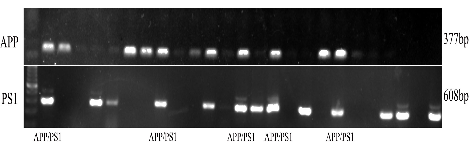
APP/PS1 double transgenic mice were bought from the Model Animal Research Center of Nanjing University [Animal license number: SCXK (su) 2012–0007, stock number: 0014406]. All time points referred to herein indicate the number of days after transferation of APP/PS1. The new transgenic AD APP/PS1 mouse model was obtained by breeding APP/PS1 AD mice, and the genotypes were confirmed by PCR of mouse tail genomic DNA using the 2*EasyTaq PCR SuperMix (Trans Technology, Lot:), according to the manufacturer’s instructions. All the experimental procedures were approved by the Animal Laboratory Administrative Center and the Institutional Ethics Committee at Xiangya Medical University and also in accordance with the National Institutes of Health guidelines. Specific primers for APP gene contained within the Sangon Biotech (Shanghai) designed (forward Neo5ʹ; 5ʹ-GACTGACCACTCGACCAGGTTCTG-3ʹ and reverse Neo3ʹ; 5ʹ-CTTGTAAGTTGGATTCTCATATCCG-3ʹ). Also, specific primers for PS1 gene contained within the Sangon biotech (Shanghai) designed (forward Neo5ʹ; 5ʹ-AATAGAGAACGGCAGGAGCA-3ʹ and reverse Neo3ʹ; 5′-GTGGATAACCCCTCCCCCAGCCTAGACC − 3). After PCR reactions, the amplified products were separated in agarose gels and analyzed. C57/BL6 as wild type were purchased from the Experimental Animal Center of Jingda [Animal license number: SCXK (xiang) 2013–0004]. There were 4 groups including C57/BL6 as wild type (control) group, APP/PS1 (model) group, APP/PS1 + LVCON307 group and APP/PS1 + LV-NLRC3 group. Thirty 12-month-old APP/PS1 transgenic mice were randomized into three groups as model group, APP/PS1 + LVCON307 and APP/PS1 + LV-NLRC3 group. Ten 12-month-old wild-type C57 mice were chosen as control group. Mice in APP/PS1 + LVCON307 and APP/PS1 + LV-NLRC3 group were injected with LVCON307 or LV-NLRC3 through intracerebroventricular injection (4 µl, 107 virus particles (VPs) /kg). Mice in control group and model group were administrated with the same volume of normal saline. All the animals were housed in barrier facilities (Experimental Animal Center, Xiangya Medical School of Central South University, temperature 22 °C, under a 12:12 h light–dark cycle light 7:00–19:00; humidity 40–60%), and food and water were freely available.
Lentiviral vectors (LV)
In this experiment, the construction and titer determination of LV-NLRC3 and negative control lentivirus (LVCON307) were constructed and purified by GeneChem Biomedical Co. Ltd (Shanghai, China). The titers were 5E + 8 and 6E + 8 transducing units (TU)/ml.
Intracerebroventricular injections
Mice were anesthetized by intraperitoneal injection of ketamine and intracerebroventricular injection with microinjector.The Bregma point was fully exposed by cutting an incision about 0.5 cm long along the median line of the head at the beginning of the operation. At 1.5 mm posterior to the point and 1 mm lateral to the right, the microinjector was used to penetrate the skull vertically at a depth of 3 mm and retained for 5–10 minutes. Then slowly inject LV-NLRC3 or LVCON307 at the speed of 1 ul/min, and then retain the needle for 5–10 minutes. Drew the needles and sutured skin. After 6 months, the mice were decapitated and executed. The brains were quickly removed on ice and sagittally cut along the median line.
Morris water maze and Behavior test
Morris water maze was used to evaluate spatial learning and memory (SLM) function of mice at age 18 month in each group, according to the protocol of van Praag et al. (1999) and Akers et al. (2014). Mice were housed and habituated for 2 h in behavioral testing rooms before tests. All tests were conducted on consecutive days in a dimly illuminated room with standard conditions of temperature and free from any stray noise. All behavioral apparatus were cleaned with 75% ethanol and dried between each animal. Behavioral outcome was recorded by 2 trained assistants who were blinded to the information of each mouse. The apparatus consisted of a circular tub (120 cm in diameter, 50 cm in height) with a black inner wall and of transparent platform (10 cm diameter) submerged 1 cm below the water surface, which was painted with distinct geometric cues. The water maintained at the range of (24 ± 1 °C). Forty mice underwent four trials per day in the learning stage. Each mouse was placed into the water and randomly started from each of four different locations facing the pool wall in the trial. The trial was terminated and the latency was recorded when the mouse found the platform within 90 s. Otherwise, the trial was terminated and the mouse was gently guided to the platform. On day 5, a probe trial was conducted to estimate the memory function of each mouse. After the platform removed, mouse was put into the pool as before to evaluate its memory ability. The swim paths were recorded with an overhead video camera and tracked with automated software (San Diego Instruments, San Diego, CA, USA). The time to reach the platform during water-maze training, number of the times the target area (former platform) was crossed, and time spent in each quadrant were recorded during the probe trial.
Histology
After the Morris water maze test, 5 mice from each group were deeply anesthetized (300 mg/kg, i.p. 10% chloral hydrate in DDW) and were perfused with ice-cold normal saline (50 ml) to remove blood from the vasculature, and then with 4% paraformaldehyde in phosphate buffered saline(50 ml). Their brains were removed and incubated overnight in 4% paraformaldehyde, and then half of each brain was dehydrated in 30% sucrose in PBS for frozen sections, while half was done routine fixation, dehydration, paraffin embedding for paraffin sections. Another 5 mice from each group were deeply anesthetized and perfused with ice-cold normal saline (50 ml), and then their brain samples were harvested and stored at -80 °C for Real time PCR and western blot analysis.
Organotypic hippocampal slice culture
Prepare surgical equipments: brushes, scissors (big and small), plastic pipette(thin, middle and short), blades, plastic plates and curved dissecting forceps and plastic knifes (long). Make sure to keep everything sterilized. Sterilize the surface of the super-clean workbench with alcohol, turn the ultraviolet light on, sterilizing for two hours. Use a container of ice to place the cutting medium. The composition of the cutting medium are (PH = 7.2): Earle’s MEM (cat.61100-06, Lot.74k4064 Gibco), Hepes 5.95 g, Tri-base 1.21 g, Glucose 1.8 g, MgCl2 6 ml (all reagents from Gibco or Sigma). Get packs of cell well dishes and place inside each of the wells an insert of Millipore. Add to the outside edge of the well 1 mL of tissue medium. Put dishes into the incubator.
Add 1 ml tissue culture medium to each well of 6-well plate, inserts of Millipore were then placed into the wells where the tissue culture medium had been added. The composition of the culture medium are(PH = 7.25, in 1L):BME(B 9638), EBSS(E7510), NaCI(S5886) 1.167 g, NaHCO3(S 5761) 0.42 g, Ascorbica cid (A 4544),Glucose(G 7021), Hepes(free acid H3375) 6.36 g, CaCl2·2H2O(C 7903) 0.0293 g, MgS04(M 2643) 0.203 g, Glutamine(G 7029), 0.39 g, Insulin (2 mg/ml stock I4011) 0.667 ml, Penicillin(P 3032) 24.8 mg,20% Horse serum (all reagents from Gibco or Sigma).Put dishes into the incubator. Prepare the chopper (LEICA VT1200S, VIBRATOME LINE), cut head of pups(postnatal 6–9 days) quickly and remove skull, membrane till you get to brain. Put brain in cutting medium with paper and slice into two pieces and get hippocampus. Use a brush to gently move the separated pieces of hippocampus with cutting medium onto chopping board. Cut the hippocampus into 400 micron slices, and gently place these sections in cutting medium. Get the spare 6-well plates containing the culture medium from the incubator. Place 2–5 pieces of dorsal hippocampal at approximalcoronal section in each pup on the Millipore inserts of each well. Excess cutting medium should be sucked. Place plate in incubator and label (name, date). After 2–3 hours, use sucker to remove old medium of the bottom in each well and add new culture medium. Feed slice culture tissue each other days about 10 days. Treatment slices culture tissue about 6–10 days. NLRC3 protein was used for 48 hours, Aβ was used 36 hours before tissue harvested.
Immunohistochemistry, Immunofluorescent and confocal microscopy
Immunohistochemistry was performed to investigate the expression of Aβ, NLRC3 and Glial fibrillary acidic protein (GFAP) in the mice brains. Briefly, brains sections were dewaxed and rehydrated in decreasing concentrations ethanol. Then, endogenous peroxidase of the sections was blocked with 3% H2O2 under room temperature for 10 minutes. Slides were washed with PBS for several times and blocked in 2% BSA with 0.3% Triton X-100 for 40 min at 37 °C. Thereafter, the sections were incubated with primary antibodies (GFAP 1:200 Millipore, Billerica, MA, USA; 6E10 1:1000 Thermo Fisher Scientific; NLRC3 1:100 abnova, Taiwan, China) overnight at 4 °C. Then, rinsed in PBS and incubated with biotinylated secondary antibody (Vector, 1:200) and streptavidin-horseradish peroxidase (Jackson, Immunoreaserch, West Grove, PA, USA) for 1 h at 37 °C, and then rinsed for another 3 min × 3 with PBS before reaction with diamino-benzidine (DAB) (Vector) solution. The sections were observed under a microscope. Nissl staining was performed to quantify neuronal density insections.
For immunofluorescent staining, the sections were boiled in citric acid buffer (pH 6.0) for 20 min in a bain marie oven. After the sections were cooled, they were treated with 0.3% Triton X-100 and 2% BSA for 1 h at room temperature. The sections were then incubated overnight at 4 °C with a primary antibody ( neuron-specific nuclear protein (NeuN) 1:200 CST, USA; NLRC3 1:100 abnova, Taiwan, China), and then with a secondary antibody (1:300 Alexa-Fluor-488-conjugated goat anti-mouse IgG2a antibody, Life Technologies, catalog number A21131; 1:300 Alexa-Fluor-555-conjugated goat anti-rabbit IgG1 antibody, Life Technologies, catalog number A31572) in PBS containing 1% BSA at room temperature for 2 h. The sections were mounted onto slides, embedded with SlowFade® Gold (Invitrogen), and covered with a coverslip.
Aβ plaques in brains were visualized using Thioflavin-S fluorescence (ThS) staining. ThS was dissolved in 50% of ethanol at 500 mM and brain sections were stained for 7 min. As a differentiation step to remove a nonspecific binding of the dye, a slide was soaked into 100, 95 and 90% ethanol solutions for 10sec each and then moved into PBS.
Quantitative real-time PCR
The level of NLRC3 mRNA was detected using Real-time qPCR kits by technicians who were blinded to the experimental groups. Total RNA was extracted from frontal cortex and hippocampus using the Trizol Plus RNA Purification System (Ambion, Invitrogen) according to the manufacturer’s instructions. RNA was quantified using the BioSpec Nano spectrophotometer (Shimadzu) and cDNA was reverse transcribed using the cDNA Synthesis kit (Thermo scientific, Lot 00285583) according to the manufacturer’s instructions. Quantitative real-time PCR was performed using the UltraSYBR Mixture (With ROX1) (CEBIO, CW2601) with the following cycling parameters: 95 °C for 10 minutes followed by 38 cycles of 95 °C for 10 seconds, 60 °C for 30 seconds, 72 °C for 32 seconds, followed by amplicon dissociation (95 °C for 15 seconds, 60 °C for 1 minute, 95 °C for 15 seconds, 60 °C for 15 seconds, increasing at 0.5 °C/ cycle until 95 °C was reached). All gene expression data were normalized to β-actin expression. All reactions were performed in triplicate. Gene expression results were calculated using the delta delta cycle threshold (two delta delta CT) method (Livak and Schmittgen, 2001). The two delta delta CT method was used to determine mean fold changes in gene expression between the control and target genes.
Western blot
Brain tissues were lysed in RIPA buffer containing phosphatase inhibitor and complete protease inhibitor cocktail. Aliquots of brain lysates were separated on SDS-PAGE, transferred to membranes and immunoblotted with primary antibodies, then horseradish peroxidase-conjugated secondary antibody (1:1000). Bands were revealed by use of an enhanced chemiluminescence (ECL) system ( Thermo Scientific, Rockford IL) and density was quantified by use of Imagequant 5.2 (Healthcare Bio-Sciences, Philadelphia, PA). The primary Abs were used as following: Iba-1 and NeuN (1:1000 dilution, CST, USA), GFAP (1:1000 dilution; Millipore, USA), NLRC3(1:1000 dilution; abnova), PI3K (1:500 dilution, CST, USA), β-actin and GAPDH (Proteintech, Chicago, USA). Secondary horseradish peroxidase–conjugated Abs were purchased from Santa Cruz Biotechnology (Santa Cruz, CA, USA) (1:1000 dilution).
Data and Statistical Analyses
The 3D image overlays were visualized with the Leica Application Suite (LAS) Advanced Fluorescence Lite software (LAS AF Lite, 2.4.1 build 6384, Leica). The ImageJ software (National Institutes of Health, Bethesda, MD, USA) was used to analyze the immunohistochemistry results. Analyses of plaque distributions were transcribed manually into the computer-acceptable format by keeping research colleagues blind. In the case of single mean comparison, Student t test were performed to analyzed the data. In case of multiple mean comparisons, ANOVA and further Newman–Keuls posttest, or two-way repeated-measures ANOVA, followed by Bonferroni tests was used. Data are expressed as means ± standard deviations of the means (SD). P value < 0.05 was considered statistically significant (Prism version 6.0 software (GraphPad), USA).