2.1. Extraction of Chrysanthemum:
The chrysanthemum of experiment is produced in Hangzhou, China, and the petals were used to prepare for water extraction. After soaking chrysanthemum petals in water that 8 times weight of materials and boiling for 0.5 hour. Keep the liquid and residue was boiled for another time. Repeat the process for 3 times and then combine all the liquid obtained. Concentrate the collection to low and high concentration of extract. (1g chrysanthemum extract obtained from 4.255g materials)[10]. The content of total flavonoids in chrysanthemum was 5.0%, according to Technical specification for inspection and evaluation of health food.
2.2. Cell experiment:
2.2.1. Cell culture:
ARPE-19 cells (China Center for Type Culture Collection) were cultured in DMEM / F12 medium (Hyclone, the USA) containing 10% fetal bovine serum, 100U/mL penicillin and 100U mg/mL streptomycin, and incubated in 5% CO2 under constant temperature of 37 ℃ and humidity. When the cells grow exponentially, it will be used in the follow-up experiment.
2.2.2. The toxicity of chrysanthemum extract to ARPE-19 cells:
The safe concentration and toxicity of chrysanthemum extract to ARPE-19 cells were detected by MTT. AREP-19 cells approximate number of 10 ×103 cells were inoculated into 96-well plates with 100 μL per well. When the cells grew exponentially, the concentrations were 0.2mg/mL, 0.4mg/mL, 0.8mg/mL, 1.0mg/mL, 1.2mg/mL, 1.5 mg/mL, 2.00mg/mL and 5.00mg/mL respectively. of chrysanthemum extract were added to 96-well plate separately and cultured for 24 hours. After the end of culture, 20μL MTT (5mg/mL) solution was added to each well and cultured in CO2 incubator for 4 hours. MTT was discarded and 150μL DMSO solution was added. After being fully dissolved, the cell activity was evaluated by measuring absorbance (A value) at wavelength of 492 nm. According to the results, the safe concentration was applied for the follow-up experiment.
2.2.3. Light induced damage of RPE cell:
After the cells were digested by trypsin, the cells were made into cell suspension and inoculated in the culture plate. The cells were irradiated directly with LED white light with the intensity of (2500 ±500) Lux for 24 hours, after the cells grew exponentially[11].
2.3. Animals:
2.3.1. Animal ethics:
Totally 32 mice of C57BL/6J of male with the age of 6 weeks were obtained from the SPF Biotechnology (Beijing, Co,.Ltd), the mice were raised in 12/12h light and dark with the temperature of 23+2℃, relative humidity of 55% in the Tianjin Eye Institute, The food and water could take freely. All the procedures were coherence with international standard of laboratory animal use and care and in accordance with the principles and guidelines of the Chinese Council Animal Care, the experiment was approved by the Institutional Animal Care and Use Committee of Tianjin Medical University. Mice were experimented or sacrificed by inhaling the isoflurane, all the steps to minimize suffering.
2.3.2. The groups division and chrysanthemum extract intervention:
Mice were randomly divided into control group (n = 8), light damaged group (LD, n = 8), low-dose chrysanthemum group (LC, n = 8) and high-dose chrysanthemum group (HC, n = 8). After concentrated and dried by water extraction, the extracts were crushed into powder and suspended with 0.9% saline when it was given to intragastric administration. In the range of effective concentration, 0.23g/kg/d for the low dose group, and 0.38g/kg/d for the high dose. 0.2ml/ per day for 8 weeks. The dose concentration was also similar with the study by Dong Lumeng et al. about the protective effect of chrysanthemum on Parkinson's mice[12].
2.3.3. Light damage method:
One day after intragastric administration, the light injury mode were as follows: light damage group(LD), low and high dose of chrysanthemum group (LC and HC)were treated with continuous white light of 9500±500lux for 7 days, 4 h /d after pupil dilated , accumulating 28 hours in total, each mouse was separated by transparent box during light exposing in order to avoid crowding. The function and morphology of retina were examined 7 days after photic injury.
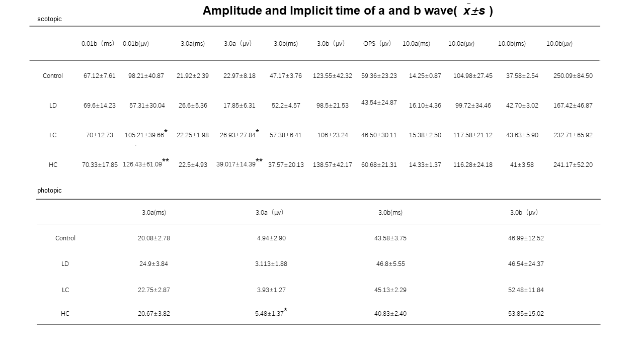
2.3.4. Electroretinogram (ERG):
12 hours after dark adaptation, mice were anesthetized by inhaling 2% isoflurane and fixed on the animal experiment table. Dilate both of eyes and certain gel to keep the cornea transparent. Connecting reference electrode to the head, annular corneal electrode to the eyeball, and the ground electrode to the tail of mouse. Visual electrophysiological apparatus (Roland Germany) was used to record the amplitude of wave a, b and Ops under the stimulating light of 0.01cds/m2 and 3.0 cds/m2 in dark. After 10 minutes of adaptation of photopic, amplitude of a and b under 3.0 cds/m2 was recorded.
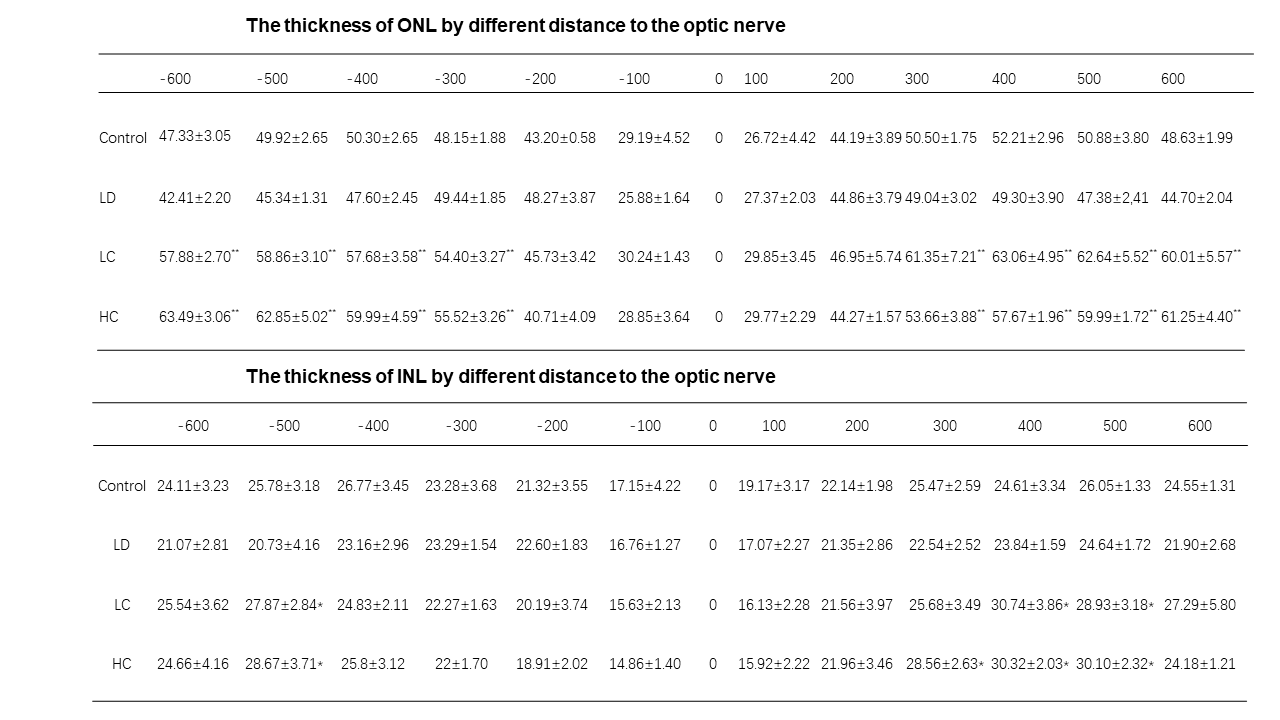
2.3.5. Optical Coherence Tomography (OCT):
Mouse was lied prone on the animal experimental platform after being anesthetized by inhaling 2% isoflurane, 0.5% tropicamide phenylephrine eye drops (Santen) were used to dilate pupils and carbomer eye gel keeping the cornea transparent. Phoenix eye testing equipment for animals (Phoenix research labs, model: Micron IV) was used to scan the retina in vivo and gain morphological imaging of each layer.
2.3.6. Fluorescein fundus angiography (FFA):
2% sodium fluorescein was injected intraperitoneally immediately after finish the OCT examination. The state of filling of the retinal vessels at arterial phase, venous phase and arteriovenous phase was observed at the same gain value. Application of angio-tool 0.6a that a software download free was to analyze the relative parameters consist of vessel area, vessel area percentage and the total number of junctions.
2.3.7. HE staining and TUNEL:
The mice were sacrificed 3 days later by Inhaling isoflurane, ophthalmic vessels blood collected, and the eyeballs were taken out at the same time. 12 hours after formalin fixation of the eyeball, part of cornea was cut off and fixed in fresh formalin for another 8 hours. After dehydration and paraffin liquid immersing at 40℃ for 6 hours, eyeballs were completely embedded Paraffin block which would be sliced into 4μm of thickness. HE and Tunel immunofluorescence staining (Tunel fluorescence kit provide by Dalian meiliun biotechnology Co.,LTD.)were performed to analyze the apoptosis rate of each retinal layer for the all groups.
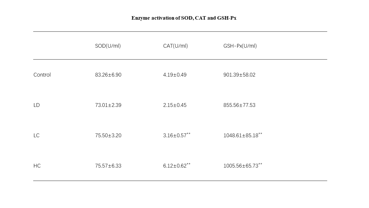
2.3.8. Anti-oxidative enzyme assay:
Superoxide dismutase (SOD), Catalase (CAT) and Glutathione (GSH-Px ) assay kit: Support by Nanjing Jiancheng Bioengineering Institute. Collected blood from ophthalmic artery was centrifugated at 4℃, 3000r/min for 10 minutes in order to obtain the serum. Keeping them at -80℃ to measure the enzyme activation of SOD, CAT and GSH-Px.
2.4. Statistical methods:
All the parameters including the amplitude of a and b wave in the examination of ERG, the vessel area, the vessel percentage and the total number of junctions measured and analyzed in FFA and angio-tool, the apoptosis rate in each layer of retina are presented in the term of χ̅ ± s , one-way ANOVA of SPSS22.0 software was to evaluate the parameter. P<0.05 has been considered as statistically significant.