Recombinant plasmid design
Briefly, having obtained, the sequences of mutated IFNβ27+101 (Kay et al. 2016) and scFv (Kehoe et al. 2013) components (VL and VH) were embedded in the C-terminal and N-terminal of the construct structure, respectively. Next, (Gly4Ser)3 sequence (Chen et al. 2013) was used as a scFv linker (connecting VL to VH) and AEAAAKEAAAKAGS sequence (Grewal et al. 2015, 2017) was applied as an IFNβ linker through which the mutated IFNβ (mIFNβ) attached to scFv. Also, human IFNβ signal peptide sequence obtained from the UniProt database (https://www.uniprot.org/uniprot/P01574) (Consortium 2019) was placed at the beginning of the construct, in order to recombinant protein secretion. Afterward, the amino acid sequence was converted to the nucleic acid sequence by the online server EMBOSS Backtranseq (https://www.ebi.ac.uk/Tools/st/emboss_backtranseq). For cloning into pcDNA3.1(+)_myc-His A vector, HindIII and EcoRI restriction sites were respectively considered at 5′ and 3′ of the recombinant construct (GenBank accession number: MN733992). Also, Myc and His6-tag sequences located in downstream of the construct were considered (Figs. 1a and 1b).
Secondary RNA structure
In silico analysis of the recombinant mRNA was accomplished by Mfold online server (http://unafold.rna.albany.edu/?q$$ =$$mfold) in order to assess the Gibbs free energy for the recombinant mRNA secondary structure.
Protein molecular weight estimation
The molecular weights including non-glycosylated and glycosylated proteins were calculated. In this regard, Protein Molecular Weight web server (https://www.bioinformatics.org/sms/prot_mw.html) (Stothard 2000) was used for calculating the non-glycosylated protein molecular weight. The N-linked glycosylated and O-linked glycosylated protein molecular weights were calculated by GlycoEP server (http://crdd.osdd.net/raghava/glycoep/submit.html) (Chauhan et al. 2013).
Protein structure
The fusion protein’s secondary structure was determined by GOR-IV web server (https://npsa-prabi.ibcp.fr/NPSA/npsa_gor4.html) (Deléage 2017). Using homology modeling, the tertiary structure was predicted by I-TASSER server (https://zhanglab.ccmb.med.umich.edu/I-TASSER/) (Yang et al. 2015; Roy et al. 2010; Zhang 2008). In addition, Ramachandran plot was drawn for the mutated IFNβ-scFv fusion protein by RAMPAGE server (http://mordred.bioc.cam.ac.uk/~rapper/rampage.php). Also, Swiss PDB Viewer version 4.1 software (http://www.expasy.org/spdbv/) (Guex et al. 1997) was applied for energy minimization of the predicted model. The solubility of the protein was evaluated by PROSO II server (http://mbiljj45.bio.med.uni-muenchen.de:8888/prosoII/prosoII.seam) (Smialowski 2012).
Recombinant plasmid construction
The recombinant vector and the vector without insert (mock) were prepared by Macrogen (Korea). The recombinant vector was drawn by Snapgene software (Fig. 1c). The recombinant gene construct was sequenced. Then, the nucleotide BLAST (https://blast.ncbi.nlm.nih.gov/Blast.cgi) was applied for approving the sequencing results. After transformation of the recombinant and mock vectors into the competent E.coli TOP10, the plasmid extraction was accomplished by Miniprep plasmid extraction kit (Genet Bio, Korea). The recombinant vector was double digested with HindIII and EcoRI restriction enzymes (Thermo Fisher Scientific, USA) and analyzed on 1% TAE (Tris-Acetate-EDTA) agarose gel electrophoresis.
Cell culture and transfection
Human embryonic kidney (HEK293) cell line was purchased from the Pasteur Institute, Iran. Cells were cultured in a Dulbecco-modified Eagle medium (DMEM, High Glucose, GlutaMAXTM) with 10% fetal bovine serum (FBS) (Biowest, France) and 1% penicillin/streptomycin (Sigma, Germany) at 37°C, 95% humidity and 5% CO2 condition. 24h before transfection, HEK293 cells were seeded in 35mm tissue culture plates up to about 60% confluency. The fresh medium was refilled, 3h prior to transfection. Then, the calcium phosphate protocol was used for transient transfection of 4µg recombinant (treated) and mock vectors in each plate (Chen 2011). Meanwhile, untransfected cells considered as a negative control. After 24h incubation of transfected and untransfected cells at 37°C, 95% humidity and 5% CO2, the fresh media were added and the cells were incubated for an extra 24h at the same conditions.
Protein purification
The cell media was poured into Ni-NTA Sepharose column. After washing with the wash buffer (20mM imidazole, 300mM NaCl and 50mM NaH2PO4; pH 7.8), the His6-tagged recombinant protein was detached by elution buffer (250mM imidazole, 300mM NaCl, 50mM NaH2PO4; pH 7.8) (Emamzadeh et al. 2006). Then, the Bradford assay was carried out for determining the recombinant protein concentration (Bradford 1976).
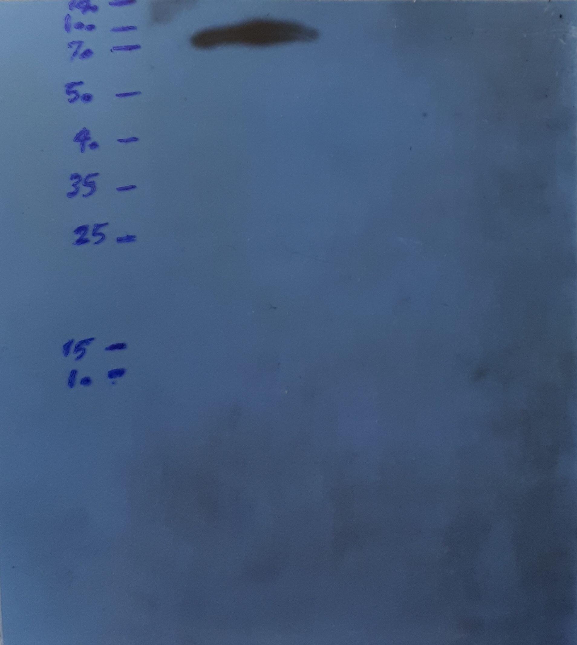
SDS-PAGE and western blotting
The 12.5% sodium dodecyl sulfate-polyacrylamide gel electrophoresis (SDS-PAGE) was used and subsequently, the gel was stained by Coomassie brilliant blue method. Western blotting technique was carried out by the monoclonal anti-polyhistidine-peroxidase antibody (Sigma, Germany). To visualize the recombinant protein, the transferred polyvinylidene difluoride (PVDF) membrane was incubated for 2min in the mixture solution of clarity™ western enhanced chemiluminescence (ECL) substrate (Bio-Rad, USA) that was prepared according to manufacturer's instruction. Next, the PVDF membrane was exposed to the X-ray film in darkness for 1 min and then, it was developed and fixed by the relative solutions for 1 and 5min, respectively.
Binding measurement of scFv in the fusion protein to human collagen type II by enzyme-linked immunosorbent assay (ELISA)
An in-house ELISA was designed and performed for confirmation of the scFv attachment to human collagen type II. To do so, the human collagen type II (EMD Millipore, USA) was coated in the test and positive control wells of a 96-well microplate (Nunc, Denmark). After blocking and washing steps, the recombinant protein and the mouse anti-type II collagen IgG2b monoclonal antibody-biotinylated (Chondrex, USA) were added to the test and negative control wells, and the positive control wells, respectively, followed by incubation and washing. Then, the monoclonal anti-polyhistidine-peroxidase antibody (Sigma, Germany) was added to the test and negative control wells, and the horseradish peroxidase (HRP) streptavidin (BioLegend, USA) was added to the positive control wells. After incubating and washing, 3,3',5,5'-tetramethylbenzidine (TMB) substrate mixture solution (BioLegend, USA) that was prepared following the manufacturer’s protocol, was added to the wells. Finally, stop solution was used and the optical density (OD) was determined by ELISA reader at 450nm.
In vitro activity determination of mutated IFNβ in the fusion protein
The biological activity of IFNβ27+101 was confirmed by MxA induction measurement (Matas et al. 2016) in human peripheral blood mononuclear cells (PBMCs) treated with the recombinant protein. With this aim, human PBMCs were isolated from whole blood of a healthy donor using lymphoprepTM (Stemcell Technologies, Canada) gradient according to manufacturer protocol. 200000 cells/well were cultured in a 12-well plate by Roswell park memorial institute (RPMI) medium (Gibco, UK) supplemented with 10% FBS at 37°C, 95% humidity and 5% CO2 condition. According to the literature, the cells were treated for 24h with 100IU/ml (3ng/ml) of Rebif®(IFNβ-1a) (Merck, Germany) and 3ng/ml of the recombinant protein as a positive control and test, respectively (Dupont et al. 2002). Also, non-treated cells were stored for 24h in the same condition as a negative control.
RNA extraction and cDNA synthesis
RNA extraction was carried out in two separate times, 48h after transfection of HEK293 cells and 24h after PBMCs treatment. Total RNA was isolated by RNeasy Mini Kit (Qiagen, Germany) following the protocol suggested by the manufacturer. Then, the RNA quantity and quality were determined by nanodrop apparatus. Next, cDNA synthesis was performed by RevertAid First Strand cDNA Synthesis Kit (Thermo Fisher Scientific, USA). Both the RNA and cDNA samples were kept at −70°C.
Real-time PCR
Real-time polymerase chain reaction (PCR) was performed using Applied Biosystems® real-time PCR device and Takara kit (SYBR® Premix Ex Taq ™ II, Tli RNaseH Plus). As a housekeeping gene, EEF1A1 was used (Dehghanian et al. 2014) and specific primers for IFNβ and MxA were designed by AlleleID7.0 and Oligo7 softwares (Table1). Real-time PCR reactions were prepared in a 20µl total volume with 0.5pmol/µl from each primer of IFNβ and EEF1A1, 0.25pmol/µl from each primer of MxA, 10µl SYBR® Premix Ex Taq ™ II, 100ng/µl cDNA, and 0.4µl of ROX Reference Dye. Real-time PCR program was carried out by primary denaturation for 2min at 95°C and 40 cycles containing 10s denaturation step at 95°C, 30s annealing step for IFNβ and MxA at 58°C and 60°C, respectively, 30s extension step at 72°C, and a final extension step for 3min at 72°C. The relative gene expression of IFNβ and MxA were calculated by the 2−ΔΔCt method using the average cycle threshold (Ct) values of three independent experiments.
Table 1
Oligonucleotide primer sequences used for real-time PCR
|
Primer
|
Sequence
|
|
IFNβ F
|
5’ GCTACAACTTGCTTGGATTC 3’
|
|
IFNβ R
|
5’ ATAGATGGTCAATGCGGC 3’
|
|
MxA F
|
5’ GCATTCCCAGACGGCATA 3’
|
|
MxA R
|
5’ CAGAGGAGTAGGATTATCACACC 3’
|
|
EEF1A1 F
|
5’ CCCTTCTGGCTTACACACT 3’
|
|
EEF1A1 R
|
5’ TGAACCAAGGCATGTTAGCAC 3’
|
Statistical analysis
Statistical analysis was carried out using SPSS16.0 software (SPSS Inc, USA) through One-way ANOVA and Wilcoxon tests. Results were shown as mean percentage ± relative standard deviation (SD). The p<0.05 was considered as meaningful.