Clinical samples and cell culture
Human samples of osteochondroma (n=20) and OS tissues (n=20) were collected from patients who underwent surgery at the Department of Orthopedic Surgery, The First Affiliated Hospital of Nanchang University (Nanchang, China), and patients have received no preoperative treatment prior to the sample collection. All procedures were approved by the Ethics Committee of The First Affiliated Hospital of Nanchang University and carried out in accordance with the Helsinki Principles. Written informed consent was provided by all patients. Fresh human samples validated by pathological diagnosis were frozen in liquid nitrogen and stored at −80 °C until RNA extraction. The human osteosarcoma cell lines 143B, HOS, MG-63, SJSA-1, Saos-2, and U2OS and non-tumor control cell line hFOB 1.19 were cultured in DMEM supplemented with 10% fetal bovine serum (FBS).
Real-time PCR and quantitative real-time PCR
Genomic DNA (gDNA) was extracted using the DNA Mini Kit (QIAGEN, Germany). Total RNA was isolated from OS cell lines and tissues using the RNeasy Mini Kit (QIAGEN, Germany) according to the manufacturer’s instructions. For the detection of circRNA, RNA samples were treated with RNase R (3 U/ug, Epicenter, Madison, WI, USA) at 37 °C for 15 min, and cDNA was synthesized using the reverse transcription kit (Takara, Japan). Quantification of mRNA, circRNA, and gDNA was performed using the SYBR Green PCR Kit (Takara, Japan) and analyzed by a Real-Time PCR System (Applied Biosystems, USA). The differences of circRNA and mRNA were normalized to the levels of b-actin, and primers are shown in the Additional file 1: Table S1.
Northern blot
The junction probe for circ-CTNNB1 was synthesized and labeled with digoxigenin, as described in our previous study (16). Briefly, 20 µg of total RNA was separated on 3-(N-morpholino) propanesulfonic acid (MOPS)-buffered 2% (w/v) agarose gel containing 1.2% (v/v) formaldehyde under denaturing conditions for 4 h at 80 V and transferred to Hybond-N+ membrane. Prehybridization was carried out at 65 °C for 30 min in DIG Easy Hyb solution (Roche). Hybridizations were performed at 65 °C for 16-18 h. Blots were washed thoroughly, detected by anti-digoxigenin (DIG) antibody staining, and recorded on X-ray films with the chemiluminescence substrate CSPD (Roche).
Western blot
Total proteins from cells were extracted with RIPA lysis buffer (Thermo Scientific, USA). Western blot analysis was performed as described before with antibodies specific for β-actin (ab125402), β-catenin (ab32572), RBM15 (ab244374), ENO1 (ab155102), GPI (ab66340), PGK1 (ab38007), Flag (ab45766, Abcam, USA), ALDOA (sc-390733), and IGF2BP1 (sc-166344, Santa Cruz Biotechnology, USA).
Plasmid construction and stable transfection
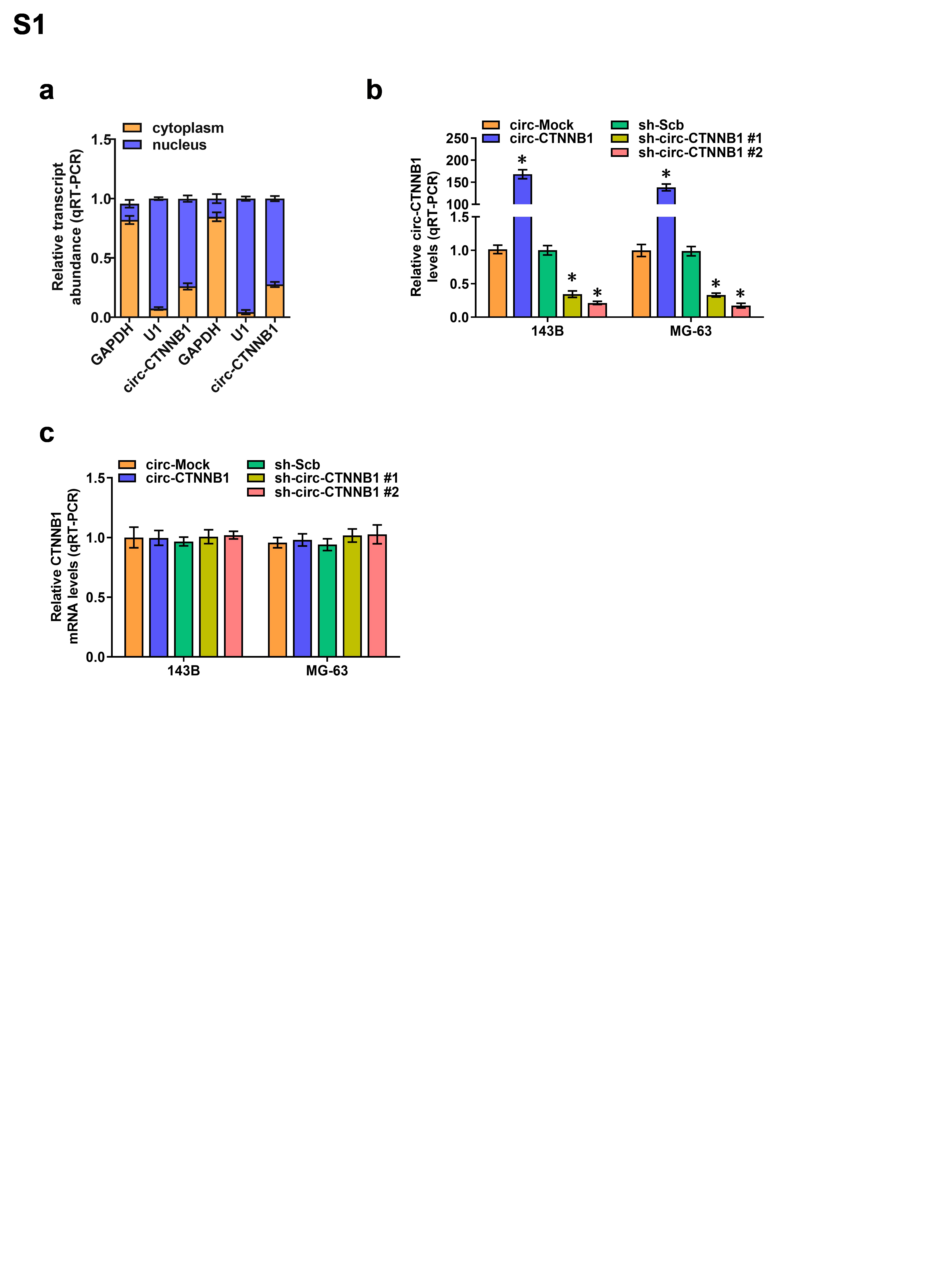
Mature linear circ-CTNNB1 (hsa_circ_0123778) was synthesized by TsingKe Biotech Company (Wuhan, China) and inserted into pLCDH-ciR vector (Geenseed Biotech, China), which contained a front circular frame and a back circular frame. Human RBM15 cDNA (2934 bp) was synthesized by TsingKe Biotech Company (Wuhan, China), and the truncations of RBM15 were obtained by PCR amplification with differential primer pairs (Additional file 1: Table S2) and subcloned into pCMV-3Tag-1A (Addgene, Cambridge, USA). Oligonucleotides specific for shRNAs against circ-CTNNB1 or RBM15 (Additional file 1: Table S2) were inserted into GV298 (Genechem Co., Ltd., Shanghai, China). Lentiviral plasmids were co-transfected with the packaging plasmids psPAX2 and pMD2G into HEK-293T cells. Infectious lentiviruses harvested from cultured cells were processed by ultracentrifugation (2 h at 120,000 g). Stable cell lines were obtained, followed by selection with 2 μg/mL puromycin for 2-3 weeks.
RNA fluorescence in situ hybridization (RNA-FISH)
A biotin-labeled antisense probe for the circ-CTNNB1 junction sequence and probes for GAPDH and U1 were synthesized as we previously described (16). The probes were hybridized using the Fluorescent In Situ Hybridization kit (RiboBio) following the manufacturer’s instructions. The nuclei of OS cells were counterstained with 4',6-diamidino-2-phenylindole (DAPI), and the images were analyzed using a Nikon A1Si Laser Scanning Confocal Microscope (Nikon, Japan).
Dual-luciferase reporter assay
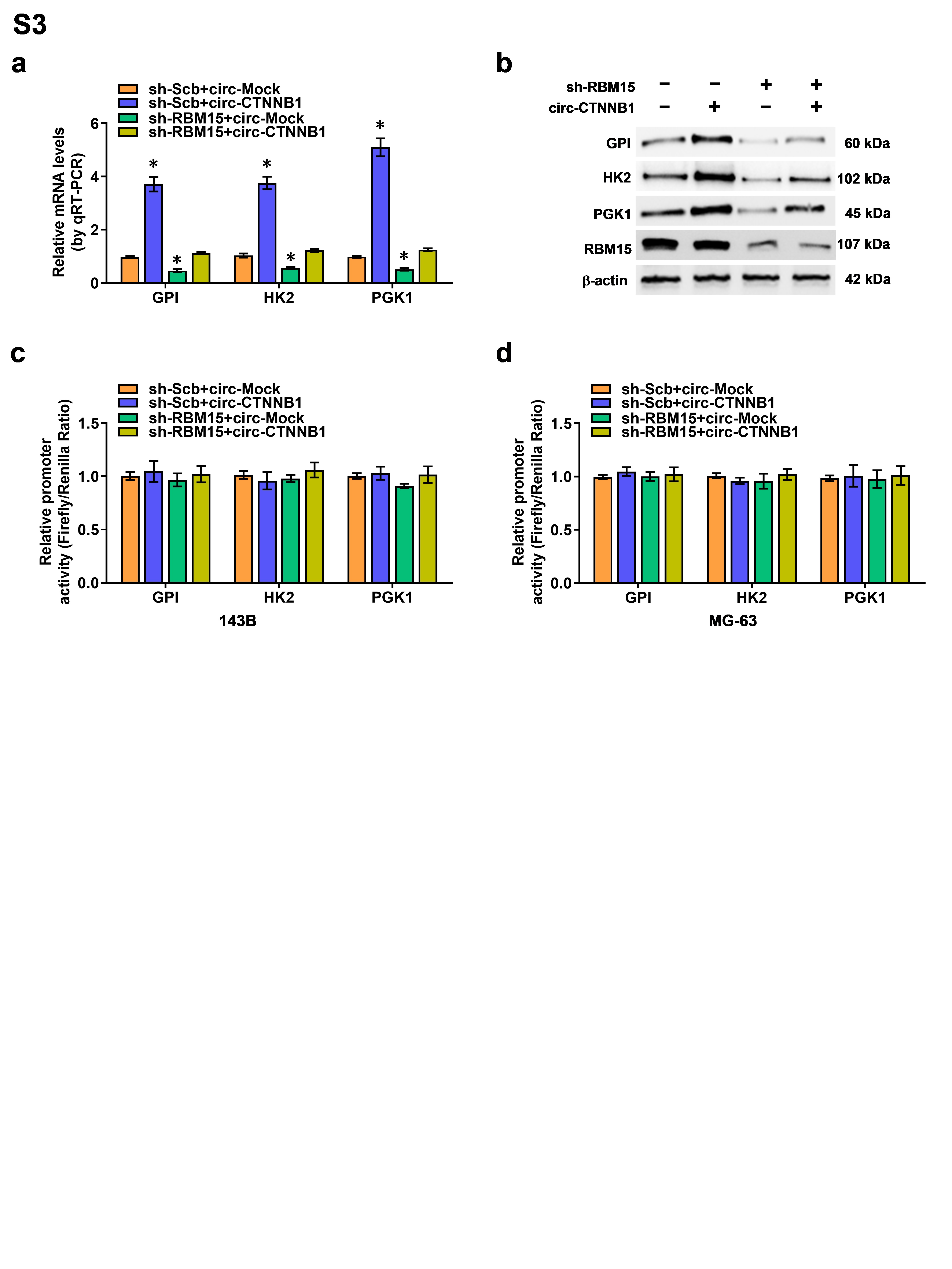
The TOP-FLASH and FOP-FLASH reporters for the activity of the canonical Wnt pathway were obtained from Millipore (Temecula, CA, USA). The promoter fragments of human HK2 (-1813/+424), GPI (-1,854/+247), PGK1 (-882/+246), and 3'-UTR of target genes amplified from genomic DNA (Additional file 1: Table S2) were subcloned into pGL3-basic and psiCHECK2. Mutations of m6A sites at the 3'-UTR were performed with the GeneTailorTM Site-Directed Mutagenesis System (Invitrogen). The dual-luciferase assay was performed according to the manufacturer’s instructions (Promega). The luciferase signal in the promoter activity assay was normalized to the firefly/Renilla ratio, while the activity of the 3'-UTR reporter was measured by the Renilla/firefly ratio.
Biotin-labeled RNA pull down and mass spectrometry analysis
The biotin-labeled RNA probe for circ-CTNNB1 was in vitro transcribed using the Biotin RNA Labeling Mix kit (Roche) and T7 RNA polymerase, as described in our previous study (22). RNA pull-down assay was performed at room temperature, and the biotinylated proteins were detected by mass spectrometry at the Wuhan Institute of Biotechnology (Wuhan, China).
Fluorescence immunocytochemical staining
OS cells were grown on coverslips, incubated with 5% milk for 1 h, and treated with antibody specific for RBM15 (ab244374, Abcam, USA) at 4 °C overnight. Then, the cells were stained with Alexa Fluor 594 IgG and DAPI. The images were photographed under a Nikon A1Si Laser Scanning Confocal Microscope (Niko, Japan).
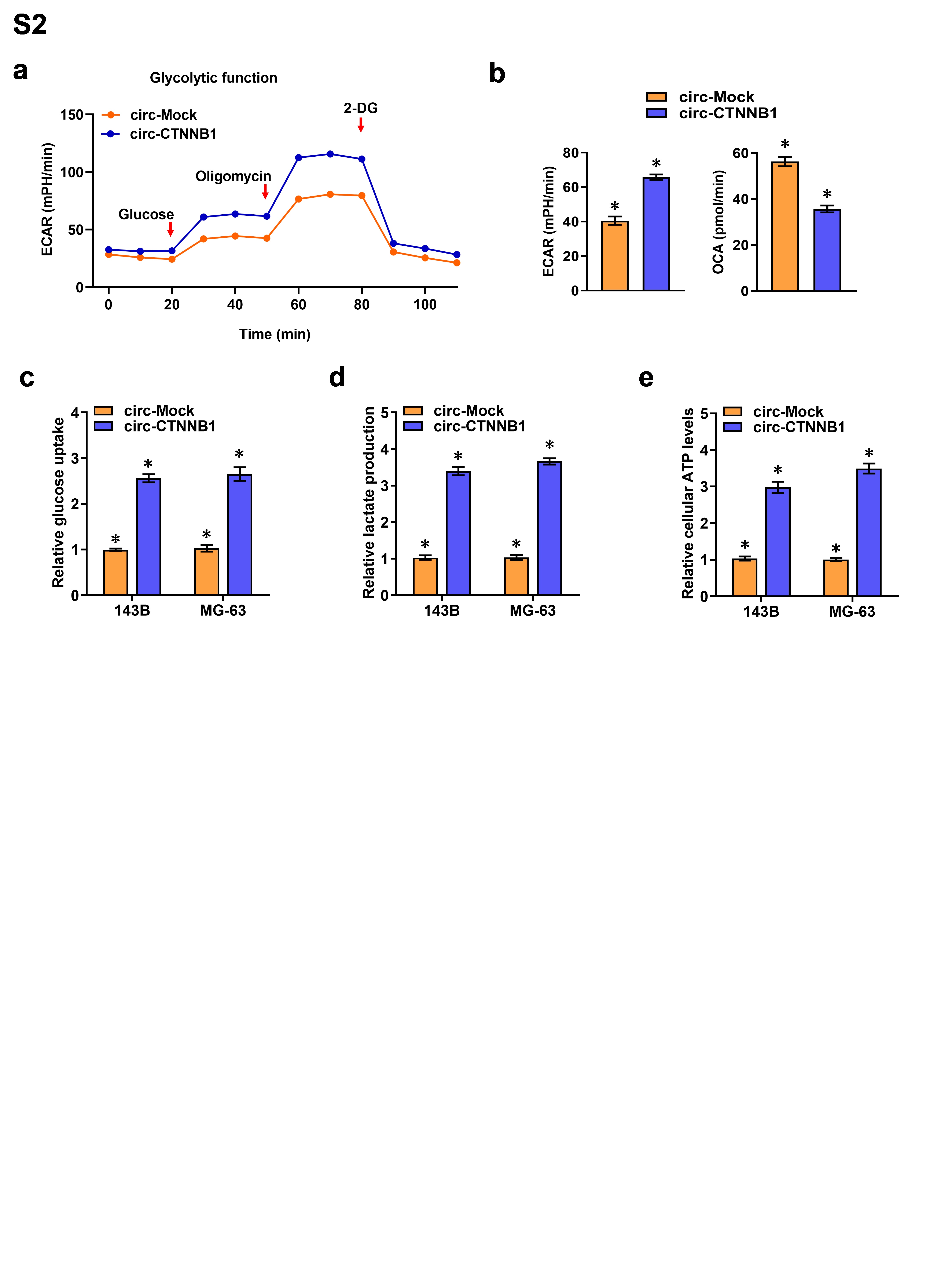
Aerobic glycolysis and seahorse extracellular flux assays
Cellular Aerobic glycolysis activity and glucose uptake, lactate production, and adenosine triphosphate (ATP) levels were detected as previously described (22). Extracellular acidification rate and oxygen consumption rate (ECAR, OCR) were measured in response to glucose (10 mM), oligomycin (2 uM), and 2-deoxyglucose (2-DG, 100 mM) under basal conditions with a Seahorse Biosciences XFe24 Flux Analyzer (North Billerica, MA).
Cross-linking RIP assay
Cells were ultraviolet light cross-linked at 254 nm (200 J/cm2) in PBS and collected by scraping, and RNA immunoprecipitation (RIP) assay was performed according to the instructions of Magna RIPTM Kit (Millipore), with antibodies specific for RBM15 (ab244374, Abcam, USA), and IGF2BP1 (sc-166344, Santa Cruz Biotechnology, USA). Co-precipitated RNAs were detected by RT-PCR or real-time quantitative PCR with specific primers (Additional file 1: Table S1).
In vitro binding assay
Five truncates of RBM15 were cloned with primers (Additional file 1: Table S1) into vectors with flag tag as we described previously (23). The Flag-RBM15 and circ-CTNNB1 complexes were pulled down using Flag beads (Sigma, USA). Circ-CTNNB1 was measured by RT-PCR with divergent primers (Additional file 1: Table S1), and protein was validated by western blot.
RNA electrophoretic mobility shift assay (EMSA)
Biotin-labeled circ-CTNNB1 probe was prepared as described above. RNA EMSA was conducted according to the instructions of LightShift Chemiluminescent RNA EMSA Kit (Thermo Fisher Scientific, Inc.)
In vitro cell viability, growth, and invasion assays
The in vitro viability, growth, and invasion capabilities of OS cells were detected by MTT colorimetry, colony formation, and matrigel invasion assays, as described previously (24).
Xenografts in mice
Circ-CTNNB1 knockdown frame was inserted into GV298 vector (Genechem Co., Ltd., Shanghai, China), and the expression of the mCherry fluorescent protein was detected and imaged by the In-Vivo Xtreme II small animal imaging system (Bruker Corporation, Billerica, MA, USA). For in vivo tumor growth studies, MG-63 cells (1×10⁷) were subcutaneously injected into the dorsal flanks of five-week-old male BALB/c nude mice (n=5 per group) in a blind, randomized fashion. The growth and weight of xenografts were detected one month later. In experimental metastasis studies, tail vein injection of MG-63 (5×10⁶) cells was performed in a blind, randomized fashion in five-week-old male BALB/c nude mice (n=5 per group). Metastasis counts and survival time of each mouse were monitored and recorded, and the xenografts were studied by hematoxylin and eosin (H&E) and IHC staining. All animal experiments were performed in accordance with the NIH Guidelines for the Care and Use of Laboratory Animals and approved by the Animal Care Committee of Tongji Medical College.
Statistical Analysis
All data are presented as the mean ± standard error of the mean (SEM) processed by GraphPad Prism 5.0 (La Jolla, USA). Student’s t-test or one-way analysis of variance (one-way ANOVA) was used to evaluate differences between groups. All statistical tests were two sided. A value of P < 0.05 was considered statistically significant.